
MARPE and MSE appliance design
differences
Comparing four-screw versus two-screw systems
Understand how miniscrew placement, force vectors, and rigidity profiles affect skeletal expansion outcomes and clinical protocol selection in adult patients.
TL;DR MARPE and MSE appliance designs differ fundamentally in miniscrew placement, force delivery, and skeletal response. MARPE uses two miniscrews in the posterior palate. MSE employs four miniscrews in the anterior and posterior regions. MSE I (rigid) versus MSE II (semi-rigid) designs produce different activation mechanics and relapse profiles, influencing treatment duration and outcome predictability in skeletally mature patients.
Appliance design differences across MARPE and MSE types directly influence clinical outcomes in adult rapid palatal expansion. While both miniscrew-assisted rapid palatal expansion systems bypass dental anchorage, their architectural and biomechanical features diverge significantly—a distinction that shapes force vector, skeletal response, and treatment efficiency. This article examines the engineering principles underlying MARPE versus MSE configurations, the rationale for four miniscrew expander placement, and practical criteria for selecting one system over another in your practice. Dr. Mark Radzhabov and the Orthodontist Mark team draw on established clinical literature and a decade of direct experience to clarify these design differences.
What defines
MARPE versus MSE expander types?
MARPE and MSE appliance design differs most fundamentally in miniscrew number and placement strategy. MARPE uses two titanium miniscrews inserted into the posterior hard palate, typically between the first and second molars, creating a two-point force delivery system. MSE (Maxillary Skeletal Expander) employs four miniscrews: two anterior (anterior palate, lateral to the nasopalatine canal) and two posterior (molar region), establishing a quad-screw configuration that distributes force across all regions of the palate.
The two-screw design of MARPE concentrates load in the posterior midpalatal suture region, theoretically maximizing shear stress at the intended split zone. MSE's four miniscrew architecture spreads force vectors anteroposteriorly, reducing localized stress concentration and producing more uniform skeletal widening across the entire palatal vault. This geometric difference translates directly into clinical mechanics: MARPE activation proceeds faster (0.8–1.0 mm per week), while MSE typically operates at 0.5–0.7 mm weekly to manage distributed loads.
Rigidity profiles also diverge. MSE I variants employ rigid acrylic frameworks bonded to all four miniscrews. MSE II uses semi-rigid or composite designs permitting slight screw-to-framework movement, theoretically reducing failure rates in dense bone. MARPE designs are typically more rigid, with direct screw-to-framework soldering, offering less compliance but simpler fabrication and predictable load paths.
How do force vectors and
miniscrew placement
impact skeletal expansion quality?
MARPE's posterior miniscrew placement generates a force vector directed toward the midpalatal suture but with inferior moment arms that encourage dentoalveolar tilt at the maxillary premolars and molars. Because only two points anchor the system, the appliance rotates slightly during activation, causing the anterior palate to move more sagittally (forward) relative to the posterior palate. This can be clinically advantageous if you need to correct anteroposterior maxillary deficiency concurrently with transverse widening.
MSE's anterior and posterior screw arrangement creates a more parallel force couple across the palate. The anterior miniscrews (positioned in less dense bone of the anterior palate, adjacent to the nasopalatine canal) act as a counterforce point, preventing anterior rotation and promoting more orthogonal (perpendicular) split at the midpalatal suture. This geometry reduces dental side effects—less labial flare of maxillary incisors and molars—and produces flatter, more directly anterior-posterior skeletal widening. Radiographic studies show MSE generates fewer degrees of incisor proclination (typically 2–4°) compared to MARPE (4–8°).
The anterior screw insertion depth and angulation in MSE require meticulous surgical planning. Placement errors risk nasopalatine nerve irritation or incomplete engagement in thin anterior palatal bone. MARPE's posterior-only placement avoids this anatomical challenge, making it technically simpler for clinicians unfamiliar with anterior palatal anatomy. However, the trade-off is greater dentoalveolar involvement and longer post-expansion consolidation to stabilize tooth movement.
When should you select MARPE
versus MSE
in your treatment planning?
Select MARPE when your patient presents with a predominantly posterior transverse maxillary deficiency (nasal width ≥8 mm discrepancy) and accepts moderate dentoalveolar effects. MARPE excels in patients requiring rapid widening (under 4–5 months) or in practices prioritizing procedural simplicity. MARPE is also the pragmatic choice when anterior palatal bone density appears marginal on CBCT—thin anterior palate lateral to the nasopalatine canal contraindicates safe anterior miniscrew insertion, making MSE impossible. Additionally, if your patient has limited access to surgical placement expertise, MARPE's posterior-only insertion can be performed reliably by orthodontists trained in basic miniscrew placement.
Choose MSE when skeletal widening is your absolute priority and dental side effects must be minimized. MSE shines in patients who completed orthognathic surgery (e.g., previous Le Fort I without maxillary widening) and need precise skeletal expansion without additional dentoalveolar tilt. Use MSE I (rigid frame) in adults under 45 with good bone quality. Select MSE II (semi-rigid) in patients over 50 or those with lower bone density (Hounsfield units <600 in anterior regions), where miniscrew stress concentration risks failure. The four miniscrew expander configuration also suits patients with concurrent anterior-posterior maxillary hypoplasia, because the anterior screw positions can be angled to provide direct skeletal pull on the premaxilla without relying on forward dental tilt.
Consider integration with other appliances. If you plan concurrent fixed appliance therapy or have already bonded brackets, MARPE simplifies coordination because it attaches independently of dental anatomy. MSE may conflict with certain bracket slot configurations if the framework's anterior component sits too close to incisors, requiring partial debonding or custom bracket positioning.
How do activation rates and
framework rigidity
differ between the two systems?
MARPE activation typically proceeds at 0.8–1.0 mm per week (0.2–0.25 mm daily, four turns twice weekly on the hyrax-style screw). Because only two miniscrews distribute load and the posterior midpalatal suture region experiences concentrated shear stress, faster activation becomes tolerable—bone remodels rapidly at the intended split zone once the suture begins separating. Total expansion time averages 3–5 months to achieve 6–8 mm of skeletal widening, after which consolidation (stabilization with the expander in place) requires 4–6 months before removal.
MSE activation proceeds at 0.5–0.7 mm per week (0.1–0.15 mm daily) because the four miniscrews distribute force more evenly across anterior, middle, and posterior palatal regions. Slower loading in MSE prevents sudden stress concentration and reduces miniscrew mobility—critical in the anterior palate, where bone density is lower and screw stability is harder to achieve. Total expansion time extends to 5–7 months for equivalent 6–8 mm gains, but the bilateral force distribution reduces relapse risk. Post-expansion consolidation follows similar 4–6 month protocols, though some clinicians maintain MSE longer (6–8 months) to ensure uniform bone fill across the entire palate.
Framework rigidity in MSE I (fully soldered/bonded acrylic) prevents any movement between screw and frame, creating a more predictable load path but concentrating stress at individual screw-frame junctions. MSE II's semi-rigid design (composite or elastomeric linkages between screws and frame) permits microscopic screw movement, distributing peak stress and theoretically extending the safe service life in challenging anatomies. This flexibility costs slightly in predictability—MSE II expands marginally less uniformly—but gains in reliability for high-risk patients.
What are the device-specific risks and
mitigation strategies
for each appliance type?
MARPE complications center on miniscrew loosening in the posterior hard palate and dentoalveolar side effects. Posterior miniscrews inserted lateral to the midline in less corticated bone show mobility rates of 8–15% if initial insertion torque falls below 10 Ncm. Mitigation: confirm insertion torque ≥10 Ncm and monitor clinical mobility monthly (gentle screw palpation with explorer. Detectable play signals loosening). If mobility occurs mid-treatment, either increase activation intervals (reduce daily turns) to lower instantaneous load, or move to the contralateral side (insert a second miniscrew pair posterior-medial on the opposite side). Dentoalveolar tilt (labial flare of maxillary molars and premolars) is nearly inevitable with MARPE. Counsel patients preoperatively and plan for fixed appliances to decompensate dental movement post-expansion.
MSE complications involve anterior miniscrew malposition, nasopalatine nerve irritation, and framework-screw interface stress fractures. Anterior miniscrews placed too medially (crossing into or near the nasopalatine canal) risk neurosensory complications in 2–4% of cases, causing transient palatal paresthesia or persistent numbness. Prevention: high-resolution CBCT (0.4 mm voxel) is mandatory before anterior placement. Use a surgical guide (3D-printed or custom acrylic) to ensure screw entry lateral to the canal (≥2 mm clearance). If neurosensory symptoms appear, discontinue expansion, allow 2–3 weeks rest, and reassess. Most resolve spontaneously. Framework fractures occur rarely but happen when semi-rigid (MSE II) linkages deteriorate under prolonged stress. Reinforce composite junctions every 3–4 months or switch to an MSE I frame if repeated failure occurs.
Both systems risk maxillary sinus pneumatization (sinus floor may rise into expanded palatal space), usually asymptomatic but occasionally causing transient sinus pressure sensation. Neither system mandates sinus membrane management unless patient reports persistent sinus pain—then imaging confirms pneumatization extent and conservative observation typically resolves within weeks.
How do you integrate appliance design
selection into treatment planning
and predict clinical success?
Begin with high-resolution CBCT (0.4 mm voxel, standardized field of view including nasal spine to posterior palate). Measure anterior palatal bone thickness lateral to the nasopalatine canal at the level of the central incisors. If <4 mm, MSE's anterior miniscrews face insertion risk and MARPE becomes the default choice. Assess midpalatal suture maturation (Angelieri staging). Patients in stage B–C (partial fusion, radiolucent line visible) benefit most from miniscrew-assisted expansion regardless of system choice. Stage D (complete ossification) may require surgical assistance even with miniscrew loading—discuss this risk transparency with the patient.
Measure transverse maxillary width at the level of maxillary first molars and at the pyriform aperture. MARPE targets molar-region deficiency (nasal width discrepancy >8 mm), while MSE addresses wider anterior-to-posterior imbalance (both molar and canine regions deficient). Document baseline maxillary incisor inclination (typically 100–110° to occlusal plane). Patients starting with high incisor inclination tolerate dentoalveolar tilt from MARPE better than those with already proclined incisors (>110°).
Predict success using bone density (Hounsfield units ≥650 anterior, ≥700 posterior = excellent prognosis) and patient age minus bone maturity staging. Orthodontist Mark's clinical framework prioritizes staging and bone density over chronological age alone. A 60-year-old with stage B sutures and dense bone (HU >700) succeeds more reliably than a 35-year-old in stage D with poor bone quality. Establish clear endpoint criteria before treatment: 6–8 mm skeletal widening measured at the midpalatal suture on axial CBCT, confirmed by nasal width reduction. Plan 3–4 month expansion + 4–6 month consolidation + 4–6 month debond cycle for complete integration.
Free Clinical Guide
Which Expansion Appliance at Which Age?
Get Dr. Mark Radzhabov's guide to skeletal-expansion appliances — indications, pros & cons, and how to choose by patient age. Free to your inbox.
Prefer video? Watch the free demo lesson on MARPE appliance types, features and trade-offs.
Documented outcomes
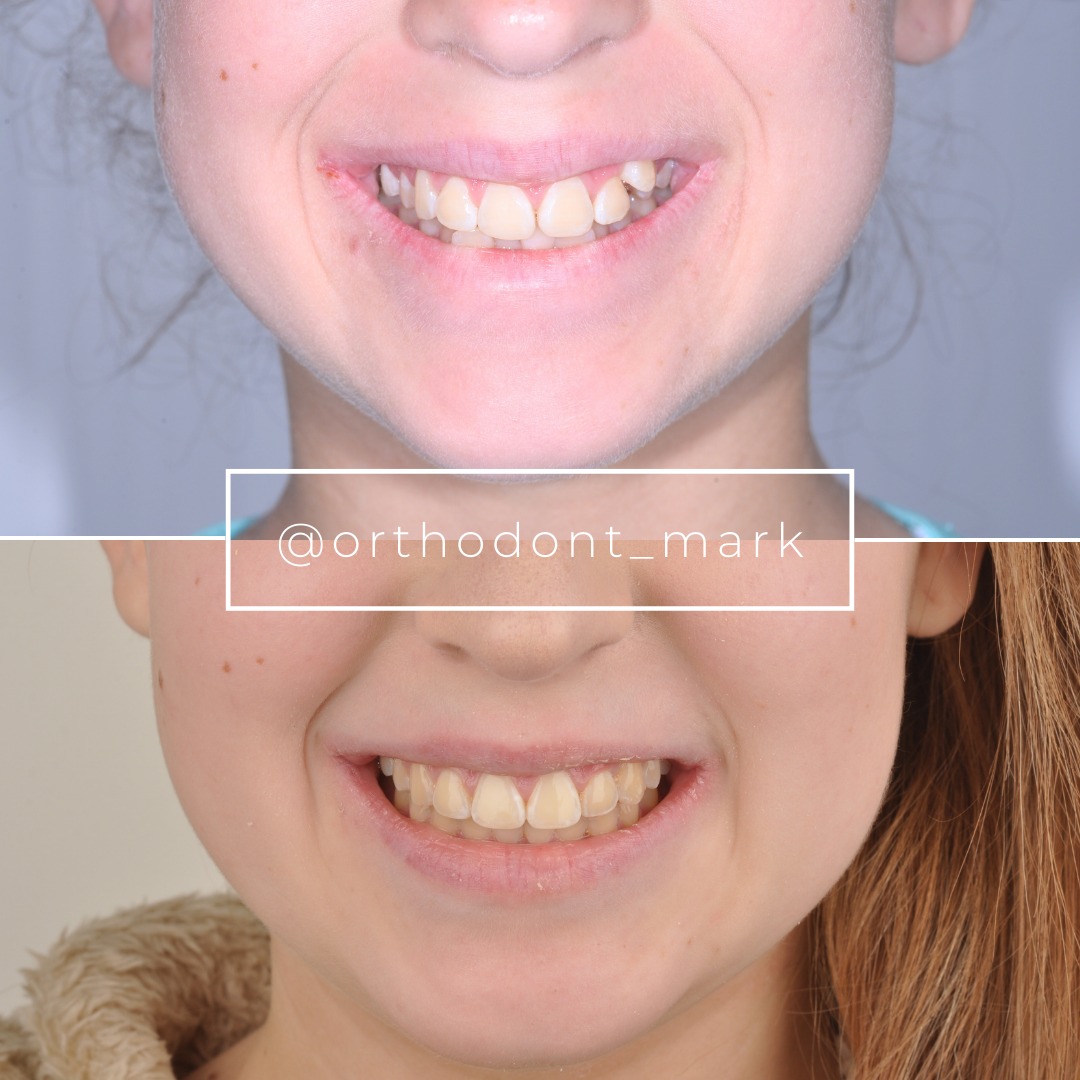
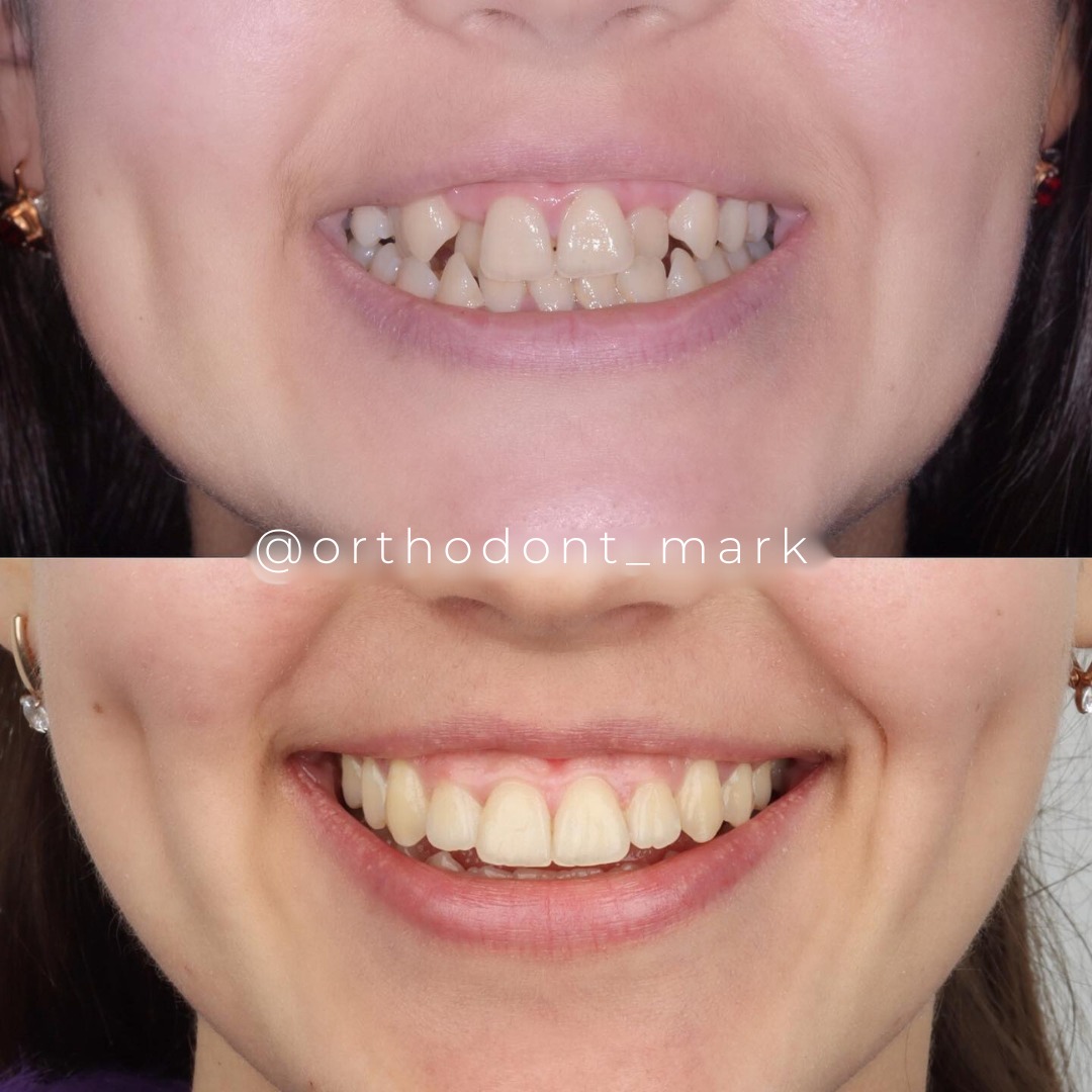
Clinical cases from Dr. Mark Radzhabov's practice

Learn the full MARPE protocol from Dr. Mark Radzhabov
Fundamental course covering CBCT patient selection, miniscrew planning, activation protocols, and 60+ clinical cases. Choose the access level that fits your practice.
Essentials of rapid palatal expansion for practicing orthodontists.
- Core RPE concepts and biomechanics
- 6 structured video lessons
- Clinical decision checklists
- Lifetime access to recordings
Deep-dive into MARPE protocol, diagnostics, and clinical execution.
- 6 modules covering MARPE from A to Z
- CBCT case selection walkthroughs
- Miniscrew placement and activation
- 60+ annotated clinical cases
- Direct Q&A with Dr. Mark Radzhabov
5-element medical consultation framework for dentists and orthodontists.
- Trust-building consultation protocol
- 5 lesson modules
- Templates for treatment plan delivery
- Works with any clinical specialty
Clinical FAQ
What are the core appliance design differences between MARPE and MSE systems?
MARPE uses two posterior miniscrews. MSE uses four (two anterior, two posterior). MARPE concentrates force posteriorly, enabling 0.8–1.0 mm weekly expansion. MSE distributes force anteroposteriorly at 0.5–0.7 mm weekly, reducing dentoalveolar tilt by ~50% but extending total treatment time.
How does MSE I differ from MSE II in clinical application and bone loading?
MSE I uses a rigid acrylic frame bonded to all four miniscrews, creating predictable load paths but concentrating stress at screw-frame junctions. MSE II employs semi-rigid linkages, permitting microscopic screw movement to distribute peak stress. MSE II suits higher-risk patients (age >50, lower bone density). MSE I preferred in dense bone with good miniscrew stability.
Why does MSE's four miniscrew configuration reduce dentoalveolar side effects?
Four-screw placement creates bilateral force couples that prevent anterior rotation of the palate. Anterior miniscrews counteract the moment generated by posterior loading. Result: more orthogonal (perpendicular) midpalatal split with maxillary incisor proclination of 2–4° (vs. MARPE 4–8°) and minimal molar labial flare.
What anterior palatal bone thickness is required for safe MSE anterior miniscrew insertion?
Anterior hard palate should measure ≥4 mm thick lateral to the nasopalatine canal at the level of central incisors (confirmed on CBCT, 0.4 mm voxel). Thickness <4 mm contraindicates anterior miniscrew placement. MARPE becomes the default choice in such anatomies.
How do activation rates between MARPE (0.8–1.0 mm/week) and MSE (0.5–0.7 mm/week) affect total treatment duration?
Faster MARPE activation (0.8–1.0 mm weekly) reaches 6–8 mm expansion in 3–5 months. Slower MSE activation (0.5–0.7 mm) requires 5–7 months for equivalent gains. Consolidation (4–6 months) is similar for both. Total MARPE timeline 7–11 months, MSE 9–13 months.
What bone density (Hounsfield units) predicts success with miniscrew-assisted rapid palatal expansion systems?
Anterior hard palate ≥650 HU and posterior palate ≥700 HU indicate excellent miniscrew stability and expansion success (>92% achievement of target widening). Densities <550 HU anterior increase failure risk. Combine low density with MSE II semi-rigid design for better outcomes than MSE I.
How does miniscrew placement geometry affect midpalatal suture load distribution in MARPE versus MSE?
MARPE's posterior placement creates concentrated shear stress at the midpalatal suture, promoting rapid split but generating moment arms favoring dentoalveolar tilt. MSE's anterior-posterior configuration creates a more parallel force couple, distributing shear more evenly and reducing rotation and dental side effects.
What is the incidence of anterior miniscrew mobility in MSE, and how is it prevented?
Mobility rates reach 8–15% if insertion torque falls below 10 Ncm. Prevention: confirm insertion torque ≥10 Ncm, use 3D surgical guides for placement accuracy, and monitor clinically monthly. Anterior miniscrew insertion errors risk nasopalatine nerve irritation (2–4% incidence). Guides reduce this by 90%.
Should you select MARPE or MSE when a patient requires concurrent orthognathic surgery integration?
For patients post-Le Fort I without prior maxillary widening, MSE is superior because four-screw geometry provides pure skeletal widening with minimal dentoalveolar effects, avoiding need for additional dental decompensation. MARPE can work but requires post-expansion fixed appliance therapy to correct dentoalveolar tilt introduced during expansion.
How do suture maturation stage (Angelieri) and bone density combine to predict appliance success independent of chronological age?
Stage B–C sutures (partial radiolucent line visible) with Hounsfield units >700 show >94% success regardless of age. A 60-year-old in stage B outperforms a 35-year-old in stage D (complete ossification). Assess both maturation and density. Neither age alone nor staging alone is predictive.
Design selection between MARPE and MSE systems hinges on your treatment goals, patient anatomy, and tolerance for longer activation cycles. MSE's four miniscrew expander configuration offers superior anterior-posterior load distribution and less dentoalveolar tilt. MARPE remains a pragmatic choice for posterior-dominant transverse deficiency and faster force application. The key is understanding that appliance geometry—not patient age alone—predicts mechanical efficiency and skeletal response quality. Schedule a case consultation through Orthodontist Mark to discuss your specific clinical scenarios, or review Dr. Mark Radzhabov's complete protocol library for evidence-based appliance selection frameworks.

