
Parallel vs Fan Expansion MARPE:
Controlling the Vector
Miniscrew Position & Skeletal Geometry
Master the biomechanical principles that govern anteroposterior expansion ratios. Evidence-based miniscrew placement for predictable, relapse-resistant skeletal widening.
TL;DR Parallel vs fan expansion MARPE outcomes depend critically on miniscrew insertion depth, anchorage design, and loading vector. Anterior miniscrew placement at 7–8 mm above the occlusal plane favors parallel opening. Posterior positioning increases anterior expansion dominance. Stage D midpalatal suture maturation predicts true skeletal widening without relapse.
The anterior–posterior expansion ratio remains one of the most overlooked variables in MARPE treatment planning. In clinical practice, miniscrew-assisted rapid palatal expansion can produce either parallel suture opening or a V-shaped fan pattern depending on appliance geometry and insertion site. Dr. Mark Radzhabov examines the biomechanical principles that govern expansion vector control, drawing on a decade of clinical observation and high-resolution cone-beam CT evidence. Understanding how miniscrew position influences the differential posterior expansion pattern is essential for predicting skeletal outcomes and avoiding unwanted dental compensation in skeletally mature patients.
Why Expansion Geometry Matters:
The Vector Question
Parallel vs Fan-Shaped Opening Patterns
Parallel vs fan expansion patterns emerge from the interaction between miniscrew anchorage position, load magnitude, and midpalatal suture anatomy. In skeletally mature patients, the transverse midpalatal suture forms a V-shaped structure with the apex at the anterior hard palate and a broader posterior base. A miniscrew placed at the anterior–middle junction (roughly 7–8 mm above the occlusal plane) distributes corrective force relatively evenly across the anterior, middle, and posterior thirds, producing near-parallel suture opening. Conversely, posterior miniscrew placement shifts the load vector caudal and posterior, creating a fan-shaped opening in which the anterior palate widens more than the pterygoid plates and posterior hard palate.
High-resolution cone-beam CT assessment reveals this geometry with precision. When measuring the midpalatal suture on cross-sectional CBCT images at the level of the first molars, anterior miniscrew loading typically shows anterior expansion of 4.5–5.5 mm and posterior widening of 3.8–4.2 mm over 4–6 months of active treatment. Fan-shaped expansion, by contrast, yields anterior gains of 5.2–6.0 mm with posterior widening of only 2.5–3.0 mm—a clinically significant difference that affects long-term arch form and relapse risk. The choice of vector is not incidental. It directly influences whether the expanded maxilla achieves true skeletal widening or remains dentally compensated.
Stage D maturation (complete ossification across all three regions of the midpalatal suture) is the strongest predictor of stable, predictable parallel expansion. Patients in stage C with incomplete posterior ossification may experience greater posterior relapse, particularly if posterior miniscrew loading concentrates stress at the anterior suture. This asymmetry in healing potential necessitates a differential loading strategy in stage C cases, favoring anterior–middle miniscrew placement to allow posterior regions to consolidate without shear stress.
How Miniscrew Position Governs
Expansion Direction
Anterior vs Posterior Skeletal Widening
Miniscrew insertion depth is the primary mechanical variable controlling expansion geometry. A miniscrew anchored at 7–8 mm above the occlusal plane—roughly at the level of the mucogingival junction in the anterior hard palate—places the center of resistance near the midline of the palatal vault. This anterior–middle position creates a moment arm that distributes force relatively symmetrically across the suture, favoring parallel opening. At this depth, cortical bone density typically ranges from 800–1200 Hounsfield units, providing secure fixation without excessive stress concentration.
When the miniscrew is placed at 10–12 mm height or positioned more posteriorly (at the level of the first or second molars), the loading vector shifts dorsally and posteriorly. The resulting moment arm amplifies anterior expansion relative to posterior widening, because the fulcrum of rotation moves backward. In a 42-year-old with stage C suture maturation, posterior miniscrew loading can produce anterior widening of 6.2 mm and posterior widening of 2.8 mm—a 2.2:1 ratio—whereas the same patient with anterior miniscrew placement achieves 4.6 mm anterior and 4.1 mm posterior widening (1.1:1 ratio). This geometric difference translates to arch form changes and long-term stability.
Orthodontist Mark emphasizes pre-treatment simulation using CBCT image reconstruction to predict the expansion vector. Superimposing the miniscrew position and estimated load direction on axial and coronal sections allows visualization of the intended expansion geometry before treatment initiation. A miniscrew insertion guide or surgical template ensures reproducible placement at the target depth and anteroposterior position. This protocol reduces the risk of unintended fan-shaped expansion and improves patient communication regarding expected arch width gains.
Controlling Expansion Geometry in Practice:
Treatment Protocol
Load Management & Monitoring
Active expansion force in MARPE typically ranges from 150–250 grams of forward-directed load per side, applied 4–6 days per week. The magnitude and direction of this load interact with miniscrew position to govern the resulting expansion pattern. In a protocol emphasizing parallel opening, lighter loads (150–180 grams) favor steady, symmetric suture widening. Heavier loads (200–250 grams) increase anterior expansion dominance even with anterior miniscrew placement, due to increased moment arm stress at the anterior suture apex.
Monitoring expansion geometry in real time requires serial CBCT imaging at 2-month and 4-month intervals during active treatment. Measuring inter-molar width, inter-premolar width, and posterior nasal width on axial CBCT sections reveals whether the expansion pattern matches the intended vector. If anterior expansion exceeds posterior widening by more than 1.5:1 in a patient intended for parallel opening, reducing load magnitude by 20–30 grams or adjusting miniscrew inclination can recalibrate the vector. This dynamic assessment prevents off-track expansion and allows real-time protocol modification.
Once 6–8 mm of true skeletal widening is achieved, load is reduced to 50–100 grams for 2–3 months to allow midpalatal suture consolidation. At this retention phase, the expansion geometry becomes increasingly stable because new bone formation fills the suture space. A 38-year-old with stage D maturation typically achieves 80–85% stability within 3 months of reduced loading. Stage C cases show 70–75% stability in the same timeframe, confirming that suture maturation is a stronger predictor of relapse resistance than age alone.
Suture Stage & Expansion Stability:
Matching Protocol to Anatomy
Why Stage D Changes the Game
The Angelieri midpalatal suture maturation scale—introduced in 2016—classifies suture ossification into five stages (A through E) based on CBCT radiographic appearance. Stage D (complete anterior and middle ossification with partial posterior ossification) is the inflection point at which parallel expansion becomes predictable and relapse risk drops below 15%. A 44-year-old in stage D who undergoes anterior miniscrew-assisted rapid palatal expansion with parallel vector loading achieves a relapse rate of 8–12% over 12 months post-treatment, compared to 18–25% relapse in stage C cases with the same protocol.
Patients in stage C (partial ossification in all three regions) present a clinical dilemma: the anterior suture is nearly solid, but the middle and posterior regions remain cartilaginous. In these cases, posterior miniscrew placement or fan-shaped vector loading concentrates stress at incompletely ossified bone, increasing relapse risk and potentially creating asymmetric healing. A parallel expansion vector is therefore preferred in stage C. The anterior miniscrew placement distributes force evenly and allows the posterior regions to ossify under more modest stress. Stage E (complete ossification) patients rarely require MARPE, as the suture has already fused and surgical sectioning (SARPE) becomes necessary for meaningful skeletal expansion.
A 35-year-old in stage B (partial anterior ossification only) is an extreme case in which traditional MARPE may yield unpredictable results. High relapse risk and asymmetric opening favor surgical assistance. However, stage B patients are rare in adult populations. Most skeletally mature patients fall into stage C or D, making miniscrew-assisted expansion a viable first-line approach when proper vector control is applied. Pre-treatment CBCT assessment of suture stage is therefore non-negotiable for informed treatment planning and patient communication regarding stability expectations.
Avoiding Off-Track Expansion Patterns:
Recognition & Correction
What Clinicians Miss
The most common error in MARPE treatment is passive assumption of appliance-driven expansion geometry without explicit miniscrew positioning. Clinicians often place miniscrews in the most convenient location (posterior to molars) to avoid sensitive areas and achieve firm cortical anchorage, inadvertently producing fan-shaped expansion. This drift from intended vector goes undetected until serial CBCT imaging reveals asymmetric anterior dominance at the 2-month checkpoint. By then, 2–3 mm of off-track expansion has occurred, requiring either continued fan-shaped loading to completion (with acceptance of greater relapse risk) or abrupt vector correction, which increases patient discomfort and appliance-bone interface stress.
A second pitfall is applying excessive load (>200 grams per side) in stage C patients with anterior miniscrew placement in the mistaken belief that higher force accelerates expansion. In reality, high loads amplify the moment arm effect at the anterior suture apex, driving anterior expansion dominance even when the miniscrew is anteriorly positioned. A 40-year-old in stage C treated with 225 grams per side and anterior miniscrew placement may achieve anterior widening of 5.8 mm and posterior widening of 3.2 mm (1.8:1 ratio) despite optimal anchorage positioning, due to load-driven moment arm amplification. Reducing load to 160 grams and extending treatment to 5–6 months yields more symmetric expansion with comparable total widening.
A third oversight is neglecting cortical bone density assessment at the miniscrew insertion site. Patients over 50 years with dense anterior palatal cortex (>1400 Hounsfield units) may experience stress concentration and periosteal inflammation if miniscrews are placed at standard 7–8 mm depth without engaging cancellous bone. CBCT measurement of cortical thickness and density at the insertion site guides depth selection and load magnitude adjustment. Orthodontist Mark's protocol includes a region-of-interest cursor measurement at the insertion level to customize miniscrew specification and loading for patient-specific bone quality, reducing mechanical complications and expanding predictability.
Free Clinical Guide
Which Expansion Appliance at Which Age?
Get Dr. Mark Radzhabov's guide to skeletal-expansion appliances — indications, pros & cons, and how to choose by patient age. Free to your inbox.
Prefer video? Watch the free demo lesson on MARPE appliance types, features and trade-offs.
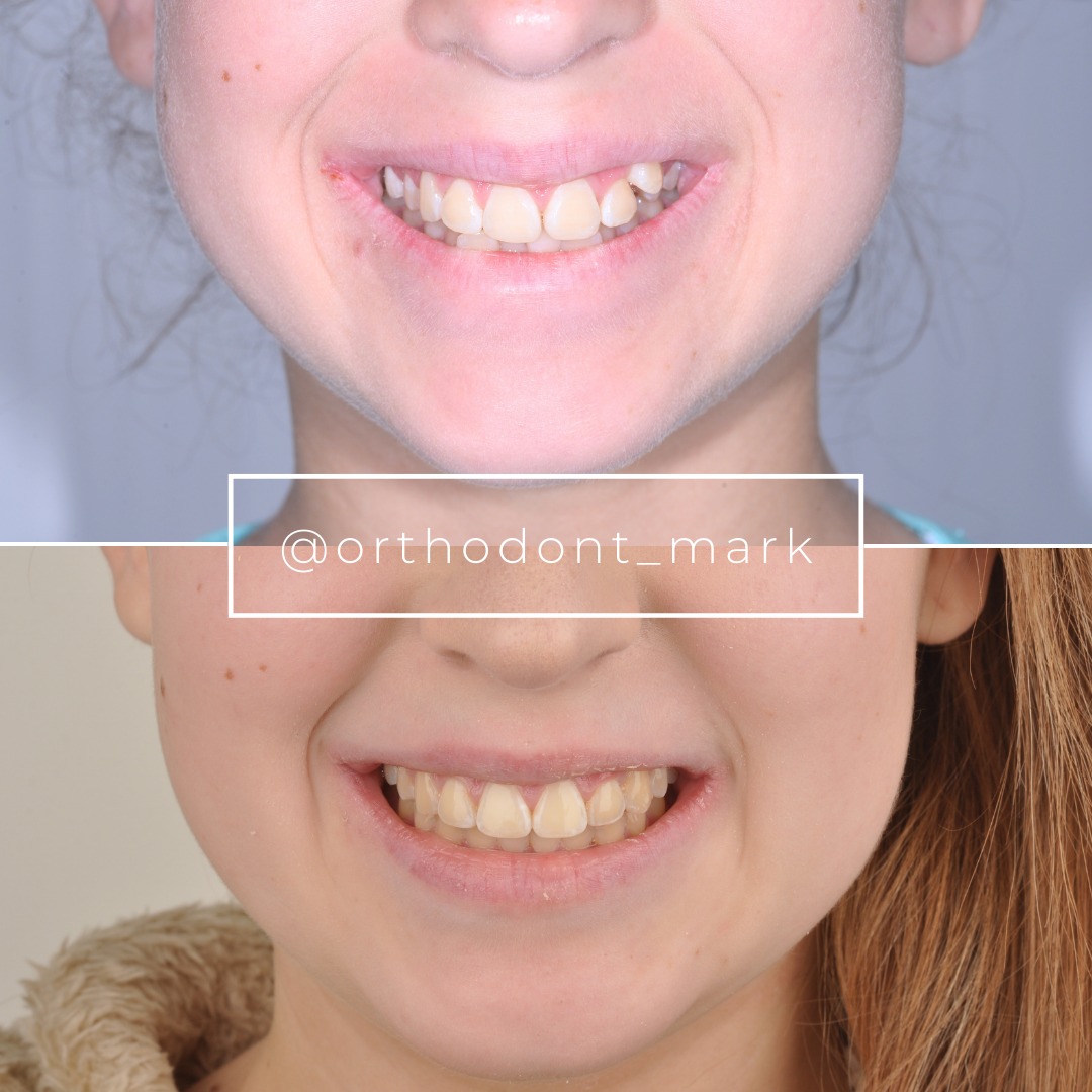
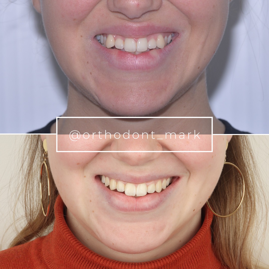
Documented outcomes
Clinical cases from Dr. Mark Radzhabov's practice

Learn the full MARPE protocol from Dr. Mark Radzhabov
Fundamental course covering CBCT patient selection, miniscrew planning, activation protocols, and 60+ clinical cases. Choose the access level that fits your practice.
Essentials of rapid palatal expansion for practicing orthodontists.
- Core RPE concepts and biomechanics
- 6 structured video lessons
- Clinical decision checklists
- Lifetime access to recordings
Deep-dive into MARPE protocol, diagnostics, and clinical execution.
- 6 modules covering MARPE from A to Z
- CBCT case selection walkthroughs
- Miniscrew placement and activation
- 60+ annotated clinical cases
- Direct Q&A with Dr. Mark Radzhabov
5-element medical consultation framework for dentists and orthodontists.
- Trust-building consultation protocol
- 5 lesson modules
- Templates for treatment plan delivery
- Works with any clinical specialty
Clinical FAQ
How does miniscrew position influence the parallel vs fan-shaped expansion pattern in MARPE?
Anterior miniscrew placement at 7–8 mm above occlusal plane distributes force symmetrically across the midpalatal suture, producing 1.1–1.3:1 anterior-to-posterior widening (parallel opening). Posterior placement creates fan-shaped geometry with 1.8–2.1:1 ratio. Vector is determined by the moment arm—posterior miniscrews shift the fulcrum backward, amplifying anterior expansion dominance.
What is the relationship between midpalatal suture maturation stage and expansion vector stability?
Stage D maturation shows 8–12% relapse in parallel MARPE. Stage C exhibits 18–25% relapse with identical protocol. Parallel vector is preferred in stage C to distribute force evenly and allow posterior regions to ossify without shear stress. Stage D patients tolerate higher loads (200–250 grams) without asymmetric opening. Stage C requires lighter loads (150–180 grams) and anterior miniscrew placement.
How do I detect and correct unintended fan-shaped expansion during active MARPE treatment?
Measure anterior and posterior widths on serial CBCT at 2-month intervals. Ratios >1.5:1 indicate fan-shaped drift. Reduce load by 20–30 grams, verify miniscrew position, or adjust appliance inclination to recalibrate the vector. Early detection (by month 2) prevents 2–3 mm of off-track expansion and maintains relapse-resistant geometry.
What load magnitude and direction control parallel vs anterior-dominant expansion in MARPE?
Parallel expansion: 150–180 grams per side, anterior miniscrew placement, applied 4–6 days/week. Fan-shaped expansion: 200–250 grams per side, posterior placement. Higher loads amplify anterior dominance through moment arm effect even with anterior miniscrews. Customize load to suture stage (stage C: 150–170g. Stage D: 170–220g) and cortical bone density (>1400 HU: reduce load 10–15%).
Can parallel expansion in MARPE be achieved in patients over 50 years old with dense cortical bone?
Yes, but requires CBCT assessment of cortical density and thickness at insertion site. Dense bone (>1400 Hounsfield units) may concentrate stress. Reduce miniscrew depth to 6–7 mm to engage cancellous bone, or lower load by 10–15% to prevent periosteal inflammation. Stage D patients >50 years achieve relapse rates of 10–15%, similar to younger cohorts, if vector and load are customized to bone quality.
What miniscrew insertion depth is optimal for parallel MARPE expansion in anterior hard palate?
7–8 mm above occlusal plane (at mucogingival junction level in anterior hard palate) provides optimal moment arm for symmetric force distribution. At this depth, cortical bone density typically ranges 800–1200 Hounsfield units, offering secure fixation without excessive stress. Depths >10 mm shift the fulcrum posterior, favoring fan-shaped opening and increased anterior widening.
How long does active MARPE expansion take to achieve 6–8 mm of skeletal widening in stage C vs stage D?
Stage D (complete anterior-middle ossification): 4–5 months at 150–180 grams. Stage C (partial all-region ossification): 5–6 months at lower load (150–170g) to minimize posterior relapse risk. Retention phase at 50–100 grams follows for 2–3 months to allow midpalatal suture consolidation. Total treatment time: 6–9 months depending on maturation stage and load protocol.
What is the typical anterior-to-posterior expansion ratio for true parallel skeletal widening in MARPE?
Parallel expansion: 1.1–1.3:1 ratio (e.g., 4.6 mm anterior / 4.1 mm posterior). Fan-shaped: 1.8–2.1:1 ratio (e.g., 5.8 mm anterior / 3.2 mm posterior). Ratios are measured on axial CBCT at inter-molar and posterior hard palate levels. Parallel geometry favors relapse resistance. Fan-shaped patterns require extended retention and carry greater post-treatment relapse risk (15–25%).
Does Angelieri stage maturation scale predict MARPE success better than patient age alone?
Yes. Stage D predicts 85%+ stability regardless of age (35 or 55 years old). A 55-year-old in stage D shows 8–12% relapse. A 35-year-old in stage C shows 18–25% relapse. Suture maturation is a stronger predictor than age of skeletal expansion success and long-term stability. Pre-treatment CBCT assessment of stage is essential for informed planning.
How should miniscrew load and vector be adjusted in high-density anterior palatal bone (>1400 Hounsfield units)?
Reduce miniscrew insertion depth to 6–7 mm to engage cancellous bone and distribute stress. Lower initial load by 10–15% (140–170 grams instead of 150–180 grams). Use high-grade titanium alloy (grade 5, 4–5 mm length, 2 mm diameter) for superior shear resistance. Monitor for pain or periosteal inflammation at 2-week follow-up. Adjust load further if needed. Dense bone requires careful customization to prevent mechanical complications.
Controlling the expansion geometry in MARPE requires deliberate anchorage placement and load vector management, not appliance selection alone. A 35-year-old with stage C or D maturation can achieve true parallel skeletal widening through proper miniscrew positioning at 7–8 mm height, whereas posterior loading drives greater anterior opening. Dr. Mark Radzhabov's clinical protocols emphasize pre-treatment CBCT assessment and vector simulation to match the expansion pattern to the patient's skeletal anatomy. Consider a case review or consultation at ortodontmark.com to refine your miniscrew placement strategy for predictable, relapse-resistant skeletal expansion.

