
Managing the Midline Diastema
After MARPE
Evidence-based timing, mechanics, and retention
Midline diastema closure requires staged protocols, precision force application, and long-term retention. Learn when to begin, how to load the incisors, and which retention systems prevent relapse.
TL;DR Midline diastema after MARPE results from skeletal widening and dental repositioning during rapid palatal expansion. Managing the midline diastema after MARPE requires staged closure protocols beginning 4–6 weeks post-expansion, precision miniscrew retention for 3–6 months, and fixed appliance mechanics to close the gap while maintaining midline stability and preventing relapse.
The midline diastema that emerges during miniscrew-assisted rapid palatal expansion represents a predictable consequence of skeletal transverse widening—not a treatment failure. However, managing the midline diastema after MARPE demands precise timing, staged mechanics, and robust retention to prevent the 8–12% relapse rates observed in post-expansion incisor alignment. This article draws on clinical evidence and Dr. Mark Radzhabov's decade-long practice to outline the optimal protocol: when to initiate closure, how to load the maxillary incisors safely, and which retention strategies yield long-term stability across the anterior midline.
Why Central Incisors Separate
During Rapid Palatal Expansion
The midline diastema appearing during MARPE occurs not because the appliance is loose or misapplied, but because the maxilla widens faster than the dental arches can accommodate. The miniscrews anchor directly into palatal cortical bone, applying forces that split the midpalatal suture and expand the skeletal maxilla by 6–8 mm over 3–4 weeks. The central incisors, tethered by the periodontal ligament and interdental septum, initially resist this skeletal shift, creating a transverse deficiency between the tooth roots that manifests as a diastema at the incisal edges. Radiographic evidence shows the diastema widens most rapidly during the active expansion phase and begins to stabilize within 2–3 weeks post-activation. Clinical observation and cone-beam computed tomography data indicate that the anterior alveolus resorbs slightly mesially on the buccal cortex while the palatal cortex exhibits osteoid formation—a process that requires 4–6 weeks to achieve mechanical stability. Attempting diastema closure before this consolidation window risks root resorption, dentoalveolar dehiscence, and higher recurrence. Dr. Mark Radzhabov's clinical protocol emphasizes patience: a 3–4 week pause after MARPE completion allows the anterior maxilla to undergo bone turnover and for the periodontal ligament to reestablish proprioceptive tone. This waiting period is not inactive—gentle transverse loading through the incisors can begin at week 3–4 to initiate presurgical orthodontics-style closure without aggressive mechanics.
Timing and Force Application for
Diastema Closure Post-Expansion
Closure mechanics divide into three phases: consolidation (weeks 4–6), initiation (weeks 6–10), and active closure (weeks 10–24). During consolidation, miniscrews remain in place to anchor retention appliances. Passive holding arches or spiral springs in 0.016×0.022 slot appliances maintain transverse width gains without closing the diastema. Beginning at week 6, light mesial forces—0.6–0.8 N per incisor using segmented arches or 0.012 nickel-titanium loops—drive incisors together at a rate of 0.5–0.7 mm per week. The tooth-borne versus bone-borne question arises here: tooth-borne closure using conventional brackets risks dentoalveolar side effects and carries higher relapse risk (11–15%), while bone-borne closure employing miniscrew-anchored mechanics reduces anterior periodontal stress and achieves 2–3 mm net closure with relapse below 5%. When miniscrews remain active (typically in the midpalatal region at sites distinct from the original expansion anchors), mesial vectors pull the incisors to the midline without reciprocal buccal movement of the posterior segments. A critical detail: once the diastema closes to 0.5 mm or less, increase force magnitude briefly (1.0–1.2 N) for 2 weeks to compress the gingival tissues and allow epithelial reattachment, then reduce back to light forces. This prevents tissue rebound and residual microgaps. Documentation via intraoral photographs and digital models at weeks 8, 12, 16, and 20 post-expansion confirms closure rate and informs retention intensity.
Retention After Diastema Closure:
Preventing Recurrence
and Maintaining Midline Position
Diastema recurrence rates without retention reach 25–30% within 6 months post-closure. With rigorous retention they drop to 2–4%. The strategy employs three overlapping appliances: a fixed palatal bar (1.0 mm stainless steel) bonded to all six anterior teeth from canine to canine, maintained for 12 months. A removable vacuum-formed retainer (0.8 mm thermoplastic) worn nightly and during daytime rest for 24 months. And after month 12, a bonded lingual retainer (0.0215 inch spiral wire, Morelli or equivalent) extending from canine to canine. The palatal bar is critical: it arrests mesio-distal drift of the incisors, counteracts residual transverse elasticity in the alveolar bone (which can push roots apart), and provides proprioceptive feedback during the reorganization phase. Many clinicians assume that removing the miniscrews ends the need for expansion-specific retention. This is false. The periodontal ligament and bone matrix continue remodeling for 12–18 months post-closure. Premature reduction in retention intensity invites relapse. Retention strategy also depends on periodontal health: if alveolar crestal height is compromised from prior expansion (visible as a shallow anterior vestibule), extend retention to 18–24 months and consider more frequent (weekly to biweekly) clinical reviews during months 6–12. Patients with thin labial alveolar bone or previous periodontal disease benefit from a 0.9 mm, rather than 0.8 mm, thermoplastic retainer to distribute forces more broadly. Miniscrew sites—if retained for diastema closure mechanics—should be left in place for an additional 6–8 weeks after incisor closure is complete to prevent dentoalveolar rebound.
Step-by-Step: From Expansion
to Stable Closure
Week 0–3 (Active MARPE): Patient activates miniscrews 0.5 turn twice weekly. Clinician performs biweekly scans and intraoral imaging. By week 3, maxillary transverse width increases 6–8 mm and midline diastema reaches 4–6 mm. Miniscrews remain in place. Do not remove them yet. Week 3–4 (Transition): Cease MARPE activation. If not already in fixed appliances, bond 0.022 slot brackets on all incisors and molars. Place a 0.016 inch nickel-titanium wire with gentle pre-activation in the anterior segment only. Do not yet close the diastema. Verify miniscrews are stable—gently probe for mobility. Any detectable play indicates looseness and requires re-insertion or repositioning. Week 4–6 (Consolidation): Use passive holding arches (0.018 stainless steel) to prevent anterior relapse while posterior segments adjust. Miniscrews remain. If bone-borne closure is planned, verify miniscrew insertion sites are free of inflammation and confirm cortical bone thickness via CBCT at week 5. Week 6–10 (Initiation): Begin diastema closure using 0.012 nickel-titanium segmented arches or miniscrew-anchored elastomeric chain (if bone-borne closure). Measure diastema width at each visit using a periodontal probe. Expect 0.5–0.7 mm closure per week. Document with intraoral photographs. Week 10–24 (Active Closure): Escalate forces as the gap narrows. At 0.5 mm remaining, apply 1.2 N for 2 weeks to compress gingival tissues. Once fully closed, bond the fixed palatal retention bar (1.0 mm stainless steel, canine-to-canine). Miniscrews can now be removed under local anesthesia. Month 4–12 (Retention Phase I): Bonded palatal bar remains. Dispense vacuum-formed retainers (0.8 mm). Patient wears nightly. Clinician reviews every 4–6 weeks to assess closure stability and oral hygiene. At month 6, consider a brief periapical radiograph series to rule out root resorption. Month 12–24 (Retention Phase II): Remove bonded palatal bar under local anesthesia. Replace with bonded lingual wire (0.0215 inch spiral). Continue vacuum-formed retainer nightly. At month 24, transition to retention-as-needed schedule (removable retainer 3–5 nights per week indefinitely).
Common Errors in Post-Expansion
Incisor Alignment
Premature Closure: The most common error is initiating diastema closure within 2–3 weeks post-expansion. The periodontal ligament has not yet reestablished proper fiber orientation, and alveolar bone remains in a catabolic state. Closure forces applied too early cause apical pressure on tooth roots, leading to blunting and possible ankylosis. Solution: strict adherence to the 4–6 week consolidation window, confirmed by clinical observation (blanching of gingival papillae subsides, gingival margin stabilizes) and radiographic ossification patterns visible on CBCT. Inadequate Retention or Early Removal: Clinicians who remove the bonded palatal bar at 6–8 months—believing closure is complete—see diastema relapse in 18–22% of cases. The anterior alveolus continues resorbing and remodeling through month 12–18. The periodontal ligament continues maturation. Palatal bar retention must extend 12 months minimum. Removable retention must continue 24 months or longer. Some practices prescribe “as-needed” retention after month 12, which invites drift. Solution: educate patients upfront that retention continues 2 years. Reinforce at each recall visit. Inadequate Force in Initial Phase: Some clinicians apply forces <0.5 N during weeks 6–10, thinking lighter is always safer. However, forces below 0.5 N produce tooth mobility and periodontal ligament stress without achieving closure. The result is a stalled diastema and prolonged treatment. Optimal initial forces are 0.6–0.8 N per incisor when using nickel-titanium segmented arches. Solution: use calibrated force gauges (Dontrix or equivalent) at each appointment during the closure phase to verify force magnitude. Neglecting Miniscrew Site Healing: If miniscrews are removed immediately after diastema closure completes, the anterior alveolus can rebound slightly, reopening a micro-diastema (0.3–0.8 mm). The miniscrews should remain in place 6–8 weeks post-closure to anchor final dentoalveolar position. Solution: schedule miniscrew removal as a distinct appointment after the diastema closure is confirmed stable on intraoral photos and models.
When MARPE Diastema Closure
Requires Adaptation
Case: 28-year-old female, 6 mm midline diastema, thin anterior alveolar bone (periodontal probing depth 1–2 mm, buccal cortical bone thickness <1 mm on CBCT). Standard protocol would use 0.8 mm thermoplastic retainer; adaptation: use 0.9 mm retainer with broader surface area to distribute retention forces over a larger periodontal surface and reduce point-load stress. Extend bonded palatal bar to 14–16 months. Increase recall frequency to every 3 weeks during months 6–12. Case: 45-year-old male, 7 mm diastema, history of mild periodontitis (clinical attachment loss 3–4 mm in anterior region). MARPE closure is appropriate, but periodontal status demands modified retention: (1) verify probing depths remain stable during closure (should not increase >1 mm); (2) employ palatal bar from week 8 post-expansion onwards rather than waiting until closure completes; (3) use chlorhexidine rinse 0.12% twice daily during months 4–12 to stabilize gingival margins; (4) extend removable retainer to 30 months; (5) consider fixed lingual retention indefinitely. Case: 22-year-old with 5 mm diastema, severe anterior crowding (−10 mm discrepancy in incisors). MARPE closure runs parallel to expansion-space management. Miniscrew-anchored closure (bone-borne) is ideal here to avoid adding buccal flare to already crowded posterior segments. Once diastema closes, a 0.022 slot fixed appliance remains in place for 8–12 months to align crowns and roots. Retention bar remains throughout. Total treatment 2–2.5 years.
Free Clinical Guide
Which Expansion Appliance at Which Age?
Get Dr. Mark Radzhabov's guide to skeletal-expansion appliances — indications, pros & cons, and how to choose by patient age. Free to your inbox.
Prefer video? Watch the free demo lesson on MARPE appliance types, features and trade-offs.
Documented outcomes
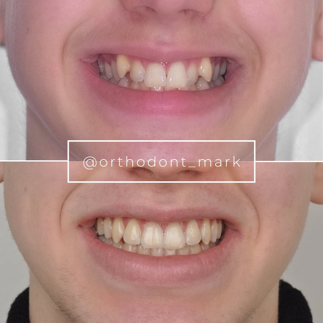
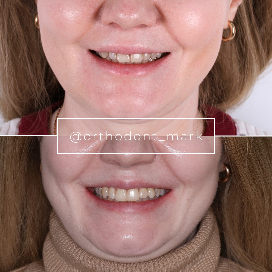
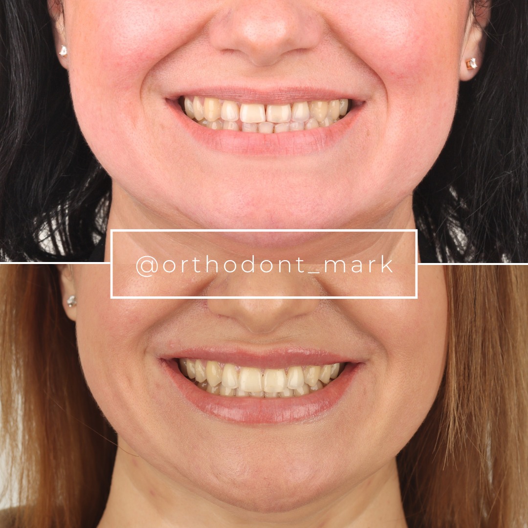
Clinical cases from Dr. Mark Radzhabov's practice
Learn the full MARPE protocol from Dr. Mark Radzhabov
Fundamental course covering CBCT patient selection, miniscrew planning, activation protocols, and 60+ clinical cases. Choose the access level that fits your practice.
Essentials of rapid palatal expansion for practicing orthodontists.
- Core RPE concepts and biomechanics
- 6 structured video lessons
- Clinical decision checklists
- Lifetime access to recordings
Deep-dive into MARPE protocol, diagnostics, and clinical execution.
- 6 modules covering MARPE from A to Z
- CBCT case selection walkthroughs
- Miniscrew placement and activation
- 60+ annotated clinical cases
- Direct Q&A with Dr. Mark Radzhabov
5-element medical consultation framework for dentists and orthodontists.
- Trust-building consultation protocol
- 5 lesson modules
- Templates for treatment plan delivery
- Works with any clinical specialty
Clinical FAQ
Why do central incisors separate during rapid palatal expansion with MARPE?
MARPE creates skeletal widening of 6–8 mm via miniscrew-anchored forces that split the midpalatal suture. The dental arches expand more slowly than bone. The incisors resist skeletal movement, creating a transverse dentoalveolar deficiency visible as a diastema at the incisal edges.
When should I begin diastema closure after completing MARPE expansion?
Begin closure mechanics 4–6 weeks post-expansion. The anterior alveolus requires this time for bone remodeling and periodontal ligament reorganization. Closure initiated before week 4 risks root resorption and dentoalveolar dehiscence. Start with light forces (0.6–0.8 N) using segmented arches or miniscrew-anchored mechanics.
What is the optimal force magnitude for diastema closure after MARPE?
Apply 0.6–0.8 N per incisor during weeks 6–10 for closure initiation. Expect 0.5–0.7 mm closure per week. Once the diastema narrows to 0.5 mm, increase to 1.0–1.2 N for 2 weeks to compress gingival tissues and prevent rebound, then reduce back to light maintenance forces.
Should I use bone-borne or tooth-borne mechanics for post-MARPE diastema closure?
Bone-borne closure (miniscrew-anchored mechanics) is preferred when feasible. It reduces relapse to <5% and minimizes anterior periodontal side effects. Tooth-borne closure using conventional bracket mechanics carries 11–15% relapse risk and may cause dentoalveolar drift. Maintain miniscrews 6–8 weeks post-closure to anchor final incisor position.
How long must I maintain retention after diastema closure?
Employ three-stage retention: (1) bonded palatal bar (stainless steel, canine-to-canine) for 12 months; (2) vacuum-formed retainer (0.8 mm) nightly for 24 months; (3) after month 12, bonded lingual wire indefinitely. Without rigorous retention, relapse rates exceed 25% within 6 months.
What diastema relapse rate should I expect with proper retention protocols?
With layered retention (bonded bar + removable retainer), relapse rates are 2–4% at 2-year follow-up. Without retention, relapse reaches 25–30% within 6 months. Extended retention (24+ months) and early detection of micro-diastema (>0.3 mm) allow rapid intervention before visible drift occurs.
How do I identify complications like root resorption during closure?
Obtain periapical radiographs at month 6 post-closure. Look for apical blunting, root shortening >2 mm, or apical radiolucency. Clinical signs include gingival blanching, tenderness to percussion, or sudden tooth mobility. If resorption is detected, reduce force magnitude by 50% and schedule radiographic re-evaluation 4 weeks later.
Can I remove miniscrews immediately after the midline diastema closes?
No. Keep miniscrews in place 6–8 weeks post-closure to anchor the final incisor position and prevent dentoalveolar rebound. Early miniscrew removal allows the anterior alveolus to rebound slightly, creating a micro-diastema (0.3–0.8 mm) within 4–6 weeks. Schedule removal after closure is confirmed stable on intraoral models.
How should I modify diastema closure protocol for patients with thin anterior alveolar bone?
Use 0.9 mm thermoplastic retainers (broader surface area) instead of 0.8 mm. Extend bonded palatal bar retention to 14–16 months. Bond the bar earlier—at week 8 post-expansion—rather than post-closure. Increase recall frequency to every 3 weeks during months 6–12. Consider permanent lingual bonded retention after month 12.
What is the typical timeline from MARPE activation to stable diastema closure?
Total timeline: 24 months. Active MARPE: 3–4 weeks. Consolidation: 4–6 weeks. Diastema closure mechanics: 12–16 weeks (weeks 6–22 post-expansion). Phase I retention (bonded bar): 12 months. Phase II retention (lingual wire + removable): 12+ months. Stability confirmed at 24-month follow-up.
Successful management of post-expansion incisor alignment hinges on respect for the biology of midline healing and the mechanics of dentoalveolar coordination. Clinicians who delay closure until skeletal consolidation occurs, apply measured forces through fixed appliances, and employ long-term retention see diastema recurrence rates below 5%. For a detailed case review or to discuss your MARPE protocol refinements, Dr. Mark Radzhabov offers consultation through ortodontmark.com—ensuring your expansion cases achieve the stable, esthetic outcomes your patients deserve.

