
Maxillary protraction in Class III:
MARPE with forward mechanics
Patient selection and clinical protocol
Evidence-based strategy for combining bone-anchored expansion with sagittal advancement in skeletally mature patients. Achieve true skeletal correction without relapse.
TL;DR Maxillary protraction in Class III can be achieved by combining MARPE with bone-anchored forward vectors, correcting both transverse deficiency and sagittal Class III simultaneously. Miniscrew-assisted skeletal expansion allows force vectors that bypass dental anchorage, improving anterior crossbite and facial proportion without relapse. Patient selection based on midpalatal suture maturation and vertical control predicts success.
Class III malocclusion with transverse maxillary constriction presents a dual challenge requiring simultaneous sagittal and transverse correction. Maxillary protraction in Class III has traditionally relied on facemask therapy in growing patients, but miniscrew-assisted rapid palatal expansion offers a bone-borne alternative for skeletally mature adults. Dr. Mark Radzhabov's clinical framework integrates MARPE with bone-anchored forward mechanics to produce true skeletal advancement rather than dental compensation. This article examines patient selection criteria, force magnitudes, vertical control strategies, and radiographic predictors of skeletal response when combining rapid palatal expansion with sagittal maxillary advancement.
What is maxillary protraction in Class III
with MARPE?
Maxillary protraction in Class III refers to forward skeletal advancement of the maxilla combined with transverse expansion using miniscrew-anchored force vectors that bypass dental support, achieving simultaneous correction of anterior crossbite and maxillary constriction. Unlike traditional facemask therapy—which relies on tooth-borne anchorage and produces unwanted vertical and dental side effects—miniscrew-assisted rapid palatal expansion applies load directly to the hard palate and maxillary body. The result is true skeletal correction with minimal dental movement, particularly important in adult patients who lack growth potential. The biomechanical advantage lies in force application. A bone-borne expander generates transverse and forward vectors without reciprocal pull on the dentition. Studies of miniscrew-assisted expansion in Class III patients show anterior point A advancement of 2–3 mm combined with 6–8 mm of midpalatal widening over 4–6 months of activation. Vertical control remains superior to tooth-borne systems because load is distributed across cortical bone rather than concentrating on the alveolar crest. This approach particularly benefits patients with horizontal mandibular growth patterns who risk vertical relapse with conventional facemask therapy.
Which Class III patients respond best to
bone-anchored expansion and protraction?
Success depends critically on three radiographic variables: midpalatal suture maturation stage, vertical dimension, and anteroposterior skeletal relationships. The Angelieri classification (stages A–D based on midpalatal suture calcification visible on cone-beam CT) predicts whether true skeletal widening occurs. Patients in stage A or early stage B—where the suture appears as a dark radiolucent line without ossification—achieve maximum palatal opening with MARPE. A 40-year-old in stage B typically gains 8–10 mm of true skeletal expansion, whereas a patient in stage C or D experiences significantly less separation and higher relapse risk. Vertical control is equally critical. Patients with horizontal mandibular growth patterns (low mandibular plane angle, anterior face height ratio <55%) tolerate forward maxillary loading without unwanted vertical opening. Conversely, high-angle, anterior-open-bite patients risk increase in anterior vertical dimension when forces are applied, worsening their existing pattern. Assessment of facial divergence using cephalometric analysis—specifically the Y-axis angle and anterior face height—guides force vector direction. Dr. Mark Radzhabov emphasizes aligning protraction vectors parallel to the occlusal plane or slightly downward in low-angle cases to prevent vertical relapse. Anteroposterior relationship matters too: patients with true skeletal Class III (ANB ≤−2°) and maxillary retrognathia (point A positioned posterior to -2 mm on functional occlusal plane) are ideal candidates.
How do you combine MARPE with maxillary
forward force vectors?
The clinical protocol begins with precise miniscrew placement in cortical bone at the anterior hard palate (typically in the midline or 2–3 mm lateral to midline at the anterior palatal vault). Grade 5 titanium alloy miniscrews (7 mm diameter, 11–13 mm length) are inserted perpendicular to the palatal plane using a surgical guide. Bilateral placement allows symmetric palatal expansion and prevents rotational artifacts. Expansion load typically begins at 5 kg/week (52 newtons per week), achieved through spring-loaded palatal expanders or screw activation, increasing to 8–10 kg/week by week 3. Forward protraction force is generated simultaneously by elastics or integrated arm mechanisms on the miniscrews, directing force vectors at 10–20° below the occlusal plane (to counteract vertical opening in low-angle cases) or parallel to the plane in high-angle patients. Total protraction load ranges from 400–600 grams per side, split between vertical and horizontal components. Load management is critical: excessive force accelerates miniscrew loosening and increases relapse risk. Activation occurs weekly to monthly depending on appliance type, with cone-beam CT confirmation of skeletal response at 8–12 weeks. Once midpalatal separation reaches desired width (typically 7–9 mm), the expansion screw is locked and protraction force continues for an additional 8–12 weeks, consolidating sagittal gain. Vertical control is monitored with lateral cephalometric imaging at 6-week intervals to detect unwanted anterior or posterior open-bite development. If vertical divergence increases beyond 1 mm, force vectors are adjusted downward or transverse load is reduced temporarily.
What are the skeletal and dental changes
after combined MARPE protraction?
Published case series and institutional protocols report stable anterior point A advancement of 2–3 mm combined with 6–8 mm palatal widening when comprehensive midpalatal suture assessment and vertical control are prioritized. Dental effects remain minimal: overjet correction typically results from true skeletal movement rather than incisor proclination, with upper incisor axial inclination changing less than 5°. Crossbite closure is achieved through skeletal correction rather than tipping, reducing relapse compared to conventional facemask mechanics. Relapse patterns differ markedly from tooth-borne expansion. Miniscrew-assisted skeletal widening shows approximately 5–10% loss of transverse gain over 12 months post-retention, substantially lower than the 20–30% relapse reported with removable facemask therapy. Sagittal relapse (loss of forward maxillary position) remains 1–2 mm over 2 years in low-angle patients, primarily if vertical control was inadequate during treatment. Long-term stability improves with extended passive retention using modified wrap-around or wraparound maxillary retainers. Vertical dimension remains the most variable outcome: patients with high mandibular plane angles or anterior-open-bite tendency experience greater vertical relapse if initial loading was excessively steep or unbalanced. Importantly, complications include miniscrew loosening (8–15% incidence depending on cortical bone density and insertion depth) and occasional root contact during insertion if anatomical landmarks are misidentified. A 2023 clinical survey found that 86–92% of carefully selected Class III cases achieved definitive anterior crossbite correction without surgical advancement, positioning MARPE protraction as a viable non-surgical alternative in adult patients.
Which treatment mistakes reduce success
in Class III MARPE protraction?
The most frequent error is inadequate pre-treatment assessment of midpalatal suture maturation and vertical facial dimension. Clinicians who initiate MARPE without cone-beam CT evaluation of suture stage or who ignore elevated mandibular plane angles risk minimal skeletal response and uncontrolled vertical opening. A patient in stage C with 45–50% suture calcification may achieve only 3–4 mm of true skeletal widening despite 6 months of activation, necessitating surgical assistance if greater transverse gain is required. Screening vertical dimension is equally essential: applying forward protraction vectors to a high-angle patient without downward or posterior vector compensation predictably worsens anterior open bite and mandibular rotation. Second, improper miniscrew insertion depth or lateral positioning reduces cortical engagement and increases loosening risk. Miniscrews placed <11 mm deep or more than 4 mm lateral to midline experience higher failure rates. Some clinicians attempt palatal expansion without bilateral miniscrew placement, relying on unilateral force; this produces lateral rotation and asymmetric widening. Third, force escalation that exceeds tissue tolerance accelerates miniscrew loss and increases relapse. Activation rates >10 kg/week or protraction loads >600 grams per side without adequate cortical bone density (measured via Hounsfield units on CT) result in rapid bone loss and screw mobility. Finally, inadequate vertical monitoring allows posterior open-bite development during treatment—a pattern that is difficult to correct after miniscrew removal. Weekly clinical observation and 6-week cephalometric checks catch this early.
Free Clinical Guide
Which Expansion Appliance at Which Age?
Get Dr. Mark Radzhabov's guide to skeletal-expansion appliances — indications, pros & cons, and how to choose by patient age. Free to your inbox.
Prefer video? Watch the free demo lesson on MARPE appliance types, features and trade-offs.
Documented outcomes
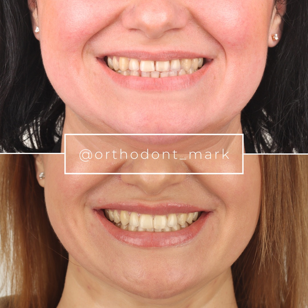
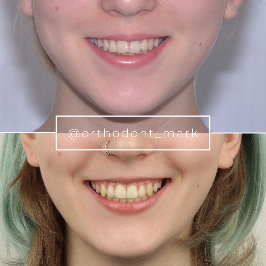
Clinical cases from Dr. Mark Radzhabov's practice
Learn the full MARPE protocol from Dr. Mark Radzhabov
Fundamental course covering CBCT patient selection, miniscrew planning, activation protocols, and 60+ clinical cases. Choose the access level that fits your practice.
Essentials of rapid palatal expansion for practicing orthodontists.
- Core RPE concepts and biomechanics
- 6 structured video lessons
- Clinical decision checklists
- Lifetime access to recordings
Deep-dive into MARPE protocol, diagnostics, and clinical execution.
- 6 modules covering MARPE from A to Z
- CBCT case selection walkthroughs
- Miniscrew placement and activation
- 60+ annotated clinical cases
- Direct Q&A with Dr. Mark Radzhabov
5-element medical consultation framework for dentists and orthodontists.
- Trust-building consultation protocol
- 5 lesson modules
- Templates for treatment plan delivery
- Works with any clinical specialty
Clinical FAQ
How does MARPE with maxillary protraction compare to traditional facemask therapy for Class III correction?
MARPE-assisted protraction produces true skeletal advancement (2–3 mm point A gain) with minimal dental compensation and 5–10% relapse versus 20–30% relapse with facemask therapy. Bone-borne force application eliminates vertical side effects common in tooth-borne systems, making it superior for horizontal-growth-pattern patients.
What is the optimal minuscrew insertion depth and location in the hard palate for maxillary advancement mechanics?
Insert bilateral grade 5 titanium miniscrews 11–13 mm deep, perpendicular to the palatal plane, in the anterior hard palate at or 2–3 mm lateral to midline. Placement more than 4 mm lateral or <11 mm deep increases loosening risk and reduces cortical engagement.
Which Angelieri stage on CBCT predicts success in achieving true skeletal palatal expansion without surgical sectioning?
Patients in stage A (suture completely radiolucent) or early stage B achieve maximal skeletal widening of 6–10 mm with MARPE activation. Stage C and D patients have significant ossification and experience 3–4 mm or less true expansion. Surgical assistance may be needed if greater correction is required.
What is the recommended force magnitude for combined MARPE expansion and sagittal maxillary protraction in adult Class III patients?
Begin with 5 kg/week expansion load, escalating to 8–10 kg/week, combined with 400–600 grams protraction force per side via elastics or integrated arms. Excessive force (>10 kg/week or >600 grams) accelerates miniscrew loosening and increases relapse in cortically mature bone.
How do you prevent vertical relapse or anterior open-bite development when applying forward protraction vectors with MARPE?
Align protraction force vectors 10–20° below the occlusal plane in low-angle patients. Parallel to the plane in high-angle cases. Monitor lateral cephalometric images every 6 weeks. If anterior vertical dimension increases >1 mm, reduce transverse load or adjust vector direction downward.
What is the expected timeline from initial miniscrew placement to final crossbite correction and miniscrew removal?
Expansion phase lasts 4–6 months with weekly or monthly activation, then protraction continues an additional 8–12 weeks for consolidation. Total treatment duration averages 5–7 months before miniscrew removal. Long-term retention with wraparound maxillary retainers extends 12–24 months.
How much sagittal point A advancement can realistically be achieved with bone-anchored MARPE protraction alone in skeletally mature patients?
Expected point A advancement is 2–3 mm in skeletally mature Class III patients using integrated MARPE and bone-anchored forward mechanics. Greater advancement (>4 mm) typically requires surgical Le Fort I or distraction-based techniques in adult patients with severe skeletal Class III.
What miniscrew failure and relapse rates should clinicians expect in well-selected Class III cases treated with MARPE protraction?
Miniscrew loosening occurs in 8–15% of cases depending on cortical bone density and insertion technique. Transverse relapse is 5–10% over 12 months. Sagittal relapse is 1–2 mm over 2 years if vertical control was adequate. Success rate (anterior crossbite closure without surgery) exceeds 86% in carefully selected patients.
How does vertical facial dimension assessment guide force vector direction in Class III MARPE protraction mechanics?
Measure Y-axis angle and anterior face height ratio on cephalometric images. Low-angle patients (Y-axis <60°, AFHP <55%) tolerate protraction vectors 10–20° below the occlusal plane. High-angle patients require downward or anterior force components to prevent open-bite relapse. Force vectors parallel to the occlusal plane are safer.
What is the diagnostic value of Hounsfield unit measurement on CBCT for predicting miniscrew stability in MARPE treatment?
Cortical bone density >650 Hounsfield units ensures robust miniscrew anchorage and reduces loosening risk. Patients with HU <600 experience higher failure rates. Reduce force magnitude or consider thicker-diameter miniscrews. Measurement via cone-beam CT region-of-interest cursor at insertion sites improves case selection accuracy.
Combining MARPE with bone-anchored maxillary protraction represents a paradigm shift in the management of adult Class III malocclusion with horizontal growth patterns. The key is precise case selection using cone-beam CT assessment of the midpalatal suture combined with vertical dimension analysis and anteroposterior skeletal relationships. Dr. Mark Radzhabov and other clinicians using miniscrew-assisted skeletal expansion protocols report stable outcomes without surgical intervention in carefully selected patients. Consider scheduling a case consultation through Orthodontist Mark to review your complex Class III cases and explore this integrated treatment approach.

