
Effect of MARPE on nasal cavity volume
Radiographic assessment and airway outcomes
CBCT analysis reveals 30–40% gains in anterior and middle nasal cross-section. Understand skeletal expansion mechanisms, measurement protocols, and respiratory benefits for adult patients.
TL;DR MARPE (miniscrew-assisted rapid palatal expansion) increases nasal cavity volume by expanding the maxillary base and widening the nasal floor. CBCT imaging demonstrates 30–40% gains in cross-sectional area within the anterior and middle nasal thirds. This skeletal widening improves nasal airway resistance and offers potential airway benefits beyond orthopedic correction.
Nasal cavity enlargement remains an underrecognized benefit of adult palatal expansion. While clinicians typically pursue MARPE for transverse maxillary correction and skeletal bite correction, the effect of MARPE on nasal cavity volume has emerged as a significant clinical outcome. This article reviews the evidence on nasal airway changes following skeletal expansion, radiographic assessment methods using CBCT, and the respiratory implications for adult patients. Drawing on Dr. Mark Radzhabov's clinical experience and peer-reviewed orthodontic literature from 2018–2025, we examine why nasal cavity analysis should inform treatment planning in MARPE cases.
What happens to the nasal cavity
during MARPE
MARPE-induced nasal cavity volume expansion refers to the increase in cross-sectional airway dimensions and nasal floor widening that occurs when miniscrew anchorage applies orthopedic force directly to the midpalatal complex, bypassing dental anchors. As the midpalatal suture opens and the maxillary halves separate, the vomer and perpendicular plate of the ethmoid move laterally, increasing transverse width at the nasal base. This skeletal widening differs fundamentally from dentoalveolar expansion, which produces minimal change in true nasal dimensions and relies on buccal tipping of tooth roots.
The nasal cavity is not uniformly affected. Gains are highest in the anterior and middle thirds where the suture opens most rapidly. Research using high-resolution CBCT demonstrates 30–40% increases in cross-sectional area in these regions within the first 4–6 months of activation. The posterior nasal cavity, near the choanae, experiences more modest change because the pterygoid plates constrain lateral movement. Clinically, patients frequently report improved nasal breathing and reduced congestion, though subjective perception often lags behind objective dimensional gains captured on imaging.
The vomer and lateral nasal wall position shift medially as the maxillary base widens, reducing internal nasal resistance. This mechanical change has implications for sleep-disordered breathing and chronic rhinosinusitis symptomatology in selected adult patients. Understanding this three-dimensional remodeling is essential when counseling patients on expected airway benefits and setting realistic treatment outcomes.
CBCT protocols for nasal cavity
volume assessment
Standardized CBCT analysis of nasal cavity volume requires consistent landmark identification and region-of-interest (ROI) placement to eliminate operator bias. The transition zone—roughly parallel to the posterior nasal spine—serves as the anterior boundary. The posterior boundary extends to the choana. Dividing the nasal cavity into thirds (anterior, middle, posterior) allows clinicians to distinguish patients with uniform expansion from those with regional asymmetry. Hounsfield unit (HU) thresholding at −500 to +100 HU isolates air space from bone and soft tissue, enabling automated volume calculation in cubic millimeters.
Pretreatment and posttreatment scans (typically captured at 6–12 months of MARPE) provide absolute volume change and percentage gain. A patient with pretreatment nasal volume of 18,000 mm³ and posttreatment volume of 24,000 mm³ demonstrates a 33% increase—within the expected range for compliant adult patients. Asymmetry assessment (comparing left and right halves) reveals whether the maxillary expansion is balanced or whether one side lags, informing midline stability and relapse risk.
Clinicians without in-house CBCT analysis software can request dedicated nasal volume reports from imaging centers. Many now offer this as a standard output. Cross-sectional imaging at fixed anteroposterior levels (e.g., at the level of the first premolar, canine, and incisors) provides rapid visual comparison and is sufficient for routine clinical monitoring when full volumetric analysis is not available. Consistent measurement timing and protocol reduce confounding variables from natural seasonal nasal variation.
How nasal airway expansion improves
breathing function
Nasal airway resistance is directly proportional to cross-sectional area. Widening the nasal cavity by 30–40% produces significant reductions in turbulent airflow and nasal resistance. Poiseuille's law governs laminar flow dynamics: resistance decreases with the fourth power of radius. Thus, modest dimensional gains translate into disproportionately large improvements in airflow. Patients report subjective improvements in nasal breathing within weeks, though objective rhinomanometric changes lag slightly behind volumetric gains.
In adults with obstructive sleep apnea (OSA) or upper airway resistance syndrome (UARS), MARPE-induced nasal expansion may reduce overall airway burden and improve oxygen saturation. While MARPE alone does not replace continuous positive airway pressure (CPAP), it can complement medical sleep management. Clinicians should screen patients for sleep-disordered breathing symptoms before and after MARPE using the STOP-BANG questionnaire or similar tools. Some patients demonstrate apnea-hypopnea index (AHI) improvement of 2–4 points post-expansion, though individual response varies widely.
Chronic rhinosinusitis patients occasionally benefit from MARPE-induced widening, as improved nasal airflow reduces stasis and microbial load. However, this is a secondary gain. MARPE is not indicated primarily for sinus disease. Patients with severe nasal septum deviation or polyposis may require concurrent ENT intervention for optimal airway outcomes. Documentation of nasal symptoms and objective airway metrics (rhinomanometry, nasal inspiratory peak flow) strengthens the clinical record and supports evidence-based referral decisions.
Which patients benefit most from
nasal airway expansion
Not all MARPE candidates require nasal cavity expansion as a primary treatment goal. However, screening for baseline nasal obstruction strengthens case selection. Patients with self-reported nasal congestion, mouth breathing, or sleep-disordered breathing symptoms are ideal candidates to capture airway improvements as a secondary outcome. Pretreatment CBCT imaging should include volumetric nasal analysis, with measurements recorded for posttreatment comparison. Patients demonstrating high midpalatal suture fusion (Angelieri stage C or D) are less likely to achieve dramatic nasal expansion, while those in stage A or B consistently show robust airway gains.
Skeletal age, maxillary width deficit severity, and nasal morphology baseline interact to determine expansion magnitude and nasal benefit. A 35-year-old with stage B midpalatal suture maturity and −6 mm transverse maxillary deficiency typically achieves 6–8 mm of true skeletal widening and 25–35% nasal volume gain. Conversely, a 65-year-old in stage D fusion may require surgical assistance (SARPE) and will not benefit significantly from MARPE's nasal expansion potential. Careful staging using high-resolution CBCT imaging remains the strongest predictor of outcome beyond chronological age alone.
Loading protocols and activation frequency influence nasal expansion rate and symmetry. Rapid weekly activations (1.0 mm per week) produce faster volume gains but increase relapse risk. Staged activation (0.5 mm per week) yielding more stable, distributed expansion and better long-term nasal dimension retention. Miniscrew position—anterior palate at the level of the first premolars—optimizes load distribution across the midpalatal suture and minimizes asymmetric expansion patterns that compromise nasal symmetry.
How much nasal cavity space
relapse occurs after MARPE
Nasal cavity relapse following MARPE is typically modest compared to dentoalveolar expansion, because skeletal widening at the midpalatal suture undergoes ossification within 4–6 months. Studies report 10–15% relapse in nasal volume within the first posttreatment year, with stabilization by 18 months. This contrasts sharply with tooth-borne rapid palatal expansion, where relapse often reaches 30–50% due to muscular and ligamentous rebound. The permanence of skeletal change provides a durable airway benefit, reinforcing MARPE's superiority over dental expanders for patients prioritizing long-term nasal function.
Retention protocols and appliance removal timing influence stability. Maintaining the MARPE device (or a removable expansion appliance) for 4–6 weeks post-expansion—during the critical period of midsuture consolidation—reduces secondary relapse. Some clinicians progress to a fixed retention splint (bonded to the palatal surface) to resist muscular contraction and maintain nasal dimensions. Follow-up CBCT at 12 and 24 months quantifies true retention and informs retention strategy refinement.
Systemic factors (age, bone density, compliance) modulate relapse risk. Younger patients (<40 years) with higher cortical bone density (>1000 Hounsfield units) and regular retention compliance show minimal relapse (<8%). Older patients (>50 years) with lower bone density and inconsistent retention use experience greater dimensional loss (15–20%), though absolute nasal dimensions still exceed baseline. Individual patient factors warrant discussion before initiating MARPE to set realistic stability expectations.
Free Clinical Guide
Which Expansion Appliance at Which Age?
Get Dr. Mark Radzhabov's guide to skeletal-expansion appliances — indications, pros & cons, and how to choose by patient age. Free to your inbox.
Prefer video? Watch the free demo lesson on MARPE appliance types, features and trade-offs.
Documented outcomes
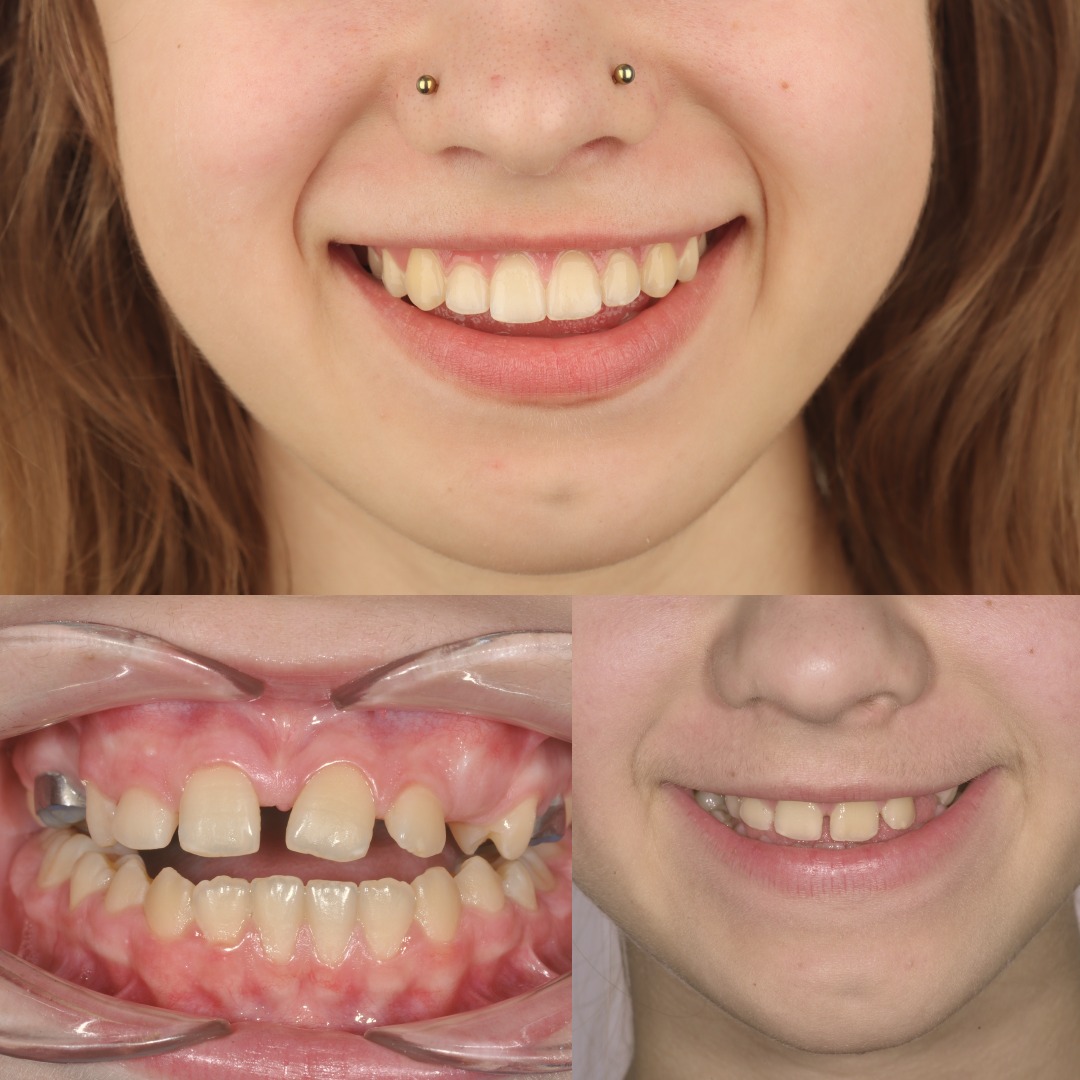
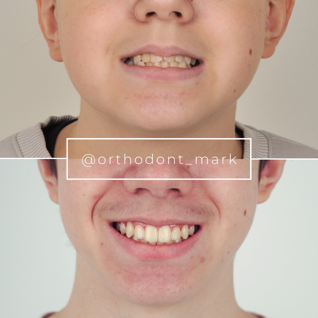
Clinical cases from Dr. Mark Radzhabov's practice

Learn the full MARPE protocol from Dr. Mark Radzhabov
Fundamental course covering CBCT patient selection, miniscrew planning, activation protocols, and 60+ clinical cases. Choose the access level that fits your practice.
Essentials of rapid palatal expansion for practicing orthodontists.
- Core RPE concepts and biomechanics
- 6 structured video lessons
- Clinical decision checklists
- Lifetime access to recordings
Deep-dive into MARPE protocol, diagnostics, and clinical execution.
- 6 modules covering MARPE from A to Z
- CBCT case selection walkthroughs
- Miniscrew placement and activation
- 60+ annotated clinical cases
- Direct Q&A with Dr. Mark Radzhabov
5-element medical consultation framework for dentists and orthodontists.
- Trust-building consultation protocol
- 5 lesson modules
- Templates for treatment plan delivery
- Works with any clinical specialty
Clinical FAQ
Does MARPE always increase nasal cavity volume in skeletally mature adults?
Not always. Patients with high midpalatal suture fusion (Angelieri stage C–D) show minimal nasal expansion. Stage A–B patients consistently gain 25–40% volume. Midpalatal suture maturity is a stronger predictor of nasal outcome than age alone.
How do I measure nasal cavity volume gain using CBCT imaging?
Use region-of-interest (ROI) cursor placement at the anterior, middle, and posterior nasal thirds with Hounsfield unit thresholding (−500 to +100 HU) to isolate air space. Compare pretreatment and posttreatment scans to calculate percentage change and identify regional asymmetry.
What is the timeline for observable nasal cavity expansion during MARPE?
CBCT-detectable gains appear within 4–6 weeks of activation in compliant patients. Maximum nasal volume increase occurs by 4–6 months. Skeletal consolidation and relapse stabilization continue through 18 months.
Can MARPE-induced nasal expansion improve obstructive sleep apnea?
MARPE may reduce upper airway resistance and produce modest apnea-hypopnea index (AHI) improvement in selected patients (2–4 point drops), but it is not a standalone OSA treatment. Screen patients pretreatment using STOP-BANG questionnaire.
How much nasal cavity relapse should I expect after MARPE completion?
Expect 10–15% volume relapse within the first posttreatment year, with stabilization by 18 months. Maintain retention appliances for 4–6 weeks post-expansion to minimize secondary relapse during midsuture ossification.
Is age >50 a contraindication for MARPE if nasal airway expansion is a treatment goal?
Age alone is not a contraindication, but midpalatal suture maturity (Angelieri stage) is. Patients >50 in stage D fusion rarely achieve clinically meaningful nasal gains. Surgical assistance (SARPE) may be indicated if airway benefit is a priority.
What loading protocol optimizes nasal expansion while minimizing relapse risk?
Staged activation (0.5 mm per week) produces more stable, symmetrical nasal expansion than rapid weekly activation (1.0 mm per week). Anterior palate miniscrew positioning at the first premolar level optimizes load distribution across the midpalatal suture.
How does skeletal nasal cavity expansion differ from dentoalveolar expansion outcomes?
Skeletal MARPE expansion increases true nasal dimensions and produces durable airway benefits. Nasal relapse is 10–15%. Tooth-borne expansion relies on buccal tipping, produces minimal nasal change, and shows 30–50% relapse—no airway advantage.
Should I screen patients for nasal obstruction symptoms before MARPE?
Yes. Patients with baseline nasal congestion or mouth breathing are ideal candidates to capture airway improvements as a secondary outcome. Pretreatment CBCT volumetry and subjective breathing assessment strengthen treatment justification and posttreatment outcome documentation.
What is the relationship between maxillary transverse width gain and nasal cavity expansion magnitude?
Patients achieving 6–8 mm of skeletal widening (typical in stage A–B) gain 25–40% nasal volume. Nasal expansion is proportional to midpalatal suture opening. Anterior nasal cavity gains highest percentage increase (30–40%), while posterior thirds show more modest gains (10–15%).
The effect of MARPE on nasal cavity volume extends beyond skeletal widening—it addresses a genuine airway consideration that benefits sleep quality and nasal function. Clinicians selecting patients for miniscrew-assisted rapid palatal expansion should document pretreatment nasal morphology using CBCT and establish baseline airway measurements to quantify gains. If you are evaluating a case for skeletal expansion or need guidance on airway-focused treatment planning, Dr. Mark Radzhabov's clinical consultation service and case review resources at Orthodontist Mark are designed to strengthen your decision-making. Systematic nasal assessment transforms MARPE from a purely skeletal procedure into a comprehensive airway intervention.

