
Radiographic follow-up protocol after MARPE
Timing, technique, and clinical interpretation
Master the CBCT sequences, occlusal landmarks, and maturation scoring that distinguish skeletal expansion from relapse risk.
TL;DR Radiographic follow-up protocol after MARPE requires high-resolution CBCT at expansion completion to confirm midpalatal suture opening, followed by occlusal radiographs at 4–6 week intervals to assess diastema formation and skeletal stability. Lateral cephalometric imaging documents transverse widening and guides retention planning in skeletally mature patients.
Completing miniscrew-assisted rapid palatal expansion is not the endpoint—it is the beginning of a critical documentation phase. Orthodontist Mark explains that radiographic follow-up protocol after MARPE demands precise timing, proper modalities, and systematic assessment to confirm skeletal rather than dental response. In this guide, Dr. Mark Radzhabov outlines when to image, what to measure, and how to distinguish true suture opening from appliance deflection or anchor loss. These protocols protect against relapse, inform retention strategy, and establish the clinical evidence your records require.
What is the baseline CBCT and why does it matter
before and after activation
A pre-activation CBCT scan—taken before miniscrew insertion—serves as the anatomical reference and risk-mitigation tool. This image documents midpalatal suture maturation stage using Angelieri's classification (stages A–D), bone density in Hounsfield units, and miniscrew insertion anatomy. Skipping the baseline is a common oversight that eliminates your ability to measure true skeletal gain against dental side effects.
Post-activation CBCT should be acquired at expansion completion—not during active loading. Timing matters: premature imaging (before full force application) shows incomplete opening and risks patient anxiety. Delayed imaging (weeks into retention) introduces measurement error from early resorption or rebound. A high-resolution protocol targeting the midpalatal region with 0.3 mm voxel size and field-of-view limited to the palate reduces radiation dose while maintaining diagnostic clarity.
On post-expansion CBCT, measure anterior, middle, and posterior opening distances in axial and coronal reconstructions. Diastema width correlates with skeletal opening but is not equivalent to it—a 3 mm diastema may represent 2.5 mm of skeletal gain if dental tipping accounts for 0.5 mm. Cone-beam computed tomography with Hounsfield analysis reveals whether bone density has recovered post-expansion, a sign of favorable healing and lower relapse risk.
Occlusal radiograph diastema expansion as a monitoring tool
serial capture at 4–6 week intervals
Occlusal radiographs—periapical or bitewing views centered on the maxillary midline—offer a rapid, low-dose screening method between CBCT scans. The diastema width, measured from distal contact point of central incisors to the midline, begins at expansion day 1 and widens predictably in the first 8–12 weeks. A diastema that stabilizes or decreases over sequential radiographs after week 10 signals suture fusion onset or incomplete opening—red flags for relapse planning.
Expected diastema progression: 0.5–1.0 mm per week during active activation, then 0.1–0.3 mm per week during consolidation. Departure from this trajectory warrants CBCT confirmation. Occlusal radiographs also reveal dental tipping (maxillary incisors tilting buccally) and miniscrew position drift, which together help clinicians separate true skeletal widening from dental compensation.
Schedule occlusal radiographs at expansion completion (day 0), then 4 weeks, 8 weeks, 12 weeks, and 6 months post-activation. This sequence is radiation-efficient and captures the critical window when suture fusion risk is highest. Compare serial images in a light box or digital viewer with fixed measurement landmarks—the palatal midline and perpendicular bisector of the central incisors—to ensure consistency.
How to assess midpalatal suture opening confirmation with CBCT
Angelieri staging and bone fill pattern
Palatal suture opening confirmation requires coronal CBCT sections through the anterior, middle, and posterior palate. In stage A (pre-fusion), the suture appears as a thin radiolucent line. Post-expansion, a complete dark gap signals bony separation. In stage B (early partial fusion), fusion is visible at the lateral aspects. Successful expansion may still occur but carries higher relapse risk. Stage C and D sutures show advanced calcification. These patients require higher load magnitudes and longer consolidation—or consideration of surgical sectioning.
Measure the gap distance in at least three locations: anterior palate (at the level of maxillary first molars), middle third (at first molars), and posterior third (at or behind second molars). The anterior palate typically shows the widest opening. The posterior shows the narrowest, especially in stage C/D cases. If the anterior opens 6 mm but the posterior opens only 2 mm, asymmetric loading or suture anatomy variation is at play—document this in your treatment record and adjust consolidation timing accordingly.
Post-expansion CBCT also reveals bone quality recovery, assessed by Hounsfield density measurement. After 3 months, Hounsfield units in the newly opened suture should approach 300–400 HU, indicating woven bone formation and lower relapse risk. Densities below 200 HU signal immature bone and warrant extended consolidation—typically 6–9 months instead of the standard 4–6 months. Orthodontist Mark's protocol integrates this density assessment into retention planning, moving beyond suture stage alone to create individualized timelines.
Lateral cephalometric MARPE imaging for documenting skeletal widening
anterior nasal spine and maxillary base widening
Lateral cephalometric radiographs provide a two-dimensional view of midpalatal suture position and maxillary depth change. While CBCT dominates for coronal assessment, a lateral cephalogram taken 2–3 days post-expansion confirms absence of anterior displacement of the maxilla (which would indicate Le Fort I level loading) and documents any sagittal relapse patterns early. The anterior nasal spine (ANS) position relative to midsagittal landmarks reveals whether expansion was coupled with forward displacement—a common occurrence in stage A and B sutures.
Measure ANS-to-midsagittal plane distance on a trace overlay. A shift of >1 mm forward may indicate incomplete transverse opening and compensatory sagittal movement. This finding warrants CBCT review to rule out asymmetric activation or miniscrew loosening. Overlay pre- and post-expansion laterals on the palatal vault and zygomatic process to confirm skeletal stability—minimal change in these bony landmarks indicates bone-borne rather than dental-borne response. This overlay technique, standard in cephalometric assessment, is radiation-efficient and integrates naturally into existing treatment records.
Schedule lateral cephalograms at three timepoints: completion of active expansion, 3 months into consolidation, and at debond. This sequence detects relapse patterns specific to maxillary dimensions and informs whether additional retention (palatal bars, fixed lingual retainers) is needed. Most orthodontists now combine a single lateral cephalogram with CBCT rather than relying on cephalometry alone, but the lateral remains valuable for documenting sagittal stability—an underreported metric in MARPE literature.
How radiographic signs guide consolidation duration and retention strategy
from CBCT maturation score to appliance selection
Radiographic follow-up findings directly determine consolidation length and retention appliance design. A patient with Angelieri stage A suture, anterior opening >6 mm, and Hounsfield density >350 HU at 3 months may safely transition to fixed lingual retention at 4–5 months post-expansion. Conversely, a stage B or C patient with asymmetric opening, anterior gap only 4 mm, and density <250 HU should extend consolidation to 6–9 months and consider a hybrid retention system (fixed + removable) to resist relapse.
Clinical decision rules based on CBCT findings: Stage A + symmetric opening + high bone density → standard 4–6 month consolidation. Stage B + moderate opening + recovering density → 6–8 month consolidation + extended palatal bar retention. Stage C/D + limited opening + low density → consider surgical intervention or 9–12 month consolidation depending on treatment goals. Diastema closure rate provides a secondary check: if diastema narrows >0.3 mm/week after week 8, suture fusion is accelerated and consolidation should extend by 2–3 months.
Occlusal radiographs taken at 6 and 12 months post-activation serve as final verification before appliance removal and debond. Stable diastema width, normal bone density, and absence of miniscrew mobility indicate readiness for retention transition. Document all imaging and measurements in the patient chart—a dated series of CBCT axial slices, occlusal radiographs with marked diastema, and cephalometric overlays creates a defensible record of treatment success and informs long-term follow-up intervals. This level of radiographic documentation is standard of care in skeletal expansion."
Free Clinical Guide
Which Expansion Appliance at Which Age?
Get Dr. Mark Radzhabov's guide to skeletal-expansion appliances — indications, pros & cons, and how to choose by patient age. Free to your inbox.
Prefer video? Watch the free demo lesson on MARPE appliance types, features and trade-offs.
Documented outcomes
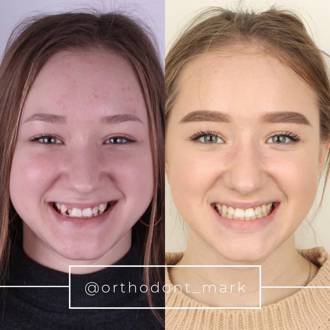
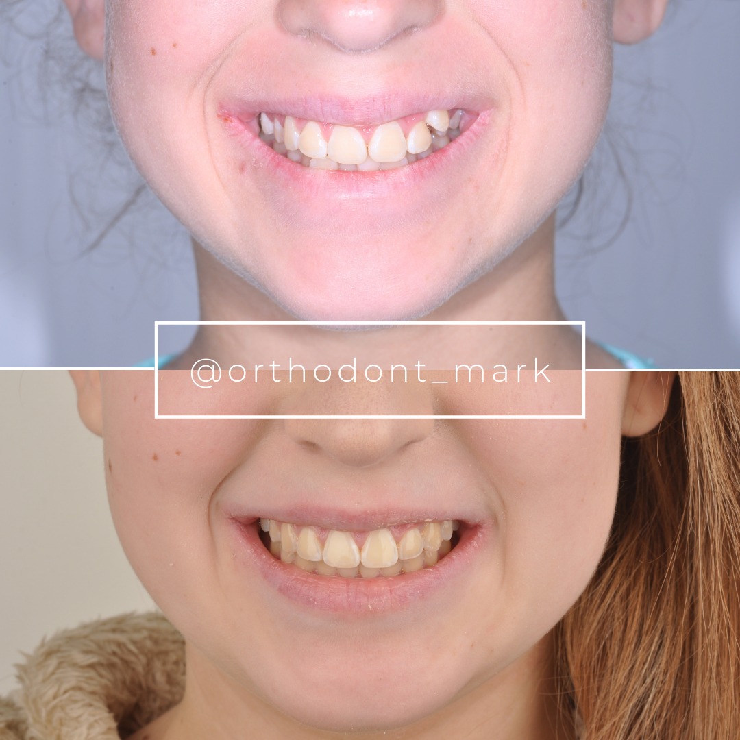
Clinical cases from Dr. Mark Radzhabov's practice

Learn the full MARPE protocol from Dr. Mark Radzhabov
Fundamental course covering CBCT patient selection, miniscrew planning, activation protocols, and 60+ clinical cases. Choose the access level that fits your practice.
Essentials of rapid palatal expansion for practicing orthodontists.
- Core RPE concepts and biomechanics
- 6 structured video lessons
- Clinical decision checklists
- Lifetime access to recordings
Deep-dive into MARPE protocol, diagnostics, and clinical execution.
- 6 modules covering MARPE from A to Z
- CBCT case selection walkthroughs
- Miniscrew placement and activation
- 60+ annotated clinical cases
- Direct Q&A with Dr. Mark Radzhabov
5-element medical consultation framework for dentists and orthodontists.
- Trust-building consultation protocol
- 5 lesson modules
- Templates for treatment plan delivery
- Works with any clinical specialty
Clinical FAQ
When should I acquire the post-expansion CBCT after MARPE completion?
Image within 7 days of final miniscrew activation. Premature imaging shows incomplete opening and introduces measurement error. Delayed imaging (>2 weeks) risks early fusion or resorption artifacts. One high-resolution CBCT at completion is standard. Repeat CBCT at 3 and 6 months if relapse is suspected.
What does successful palatal suture opening look like on CBCT?
In stage A/B sutures, expect a clear dark radiolucent gap in anterior palate measuring 4–8 mm, narrowing posteriorly to 2–4 mm. Stage C/D gaps are typically smaller (2–4 mm anterior). Symmetry between left and right halves indicates balanced loading. Asymmetry suggests miniscrew drift or uneven force distribution.
How do I differentiate true skeletal expansion from dental tipping on radiographs?
CBCT coronal view shows buccal flaring of maxillary incisors if tipping occurs. Measure root angulation and incisor tip position. Compare pre- and post-expansion dental casts or 3D models: true skeletal expansion shows no change in incisor-to-suture distance, while dental tipping increases this measurement. Lateral cephalogram overlay confirms palatal vault stability.
What occlusal radiograph timing protocol minimizes dose while capturing diastema progression?
Image at expansion day 0, then 4, 8, 12, and 24 weeks post-activation. This five-point protocol is radiation-efficient and captures the critical 8–12 week window when suture fusion risk peaks. Diastema stabilization below 0.1 mm/week after week 12 signals relapse onset and guides consolidation extension.
How does Angelieri maturation stage affect consolidation duration post-MARPE?
Stage A sutures typically consolidate in 4–6 months. Stage B in 6–8 months. Stages C–D require 8–12 months or surgical intervention. Combine stage with measured bone density (Hounsfield units >300 indicates faster healing). A stage A patient with slow diastema closure still needs extended consolidation, emphasizing that stage is one factor among many.
What is the significance of Hounsfield unit measurement in post-expansion CBCT?
Hounsfield density at 3 and 6 months post-expansion reflects bone maturity and mineralization. Values >300 HU indicate mature woven bone, supporting consolidation closure at standard timelines. Density <200 HU signals immature bone and requires 2–3 month extension to reduce relapse. Measure at anterior, middle, and posterior suture regions for regional variation.
How do I interpret asymmetric opening on CBCT, and what is the clinical response?
Asymmetry (>2 mm difference between left and right at same anteroposterior level) suggests miniscrew migration, uneven activation, or lateral wall resistance. Review miniscrew position on CBCT and check for loosening clinically. If miniscrews are stable and loading is balanced, asymmetry may reflect patient-specific anatomy. Document and extend consolidation by 2–3 months.
Should I use lateral cephalometric imaging in MARPE, or is CBCT sufficient?
CBCT is primary. Lateral cephalogram is supplementary. A single lateral cephalogram at expansion completion and at 6 months detects ANS forward displacement and sagittal relapse—metrics CBCT alone may miss. Overlay technique on palatal vault is radiation-efficient and complements CBCT coronal assessment for complete documentation.
What diastema measurement and progression rate indicates healthy suture response?
Expected progression is 0.5–1.0 mm/week during active activation. At week 4 post-activation, diastema should exceed 2–3 mm. Stabilization at 0.1–0.3 mm/week by week 8–10 is normal; <0.1 mm/week signals early fusion or incomplete opening. Diastema closure after week 12 requires CBCT review and likely consolidation extension.
How do I document radiographic findings to defend MARPE outcomes in a chart?
Archive dated CBCT axial slices at anterior, middle, posterior levels with measurements annotated. Include occlusal radiographs with marked diastema width and measurement landmarks. Add cephalometric overlays on palatal vault. Record Angelieri stage, Hounsfield density, and miniscrew position on a standard form. This 3D + 2D + clinical summary creates defensible, reproducible documentation of skeletal response.
Rigorous radiographic documentation transforms clinical impression into measurable proof of skeletal change. The radiographic follow-up protocol after MARPE is not administrative—it directly informs whether to transition to fixed retention, extend the consolidation phase, or plan surgical intervention in cases of incomplete opening. Dr. Mark Radzhabov's evidence-based approach integrates CBCT maturation scoring with occlusal findings to create a defensible, reproducible pathway. Schedule a case consultation at Orthodontist Mark or enroll in our MARPE protocol course to master these imaging sequences.

