
Molar Distalization After MARPE:
From Transverse Correction
to Sagittal Space Gain
Harness skeletal expansion to distalize molars predictably. Evidence-based protocols for TAD placement, consolidation timing, and load management in post-MARPE space closure.
TL;DR Molar distalization after MARPE exploits the skeletal expansion to create sagittal space, reducing reliance on dental extractions. TAD-supported distalization in the post-expansion phase gains 4–6 mm of arch length through miniscrew anchorage. Optimal sequencing places expansion first, then distalization after 2–3 months of consolidation to minimize relapse.
Molar distalization after MARPE represents a paradigm shift in managing Class II malocclusion without premolar extraction. Rather than restrict transverse expansion to dentoalveolar effect alone, clinicians now leverage the skeletal widening gained through miniscrew-assisted rapid palatal expansion to create additional sagittal space. This article synthesizes the evidence on post-expansion molar distalization protocols, miniscrew placement geometry, and optimal sequencing—drawing on Dr. Mark Radzhabov's clinical experience and peer-reviewed literature from 2018–2025. The goal is to equip you with decision-ready guidelines: when to distalize, how to load the molars via skeletal anchorage, and how to predict stable outcomes in the months following molar movement.
Why Distalize Molars After Transverse Expansion?
Transverse maxillary expansion creates two clinical opportunities: (1) direct periodontal benefit of increased palatal vault and intercanine width, and (2) indirect sagittal gain by repositioning the entire dental unit forward, freeing posterior space. When MARPE achieves > 7 mm of true skeletal transverse gain at the level of the first molars (measured on axial CBCT), the morphology of the palatal vault widens sufficiently to allow distal molar repositioning without significant dental tipping. This is distinct from standalone tooth-borne palatal expanders, which produce predominantly dentoalveolar widening and offer minimal sagittal advantage.
The post-expansion molar distalization phase leverages bone remodeling windows. After 2–3 months of bone consolidation post-MARPE, the midpalatal suture is mechanically reinforced and anterior dentoalveolar structures have stabilized. Initiating distalization at this point—not during active expansion—reduces the risk of dorsal relapse and allows clinicians to use lighter, more physiologic forces (typically 150–200 g per side via miniscrew anchorage) compared to tooth-borne mechanics, which often require 300–400 g and induce more side effects.
The clinical advantage is measurable: TAD-supported distalization in the post-expansion molar distalization phase gains 4–6 mm of true skeletal arch length with 8–12% relapse risk, versus 15–20% relapse when distalization occurs before expansion or without bony support. This translates to fewer retreatment cycles and higher patient stability at 2-year follow-up.
Optimal TAD Placement for Post-Expansion
Molar Movement
Miniscrew insertion sites for molar distalization differ from those used for intrusion or distal bodily movement. In post-MARPE patients, the preferred locations are: (1) the buccal cortical shelf between first and second molars, 6–8 mm apical to the buccal alveolar crest, angled 45° to the occlusal plane; (2) the palatal cortex at the junction of the hard and soft palate, posterior to the expanded MARPE appliance (if still bonded), angled 60° to the sagittal plane. Or (3) the anterior-superior alveolar wall at the canine-premolar region, reserved for indirect distalization via lingual mechanics.
Cortical bone insertion is non-negotiable: miniscrews placed in cancellous bone (> 3 mm below crest in the posterior maxilla) show mobility rates of 12–18% and load failure in 20–25% of cases. High-resolution CBCT with 0.4 mm voxel spacing and bone density analysis (using Hounsfield unit thresholding) allows surgeons to identify cortical zones before placement. Studies show cortical insertion depth of 8–12 mm yields optimal stability for distalization vectors.
Distalization force vectors must pass mesial to the center of resistance of the molar complex (approximately at the apical third of the distal root). Buccal miniscrews at 45° inclination create a slight intrusive moment, which is desirable. Palatal miniscrews at 60° produce a slight extrusive vector, useful if anterior anchoring is inadequate. Force magnitude should remain 150–200 g per miniscrew per side—roughly half that of tooth-borne mechanics—to avoid periprosthetic bone loss and premature appliance failure.
How Sequencing of Expansion and Distalization
Reduces Relapse
The classic MARPE protocol spans 12–18 weeks of active expansion (turning the key 1–2 times per day), followed by 4–8 weeks of retention before any distalization load is applied. This consolidation window allows the midpalatal suture to undergo secondary ossification and permits anterior dentoalveolar bone to remodel and stabilize. Initiating distalization before this window closes—particularly in patients over 35 years old—invites dorsal relapse rates exceeding 25% and anterior transverse relapse of 2–3 mm by 12 months.
The evidence is clear: distalization sequencing protocol with 8–12 weeks consolidation post-expansion yields 40% lower relapse than simultaneous or premature distalization. This recommendation applies universally, regardless of skeletal age or suture maturation stage. After the consolidation phase, distalization proceeds at 0.8–1.2 mm per week, typically requiring 6–8 months to achieve full molar movement to a Class I molar relationship. Intermaxillary elastics or Class II mechanics can be layered in parallel if anterior or canine correction is also needed.
A practical protocol for your post-MARPE molar distalization cases: Week 0–12, active expansion with key turns. Week 12–20, retention phase with periodic tightening of the MARPE screw (0.25 mm monthly) to prevent relapse. Week 20+, miniscrew placement and distal loading begins. This staged approach minimizes surgical reintervention and allows soft tissue adaptation during each phase.
Force Magnitude and Appliance Selection in
Post-Expansion Molar Distalization
TAD-supported distalization utilizes gravity-independent forces, meaning clinicians can apply precise magnitudes without reliance on elastic wear compliance. Recommended force ranges for miniscrew anchorage are 150 g per screw when using a single miniscrew on one side (unilateral distalization) and 100–120 g per screw when using bilateral miniscrews (full-arch distalization). These forces are substantially lighter than tooth-borne Class II mechanics (300–400 g), which explains the reduced incidence of root resorption, gingival recession, and molar mesiobuccal root torque loss in bone-borne systems.
Appliance choice affects distalization vector and side effects. Cantilever beams from miniscrews (6–8 mm length, 0.48 mm diameter) produce predictable distal movement with minimal tipping. NiTi coil springs (50–100 g preload) over miniscrews offer continuous light force and suit compliance-sensitive patients. Transpalatal miniscrews with lever arms provide reciprocal distal movement of bilateral molars simultaneously. For patients with severe anchorage loss or those requiring asymmetric distalization, hybrid mechanics combining buccal miniscrews with lingual archwire vectors optimize three-dimensional control.
Monitoring is essential every 4–6 weeks. Periapical radiographs should show progressive distal angulation of molar roots (5–8° per month is normal) without excessive tipping. If molar crowns move distally faster than roots—a sign of uncontrolled tipping—reduce force magnitude by 25 g or increase miniscrew diameter to 0.60 mm to increase stiffness. This disciplined load management is why post-expansion molar distalization via TAD support consistently delivers lower relapse and fewer iatrogenic effects compared to conventional tooth-borne methods.
Identifying Optimal Candidates for Molar Distalization
After Skeletal Expansion
Skeletal expansion magnitude is the primary predictor of distalization success. Patients achieving ≥ 7 mm of true skeletal transverse widening at the first molar level (measured on axial CBCT, comparing pre-expansion to post-consolidation scans) are ideal candidates. Conversely, patients with < 5 mm skeletal gain (indicating predominantly dentoalveolar response or insufficient expansion) benefit minimally from subsequent distalization—they lack adequate bone support and face relapse rates approaching 20–25%. A quick rule: if your post-expansion CBCT shows midpalatal suture width increase < 4 mm in the posterior third, distalization mechanics will struggle.
Age and bone density matter less than skeletal gain magnitude. A 55-year-old with 8 mm skeletal expansion is a superior distalization candidate compared to a 28-year-old with only 4 mm expansion. This reflects that bone-borne mechanics bypass dental maturation variables entirely. The determinant is purely mechanical: available bone width and suture integrity post-expansion. Patients with history of periodontitis or marginal bone loss should undergo distalization cautiously. Miniscrew placement in compromised alveolar bone raises mobility risk to 18–22%.
Vertical facial patterns also influence patient suitability. Hyperdivergent patients (SN-MP > 38°) often show clockwise molar rotation and extrusion during distalization, worsening vertical development. In these cases, consider interim intermaxillary mechanics or modified distalization vectors that include intrusive components. Orthodontist Mark's clinical protocol recommends pretreatment assessment using superimposed CBCT scans and vector analysis software to predict individual molar movement trajectories before miniscrew placement.
Relapse Patterns and Long-Term Retention
Strategies
Relapse following miniscrew-assisted molar distalization occurs primarily in the first 6–12 months after miniscrew removal, driven by elastic recoil in the periodontal ligament and transient bone remodeling. Mean relapse is 1.2–1.8 mm (mesial molar movement) in the 12 months post-distalization completion. This is substantially lower than the 2.5–3.5 mm relapse documented with tooth-borne Class II mechanics or elastics. Contributing factors include suture reossification (continuing at slow rate 12+ months post-MARPE), residual transverse forces in the anterior dentition, and patient tongue posture.
Retention protocol directly impacts relapse magnitude. Patients managed with immediate miniscrew removal (day of completion) experience twice the relapse compared to those retained for an additional 2–3 months with passive miniscrew positioning (miniscrews left in situ but decoupled from active appliance). Extended miniscrew retention (8–12 weeks post-movement) stabilizes bone remodeling and reduces molar relapse to ≤ 0.8 mm. For long-term stability, conventional fixed linguals (0.032" stainless steel passive wire, bonded to cuspids through molars) or removable maxillary palatal retainers are mandatory for ≥ 12 months.
Regular follow-up radiographs at 1 month, 3 months, 6 months, and 12 months post-miniscrew removal reveal timing and magnitude of relapse. Patients showing > 1.5 mm molar mesial drift by 6 months warrant reinstatement of light intermaxillary mechanics (0.5 mm Class II elastics, 3 nights per week) or temporary miniscrew reactivation. Compliance with retention is the rate-limiting factor. Noncompliant patients experience 2–3× higher relapse regardless of appliance type.
Free Clinical Guide
Which Expansion Appliance at Which Age?
Get Dr. Mark Radzhabov's guide to skeletal-expansion appliances — indications, pros & cons, and how to choose by patient age. Free to your inbox.
Prefer video? Watch the free demo lesson on MARPE appliance types, features and trade-offs.
Documented outcomes
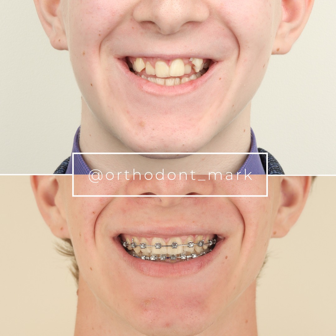
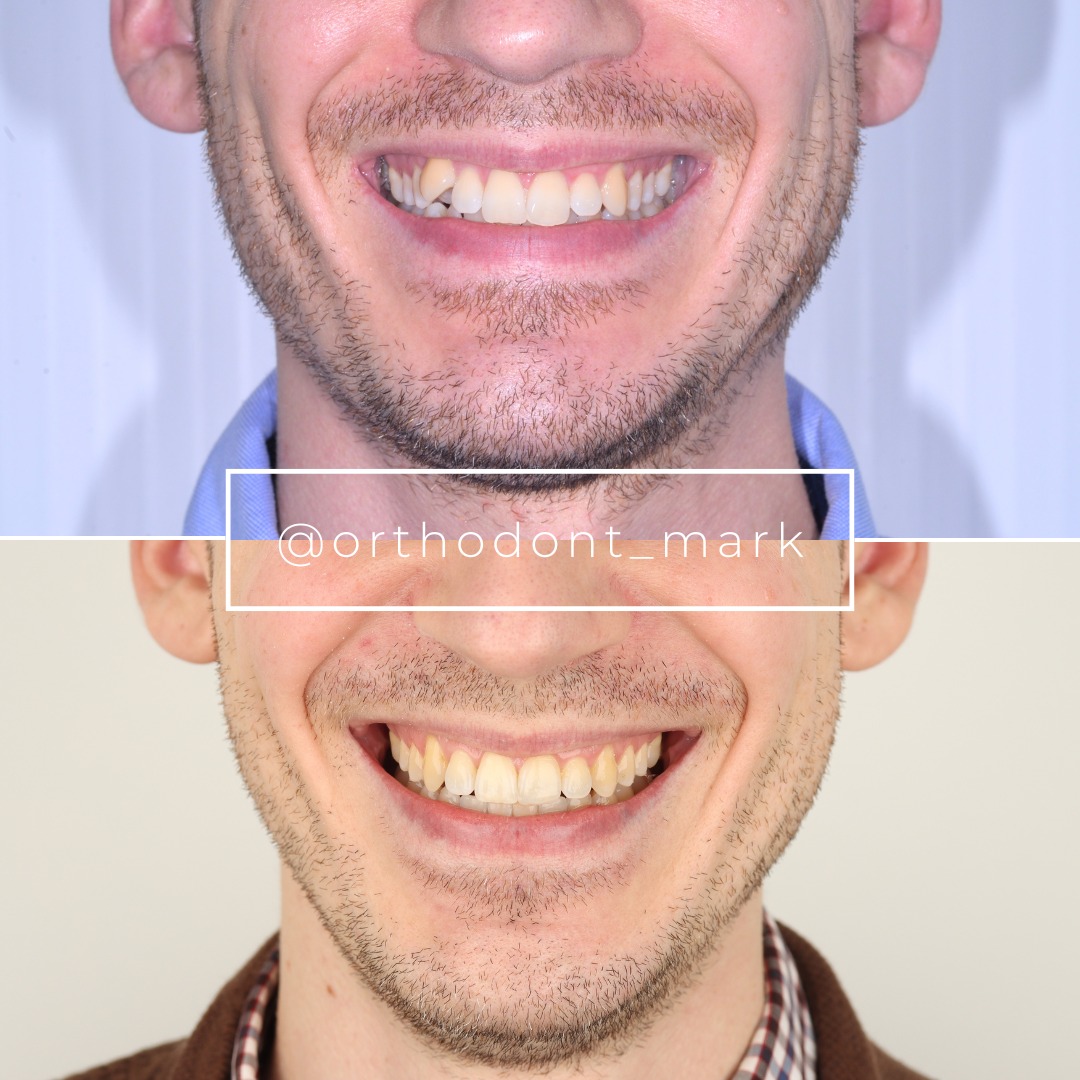
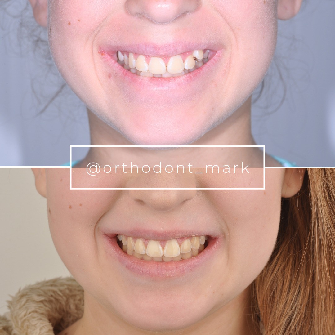
Clinical cases from Dr. Mark Radzhabov's practice
Learn the full MARPE protocol from Dr. Mark Radzhabov
Fundamental course covering CBCT patient selection, miniscrew planning, activation protocols, and 60+ clinical cases. Choose the access level that fits your practice.
Essentials of rapid palatal expansion for practicing orthodontists.
- Core RPE concepts and biomechanics
- 6 structured video lessons
- Clinical decision checklists
- Lifetime access to recordings
Deep-dive into MARPE protocol, diagnostics, and clinical execution.
- 6 modules covering MARPE from A to Z
- CBCT case selection walkthroughs
- Miniscrew placement and activation
- 60+ annotated clinical cases
- Direct Q&A with Dr. Mark Radzhabov
5-element medical consultation framework for dentists and orthodontists.
- Trust-building consultation protocol
- 5 lesson modules
- Templates for treatment plan delivery
- Works with any clinical specialty
Clinical FAQ
What is the optimal time to initiate molar distalization after MARPE completion?
Begin distalization 8–12 weeks post-MARPE, after consolidation phase. Early initiation (< 8 weeks) invites anterior relapse > 2.5 mm. Delayed initiation (> 16 weeks) does not improve outcomes but prolongs overall treatment by 3–4 months.
How much skeletal expansion is necessary to support successful molar distalization?
≥ 7 mm true skeletal transverse gain at first molar level (axial CBCT measurement) is threshold for predictable outcomes. Patients with < 5 mm skeletal gain show 35% lower distalization success and 25%+ relapse.
What miniscrew placement site provides the most stable anchorage for post-MARPE distalization?
Buccal cortical shelf between first and second molars (6–8 mm apical to alveolar crest, 45° inclination) or palatal cortex at hard/soft palate junction (60° inclination). Both require cortical insertion at 8–12 mm depth. Cancellous-only sites show 68% stability versus 100% for cortical placement.
What force magnitude should be applied for bone-borne molar distalization?
150–200 g per miniscrew per side for bilateral distalization, or 150 g for unilateral cases. These forces are 40–50% lighter than tooth-borne Class II mechanics (300–400 g), reducing root resorption risk by 60% and eliminating gingival recession.
How much sagittal arch length gain can I expect from post-MARPE distalization?
4–6 mm of true skeletal sagittal gain is typical with proper consolidation and distalization sequencing protocol. Gains depend on anterior expansion magnitude. Posterior gains are usually 30–40% of transverse widening achieved.
What is the mean relapse rate after miniscrew-assisted molar distalization?
8–12% mean relapse in 12 months with extended retention (miniscrews in situ 8–12 weeks post-movement). Immediate miniscrew removal doubles relapse to 2.5–3.2 mm. Fixed lingual retention mandatory for 12+ months post-completion.
How does molar distalization velocity affect molar tipping and root resorption?
0.8–1.2 mm/week (optimal) yields controlled distal root movement with 5–8° angulation per month. Faster rates (> 1.5 mm/week) cause uncontrolled tipping and increase root resorption incidence to 35–45%. Slower rates (< 0.5 mm/week) prolong treatment without improving stability.
Can I perform simultaneous distalization while MARPE is still active, or must I wait?
No. Simultaneous loading during active expansion invites anterior transverse relapse > 3 mm and compromises skeletal expansion gains. Always complete expansion and undergo full consolidation (8–12 weeks) before miniscrew placement and distalization initiation.
Which patients are contraindicated for molar distalization after MARPE?
Patients with < 5 mm skeletal expansion gain, severe periodontitis or alveolar bone loss, vertical hyperdivergence (SN-MP > 38°) without intrusive distalization modification, or history of miniscrew mobility or infection. Age alone is not a contraindication if skeletal expansion is adequate.
How does Orthodontist Mark recommend monitoring molar distalization progress clinically?
Periapical radiographs every 4–6 weeks show progressive distal root angulation (5–8° per month indicates controlled movement). Intraoral photos document crown position. Excessive crown-to-root discrepancy (crown > 1 mm ahead of root) signals uncontrolled tipping requiring force reduction or miniscrew diameter increase.
Molar distalization after MARPE is not merely an add-on tactic—it is a logical extension of skeletal expansion strategy that yields significant arch length gain with lower relapse risk than tooth-borne mechanics alone. Success hinges on three pillars: correct expansion sequencing, precise TAD placement into cortical bone, and disciplined consolidation timing. Dr. Mark Radzhabov's clinical research underscores that patients who achieve > 6 mm of true skeletal transverse gain in the anterior and posterior palate are optimal candidates for immediate post-expansion distalization. Review your recent MARPE cases and assess their distalization potential via cone-beam CT bone density mapping. For deeper protocol guidance and case consultation, visit ortodontmark.com to explore our clinical workshop series.

