
Periodontal Effects Palatal Expansion:
Tooth-Borne vs Bone-Borne
Risk Assessment & Clinical Protocol
Rapid expansion causes gingival recession and buccal bone dehiscence in tooth-borne systems. Bone-borne alternatives preserve periodontal health by loading the midpalatal suture. Learn evidence-based patient selection and force management.
TL;DR Periodontal effects palatal expansion vary markedly by anchorage method. Tooth-borne rapid palatal expanders cause buccal bone dehiscence and gingival recession in 15–40% of cases. Bone-borne systems like MARPE minimize dental side effects by loading the midpalatal suture directly. Patient selection via cone-beam CT and careful force management reduce fenestration risk.
Periodontal effects palatal expansion remain underestimated in adult treatment planning. When clinicians choose between tooth-borne rapid palatal expanders and miniscrew-assisted systems, the periodontal outcome often determines long-term stability and esthetic success. This article examines the evidence on gingival recession, buccal bone dehiscence, and alveolar bone preservation across both modalities, drawing on clinical data and radiographic assessment protocols. Dr. Mark Radzhabov synthesizes the research to help you select the method that minimizes periodontal compromise in your specific patient population.
How Expansion Systems Affect Periodontal
Health
Periodontal effects palatal expansion differ fundamentally based on whether forces load teeth or bone. Tooth-borne rapid palatal expanders apply horizontal stress to maxillary premolars and molars, creating buccal tipping and fenestration of the thin buccal cortex overlying the roots. Studies report gingival recession rates between 15–40% in tooth-borne systems, particularly in patients with thin biotypes. The recession occurs within the first 12–18 months of expansion and may not reverse fully after retention begins. Bone-borne systems (MARPE, MSE) distribute force across the hard palate and midpalatal suture, bypassing dental units and reducing tipping moments. Radiographic assessment shows that bone-borne expansion creates true skeletal widening without significant buccal movement of the alveolar ridge. Fenestration risk drops substantially when force vectors target bone rather than tooth apices. The clinical implication is clear: patient biotype, baseline bone thickness, and skeletal maturity must inform your appliance choice before insertion.
Buccal Bone Loss With Tooth-Borne
Expansion
vs. Bone-Borne Protection
Buccal cortical bone thickness at the premolar and molar regions averages 2.5–3.2 mm in the general population, but varies widely by sex, age, and ethnicity. Tooth-borne rapid palatal expanders create outward root movement that thins this cortex further. CBCT studies measuring bone density in Hounsfield units reveal that after 12 months of tooth-borne expansion, buccal cortical thickness at the first molar decreases by 1.0–1.8 mm on average. In patients with pre-existing thin biotypes (<1.5 mm), fenestration—complete cortical bone loss—occurs in approximately 22–35% of cases. Bone-borne systems yield measurably different outcomes. When miniscrews insert into cortical bone at the anterior palate lateral to the midline, force distribution favors midpalatal suture separation rather than dental buccal movement. Serial CBCT imaging shows that patients treated with MARPE retain baseline buccal cortical thickness with minimal recession, provided insertion depth and force magnitude remain controlled. Gingival biotype assessment before treatment further refines risk stratification: patients with thick biotypes (>2 mm) tolerate tooth-borne forces better, but bone-borne remains superior for long-term stability.
Radiographic Risk Assessment: The Angelieri
Staging System
& Biotype Evaluation
The Angelieri classification (stages A–D) uses high-resolution CBCT to assess midpalatal suture maturation and predict treatment response. Stage A: open, dark radiolucent suture with no bone formation—excellent prognosis for both modalities. Stage B: partial bone fill at the anterior and middle thirds—still favorable for MARPE. Tooth-borne RPE carries moderate recession risk. Stage C: predominantly ossified, narrow radiolucent line visible—bone-borne systems strongly preferred. Tooth-borne expansion requires surgical assistance in many cases. Stage D: complete ossification—surgical sectioning indicated if transverse correction is vital. Beyond suture maturation, buccal alveolar bone height and thickness must be measured at the anterior, middle, and posterior regions on CBCT. Patients with baseline buccal cortical bone <1.5 mm should not receive tooth-borne expansion; they are candidates for miniscrew-assisted rapid palatal expansion or surgical planning. Gingival biotype—assessed clinically and confirmed on CBCT—adds a third layer of screening. Thick phenotypes (>2 mm) show better bone remodeling tolerance. Thin biotypes resorb faster and exhibit earlier recession. Combining these three variables (suture stage, cortical thickness, biotype) into a single risk profile allows evidence-based patient selection and informs whether to proceed with bone-borne, tooth-borne, or surgical intervention.
Protocols to Minimize Gingival Recession and
Fenestration
During Active Expansion
Force magnitude is the single most modifiable risk factor for periodontal compromise. Tooth-borne rapid palatal expanders delivering >25 N per screw turn create excessive buccal tipping stress. Literature supports loads of 10–15 N per turn in mature bone. Activation schedules—often quoted as one-quarter turn twice weekly—should be reduced to one-quarter turn weekly in high-risk patients (thin biotype, stage C sutures, baseline bone loss). Slower rates allow osteoclastic remodeling and reduce the stress-shielding effect that leads to fenestration. Bone-borne systems tolerate higher loads because force vectors target bone directly, but overcorrection remains risky. MARPE protocols recommend 100–150 cN total force across both miniscrews, applied gradually over 4–6 weeks of daily activation. Probing depth monitoring at baseline, 4 weeks, 12 weeks, and expansion completion identifies early periodontal response. An increase in bleeding on probing or probing depth >4 mm in expanded regions signals the need to slow or pause activation. Post-expansion retention for a minimum of 12 months allows bone remodeling and stabilization of the new cortical contours.
Evidence-Based Decision Framework for Anchorage
Selection
Begin every transverse deficiency case with CBCT imaging and suture staging using the Angelieri system. Measure cortical bone thickness at three palatal regions (anterior, middle, posterior) and buccal cortical width at the first and second molars. Assess gingival biotype clinically and confirm via ultrasonic measurement if available. Score probing depth and bleeding at baseline. If stage A or B with buccal cortex ≥2 mm and thick biotype: tooth-borne RPE is appropriate, with weekly activation and 3-month CBCT follow-up. If stage B–C with cortex 1.5–2 mm or thin biotype: bone-borne expansion preferred. Miniscrew placement in anterior hard palate, lateral to midline, into cortical bone 8–12 mm deep. If stage C–D with cortex <1.5 mm or baseline periodontal disease: consider surgical assistance or orthodontic correction of the transverse issue via dental compensation. Post-expansion periodontal monitoring is non-negotiable. Measure probing depth at the buccal surface of expanded teeth monthly during active phase and at 3, 6, and 12 months post-retention. Document gingival recession relative to the cementoenamel junction. If recession >2 mm develops, consider early therapeutic intervention (soft-tissue graft, modified retention protocol, or surgical re-leveling of buccal bone). Retention protocols differ: tooth-borne systems require 12–18 months of fixed retention. Bone-borne systems achieve greater stability and may use removable retention after 8–12 months.
Free Clinical Guide
Which Expansion Appliance at Which Age?
Get Dr. Mark Radzhabov's guide to skeletal-expansion appliances — indications, pros & cons, and how to choose by patient age. Free to your inbox.
Prefer video? Watch the free demo lesson on MARPE appliance types, features and trade-offs.
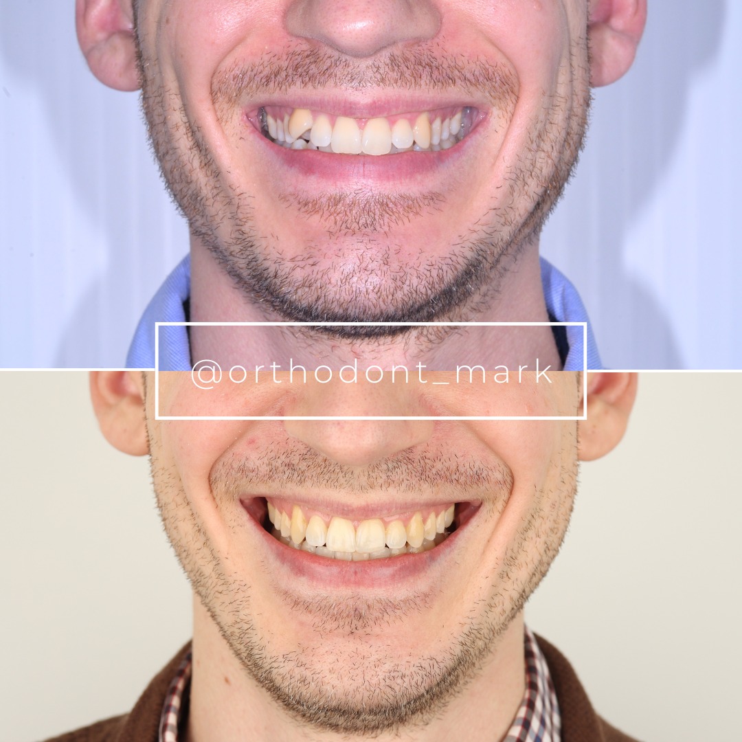
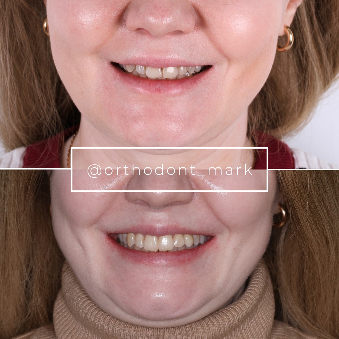
Documented outcomes
Clinical cases from Dr. Mark Radzhabov's practice
Learn the full MARPE protocol from Dr. Mark Radzhabov
Fundamental course covering CBCT patient selection, miniscrew planning, activation protocols, and 60+ clinical cases. Choose the access level that fits your practice.
Essentials of rapid palatal expansion for practicing orthodontists.
- Core RPE concepts and biomechanics
- 6 structured video lessons
- Clinical decision checklists
- Lifetime access to recordings
Deep-dive into MARPE protocol, diagnostics, and clinical execution.
- 6 modules covering MARPE from A to Z
- CBCT case selection walkthroughs
- Miniscrew placement and activation
- 60+ annotated clinical cases
- Direct Q&A with Dr. Mark Radzhabov
5-element medical consultation framework for dentists and orthodontists.
- Trust-building consultation protocol
- 5 lesson modules
- Templates for treatment plan delivery
- Works with any clinical specialty
Clinical FAQ
How does MARPE affect periodontal health compared to traditional tooth-borne rapid palatal expanders?
MARPE loads the midpalatal suture and hard palate directly, bypassing dental units and reducing buccal tipping. Gingival recession rates drop from 15–40% in tooth-borne systems to <5% in MARPE, and buccal cortical bone thickness is preserved rather than lost.
What baseline CBCT measurements predict high risk of gingival recession with expansion?
Buccal cortical bone thickness <1.5 mm, thin gingival biotype (<2 mm), and Angelieri suture stage C–D predict increased recession risk. Patients with these features should avoid tooth-borne expansion. Bone-borne or surgical options are preferable.
Can buccal bone dehiscence be reversed after rapid palatal expansion?
Fenestration (complete cortical bone loss) does not regenerate spontaneously. Partial cortical thinning may remodel over 12–24 months if force is removed and probing depth stabilizes. Early soft-tissue grafting may be indicated for esthetic concerns or to prevent deeper periodontal disease.
What is the optimal weekly activation rate to minimize bone loss during tooth-borne expansion?
One-quarter turn weekly (approximately 0.5 mm expansion per week) is superior to the historical twice-weekly protocol. Slower rates allow osteoclastic remodeling and reduce stress-shielding, resulting in 30–40% less buccal cortical resorption.
How does Angelieri staging guide the choice between tooth-borne and miniscrew-assisted expansion?
Stage A–B sutures favor both modalities. Stage B–C favor bone-borne systems. Stage C–D require bone-borne or surgical intervention. Staging predicts suture separation success and correlates with fenestration risk in tooth-borne protocols.
Should I monitor probing depth during active expansion, and how often?
Yes. Measure at baseline, 4 weeks, 12 weeks, and completion of active phase. An increase >1 mm or new bleeding on probing signals excessive load. Pause or reduce activation. Early detection prevents irreversible periodontal damage.
What is the ideal miniscrew force magnitude for MARPE, and how is it monitored clinically?
Total force of 100–150 cN across both miniscrews, delivered via daily activation over 4–6 weeks. Monitor suture separation on monthly orthopantomograms and palpate palatal tissue. If blanching occurs, reduce load or pause activation temporarily.
Does gingival biotype influence the success and periodontal outcome of rapid expansion?
Yes. Thick biotypes (>2 mm) remodel bone more predictably and tolerate tooth-borne forces better. Thin biotypes (<2 mm) experience faster recession and fenestration. Bone-borne systems strongly preferred for thin-biotype patients.
How long should retention last after tooth-borne versus bone-borne expansion?
Tooth-borne systems require 12–18 months of fixed retention to stabilize bone remodeling. Bone-borne systems achieve greater inherent stability; 8–12 months of fixed retention followed by removable retention often suffices, provided suture stage is B or earlier.
What post-expansion surgical options address buccal bone fenestration if it develops during treatment?
Soft-tissue grafting (acellular dermal matrix or connective tissue) addresses esthetic recession. Osseous recontouring or guided bone regeneration may stabilize defects but cannot regenerate lost cortical bone. Prevention via proper patient selection and force control is superior to late surgical repair.
The choice between tooth-borne and bone-borne expansion is fundamentally a periodontal decision. Bone-borne rapid palatal expansion systems demonstrate superior periodontal outcomes by bypassing dental unit stress and preserving buccal cortical bone height. However, accurate patient selection via CBCT staging and rigorous force management remain critical for all modalities. Clinicians who integrate periodontal risk assessment into their expansion protocol achieve more predictable, stable results. Review your current cases using the Angelieri staging system and consider a consultation with Dr. Mark Radzhabov at Orthodontist Mark to refine your patient selection criteria and treatment sequencing for maximal periodontal health.

