
Rapid Versus Slow Palatal Expansion
Protocols
Which Loading Rate Optimizes Skeletal Outcomes?
The velocity of midpalatal suture loading determines suture physiology, bone remodeling pattern, and stability. Learn evidence-based protocol selection for rapid maxillary expansion.
TL;DR Rapid palatal expansion protocols deliver 0.8–1.0 mm per week of skeletal widening, whereas slow expansion protocols (0.25–0.5 mm weekly) favor greater parallel separation and reduced dentoalveolar tipping. The choice depends on suture maturation stage, patient age, and desired skeletal-to-dental ratio. Rapid versus slow palatal expansion protocols differ fundamentally in their effect on midpalatal suture physiology and long-term stability.
The rate of palatal expansion fundamentally alters how the midpalatal suture responds—determining whether the result is primarily skeletal opening or compensatory dental tipping. Rapid versus slow palatal expansion protocols have been studied extensively since 2018, yet clinicians lack a unified framework for protocol selection. Dr. Mark Radzhabov synthesizes the biomechanical evidence and clinical outcomes of each approach, examining suture physiology, bone remodeling timelines, and the trade-offs between speed and stability. This reference addresses when to choose fast loading, when to apply gentle sustained pressure, and which radiographic markers predict optimal skeletal response across the spectrum of patient maturity.
What Defines Rapid Versus Slow
Expansion Protocols?
Rapid palatal expansion protocols apply 0.8–1.0 mm per week of skeletal widening, typically using miniscrew-assisted systems or high-force tooth-borne appliances. Slow expansion protocols, by contrast, deliver 0.25–0.5 mm weekly over extended intervals—3 to 6 months or longer—producing what clinicians observe as gradual suture opening with minimal dental anchorage loss. The physiological distinction is critical: rapid loading causes the midpalatal suture to open in a parallel, tent-like fashion, with new bone deposition occurring symmetrically across the anterior, middle, and posterior thirds. Slow expansion, by its sustained low-force nature, permits greater cellular reorganization within the suture and surrounding cortical plates, often resulting in a more uniform separation without the stress-induced resorption patterns seen in fast protocols. In skeletally mature patients, the choice between rapid and slow expansion determines not only the magnitude of skeletal gain but also the relapse risk, dentoalveolar compensation, and patient comfort during active treatment. A 45-year-old with stage C midpalatal suture maturation may achieve 6–8 mm of true skeletal widening with rapid loading over 3–4 months, whereas the same patient treated with slow expansion might require 6–9 months to achieve equivalent skeletal change but with lower dentoalveolar side effects.
Why Does Loading Rate Affect
Bone Formation?
Rapid palatal expansion loading initiates a cascade of stress-mediated osteoclast activation along the suture margins, followed by acute bone resorption and then robust osteoid deposition within 4–6 weeks. The high strain environment—created by forces exceeding 200 grams per miniscrew—triggers hyalinization (temporary necrosis of suture tissue), a process that paradoxically accelerates subsequent new bone formation as healing occurs. Slow expansion avoids hyalinization by applying sustained, moderate force (50–100 grams per miniscrew or distributed across tooth roots) that allows incremental osteoid formation without acute tissue death. Histological studies show that slow protocols preserve blood supply within the suture, maintaining osteoblast viability and reducing the risk of asymmetric bone resorption. The palatal expansion force application strategy also differs: rapid protocols often use daily or twice-daily activation, whereas slow systems rely on continuous fixed-force devices or very low weekly activation (0.25 mm per turn, once per week). Clinicians using bone-borne versus tooth-borne force paths report that bone-borne systems (MARPE, MSE) tolerate rapid loading better than tooth-borne RPE, which often produces maxillary dentoalveolar tipping and anchorage loss at loading rates above 0.5 mm weekly.
When Should You Choose Rapid
Expansion Timing?
Rapid expansion protocols excel in skeletally immature patients (cervical vertebral stage 3–4) and those with Angelieri stage A or B midpalatal suture maturation. Young patients tolerate rapid loading well, achieve predictable skeletal widening with minimal relapse, and benefit from shorter treatment duration. A 14-year-old with stage A suture and transverse maxillary constriction responds optimally to 0.8–1.0 mm weekly expansion, completing skeletal correction in 8–12 weeks. In adults over 40, rapid expansion protocols become more nuanced. Stage C and D sutures (partially fused, heavily ossified) present higher resistance to parallel opening. Rapid loading may produce uneven suture separation or stress concentration at the anterior suture. However, miniscrew-assisted rapid palatal expansion (MARPE) systems, by anchoring directly into cortical bone lateral to the suture, bypass dental anchorage and deliver predictable rapid loading even in mature sutures. A 52-year-old with stage C maturation can tolerate 0.8 mm weekly when force is applied to bone-borne miniscrews, whereas the same patient treated with tooth-borne RPE would risk significant maxillary dentoalveolar tipping and anchorage loss. Slow expansion protocols are preferred when: (1) the patient has stage D complete fusion, making parallel suture opening unlikely; (2) dentoalveolar side effects or patient comfort is a primary concern; (3) the goal is maximum skeletal-to-dental ratio without dental compromise. Or (4) the patient cannot tolerate weekly clinical activations.
Does Rapid Expansion Risk
Greater Relapse?
Relapse patterns differ significantly between rapid and slow protocols. Rapid expansion in young patients with stage A sutures produces 8–15% relapse over 12–24 months of retention, primarily due to elastic recoil of stretched periodontal ligament and incomplete mineralization of new suture bone. However, the absolute skeletal gain (6–8 mm) typically exceeds relapse (0.5–1.0 mm), leaving a net correction of 5–7 mm. Slow expansion shows lower percentage relapse (5–10%) because the gradual loading allows continuous bone remodeling and adaptive dentoalveolar repositioning. A patient who gains 4–5 mm over 9 months of slow loading may lose only 0.2–0.4 mm during retention, for a net stability of 3.6–4.8 mm. This advantage is most evident in stage C–D sutures, where slow loading achieves greater parallel bone formation and faster secondary mineralization. Dentoalveolar side effects—maxillary incisor proclination, buccal alveolar plate thinning, root resorption—occur more frequently with rapid tooth-borne RPE and less with rapid bone-borne MARPE or slow expansion. Rapid RPE in adults produces 3–4 mm of unwanted dentoalveolar tipping. Rapid MARPE produces <1 mm. The expansion suture physiology thus drives clinical decision-making: bone-borne rapid protocols offer speed with stability, whereas slow tooth-borne expansion minimizes side effects but extends active treatment.
How Do You Tailor Expansion Rate
to Your Patient?
Begin with high-resolution cone-beam CT imaging and cervical vertebral staging to determine suture maturation and skeletal growth status. In patients under 15 (stage 3–4 cervical vertebrae, stage A–B suture), rapid expansion is standard: activate 0.8–1.0 mm weekly, expect 8–12 week active phase, and retain for 12–24 months to allow secondary mineralization. In adults 25–40 (stage 4–5 cervical vertebrae, stage B–C suture), consider bone-borne rapid expansion (MARPE) if true skeletal widening is the primary goal and dentoalveolar side effects must be minimized. This cohort tolerates 0.8–1.0 mm weekly loading when force is applied to miniscrews, achieving 6–8 mm net skeletal gain in 3–4 months. If tooth-borne RPE is selected instead, reduce loading to 0.5–0.6 mm weekly to limit maxillary dentoalveolar tipping to acceptable levels (<2 mm). In patients over 50 (stage 5–6 cervical vertebrae, stage C–D suture), rapid loading becomes problematic: suture resistance increases, and the stress is transferred to alveolar bone. A slow expansion protocol (0.25–0.5 mm weekly) over 6–9 months yields greater parallel suture opening and lower risk of asymmetric resorption. For this cohort, consider discussing whether true skeletal expansion is achievable without surgical assistance, as stage D fusion may preclude >4–5 mm of skeletal gain regardless of protocol choice.
Free Clinical Guide
Which Expansion Appliance at Which Age?
Get Dr. Mark Radzhabov's guide to skeletal-expansion appliances — indications, pros & cons, and how to choose by patient age. Free to your inbox.
Prefer video? Watch the free demo lesson on MARPE appliance types, features and trade-offs.
Documented outcomes
Clinical cases from Dr. Mark Radzhabov's practice
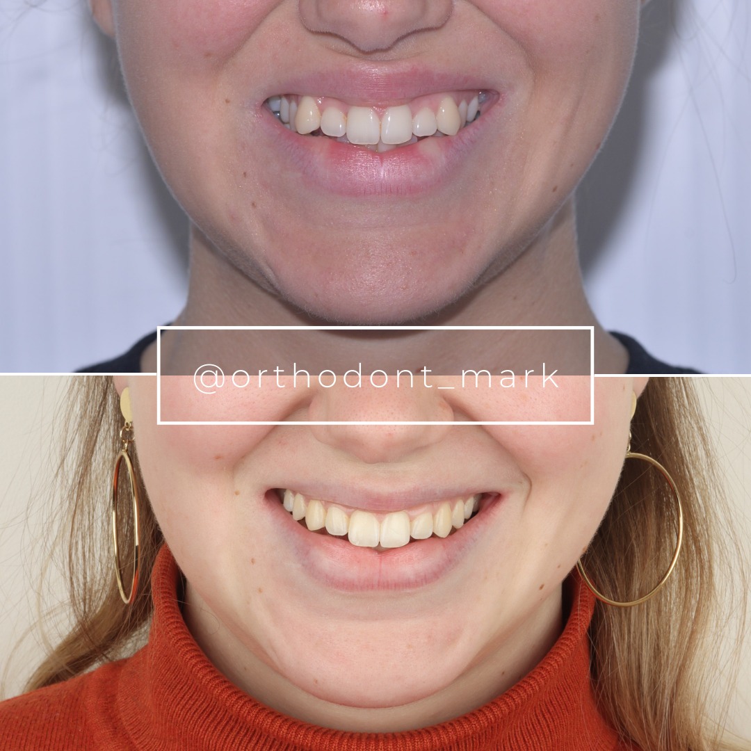

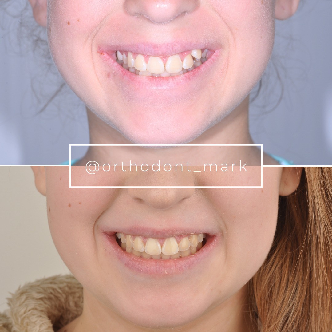
Learn the full MARPE protocol from Dr. Mark Radzhabov
Fundamental course covering CBCT patient selection, miniscrew planning, activation protocols, and 60+ clinical cases. Choose the access level that fits your practice.
Essentials of rapid palatal expansion for practicing orthodontists.
- Core RPE concepts and biomechanics
- 6 structured video lessons
- Clinical decision checklists
- Lifetime access to recordings
Deep-dive into MARPE protocol, diagnostics, and clinical execution.
- 6 modules covering MARPE from A to Z
- CBCT case selection walkthroughs
- Miniscrew placement and activation
- 60+ annotated clinical cases
- Direct Q&A with Dr. Mark Radzhabov
5-element medical consultation framework for dentists and orthodontists.
- Trust-building consultation protocol
- 5 lesson modules
- Templates for treatment plan delivery
- Works with any clinical specialty
Clinical FAQ
What is the optimal rate for palatal expansion in skeletally immature patients?
Rapid palatal expansion loading of 0.8–1.0 mm weekly is optimal in patients under 15 with cervical vertebral stage 3–4 and Angelieri stage A–B suture. Expected skeletal gain is 6–8 mm over 8–12 weeks with relapse of 8–15% (0.5–1.0 mm absolute).
How does rapid palatal expansion bone remodeling differ from slow protocols?
Rapid loading induces transient suture hyalinization and stress-mediated osteoclast activation, followed by robust osteoid formation. Slow loading (0.25–0.5 mm weekly) preserves vascular perfusion within the suture, allowing incremental bone formation without acute tissue death and reducing relapse risk.
When should I choose MARPE over RPE in adults?
MARPE is preferred in adults 25–50 with stage B–C midpalatal sutures when true skeletal expansion is the goal. Bone-borne miniscrew anchorage tolerates rapid loading (0.8–1.0 mm weekly) without dentoalveolar tipping. Tooth-borne RPE at the same rate produces 3–4 mm unwanted maxillary dentoalveolar compensation.
Does rapid maxillary expansion rate produce greater relapse?
Rapid expansion produces 8–15% relapse compared to 5–10% for slow protocols, but absolute relapse (mm) is often lower due to greater initial skeletal gain. Rapid MARPE in stage A–B sutures achieves net stable widening of 5–7 mm. Slow expansion gains 3.6–4.8 mm with lower percentage loss.
What expansion protocol bone formation occurs in stage C–D mature sutures?
Stage C–D sutures show delayed and asymmetric bone formation with any protocol. Slow expansion (0.25–0.5 mm weekly) produces greater parallel separation than rapid loading. Consider whether 4–5 mm skeletal gain is achievable without surgical intervention in this cohort.
How do I activate miniscrew-assisted rapid palatal expansion MSE correctly?
Insert grade 5 titanium miniscrews 8–10 mm into cortical bone lateral to the midpalatal suture at the level of the first premolar and molar. Activate 0.8–1.0 mm weekly (quarter-turn per day or half-turn every 2 days) for 3–4 weeks until target expansion is achieved, then maintain for 2–4 weeks before deactivation.
What skeletal expansion orthodontics outcome can I expect in a 45-year-old patient?
A 45-year-old with stage C suture and MARPE can achieve 6–8 mm skeletal gain over 3–4 months of rapid loading (0.8–1.0 mm weekly). Slow expansion yields 4–5 mm over 6–9 months with lower relapse. Clinical response depends on suture staging. Assess CBCT before treatment to confirm feasibility.
How long should retention last after rapid or slow palatal expansion?
Retain for 12–24 months after rapid expansion to allow secondary mineralization and reduce relapse. After slow expansion, retain for 9–12 months. Use fixed or removable retention. Consider slow, light forces (acrylic splint) to resist relapse during early retention phases.
What dentoalveolar side effects occur with rapid versus slow RPE?
Rapid tooth-borne RPE produces 3–4 mm maxillary incisor proclination, buccal alveolar thinning, and risk of root resorption. Slow RPE (0.25–0.5 mm weekly) produces <1 mm tipping. Rapid MARPE shows minimal dentoalveolar tipping (<1 mm) regardless of speed because force bypasses dental roots.
What midpalatal suture physiology marker predicts treatment success?
Angelieri staging on high-resolution CBCT predicts outcome better than age alone. Stage A–B sutures respond to rapid loading with parallel opening. Stage C shows mixed response. Stage D rarely achieves >4–5 mm true skeletal widening. Combine staging with cervical vertebral assessment to refine protocol selection.
Protocol selection is not arbitrary: the velocity of expansion force directly determines whether bone forms in parallel laminae or whether the suture fails to open uniformly, risking relapse and asymmetry. Clinicians who match expansion rate to suture maturation stage and patient skeletal status achieve superior long-term outcomes and lower retreatment burden. Dr. Mark Radzhabov's clinical research demonstrates that evidence-based protocol decisions—informed by cone-beam CT imaging and cervical vertebral staging—yield predictable skeletal widening without unwanted dental side effects. Review your next case with these principles and consider consulting Orthodontist Mark's protocol library to refine your own expansion strategy.

