
Diagnosing Transverse Maxillary Discrepancy:
Clinical, Radiographic, and CBCT Integration
A Evidence-Based Protocol for Adult Expansion Planning
Master the clinical, cephalometric, and CBCT methods to differentiate skeletal transverse deficiency from dental compensation—and select the optimal expansion strategy.
TL;DR Diagnosing transverse maxillary discrepancy requires integrating clinical posterior crossbite assessment with cephalometric transverse measurements and CBCT analysis of midpalatal suture maturation. A skeletal transverse deficiency is confirmed when maxillomandibular width analysis shows ≥7.5 mm posterior maxillary constriction relative to mandibular width, combined with radiographic evidence of skeletal rather than dental compensation.
Transverse maxillary discrepancy remains underdiagnosed in adult orthodontics, often masked by dental compensation or attributed solely to dento-alveolar crossbite. Accurate diagnosis of skeletal transverse deficiency determines whether treatment should employ tooth-borne rapid palatal expanders, miniscrew-assisted systems, or surgical intervention. This guide synthesizes clinical examination protocols, cephalometric transverse measurements, and CBCT interpretation—drawing on Dr. Mark Radzhabov's evidence-based approach—to help orthodontists distinguish true skeletal transverse maxillary deficiency from purely dental findings and select the most appropriate expansion strategy.
What Is Transverse Maxillary Discrepancy?
Clinical Reality
Transverse maxillary discrepancy is a skeletal or dento-alveolar mismatch in which the maxillary width is insufficient relative to mandibular width, resulting in posterior crossbite and functional compromise. Unlike simple dento-alveolar narrowing, a true skeletal transverse deficiency reflects a fundamental mismatch in bone morphology that cannot be resolved through dentoalveolar tipping alone.
In contemporary practice, clinicians often encounter patients with posterior crossbite who exhibit mixed skeletal–dental patterns. A 38-year-old may present with unilateral right crossbite, yet maxillomandibular width analysis reveals ≥7.5 mm skeletal discrepancy masked by lingual root torque and dentoalveolar compensation. The stakes are high: treating such cases with conventional tooth-borne rapid palatal expanders risks anchorage loss and relapse, whereas recognizing the skeletal component opens the door to more stable expansion methods.
Accurate diagnosis requires integrating three data streams: clinical posterior crossbite assessment, cephalometric transverse measurements (inter-molar widths, palatal vault dimensions), and CBCT analysis of skeletal anatomy and midpalatal suture maturation. No single tool suffices. This multi-modal framework prevents misclassification and guides selection between conventional expansion, miniscrew-assisted systems, or surgical correction.
Identifying Posterior Crossbite:
Beyond the Bite
Clinical red flags for skeletal transverse deficiency
Clinical assessment begins with functional assessment in CO and CR. A patient exhibiting unilateral or bilateral posterior crossbite with a functional midline shift toward the narrower side often has underlying skeletal transverse deficiency rather than pure dental malocclusion. Palpate for mandibular deflection during closure. A pronounced shift >2 mm correlates with significant skeletal constraint.
Next, evaluate palatal vault morphology and maxillary incisor inclination. A high, narrow palate combined with severely retroclined maxillary incisors suggests long-standing dentoalveolar compensation for skeletal constriction. Measure inter-canine and inter-molar widths (deciduous and permanent) using digital calipers. Maxillary first molar width <31 mm in an adult signals skeletal involvement; widths <29 mm strongly suggest transverse maxillary deficiency requiring skeletal correction. Mandibular inter-molar width typically remains stable (36–38 mm adult range), so a discrepancy >6–7 mm between maxillary and mandibular molars is clinically significant.
Assess buccal corridor fullness and smile arc symmetry. Patients with unilateral crossbite often display asymmetric buccal corridors. Bilateral narrowing with loss of corridor volume frequently reflects true skeletal constriction rather than unilateral muscular adaptation. These observations flag the need for radiographic confirmation but do not diagnose skeletal pathology alone.
Cephalometric Analysis for Palatal Width Assessment
Quantifying Skeletal Discrepancy
Lateral cephalometrics provide essential cephalometric transverse measurements via indirect proxies. While frontal cephalograms yield more direct inter-molar and inter-canine widths, they are not standard in many practices. Lateral radiographs allow measurement of palatal depth and approximation of maxillary width constraints. Measure maxillary intermolar width (MMW) and mandibular intermolar width (MMdW) from the mesiobuccal cusp tip of maxillary first molars to the mesiobuccal cusp of the contralateral maxillary first molar (and analogously for the mandible). A discrepancy of ≥7.5 mm (maxillary narrower) is consistent with skeletal transverse deficiency.
Palatal vault depth, measured as the perpendicular from the hard palate midline to a line connecting the first molar widths, typically increases when skeletal narrowing is present, creating a characteristic high, narrow vault. Measure intercanine widths at the mesial contact point. Maxillary canine width <25.5 mm in females or <27 mm in males may signal transverse constraint. These linear measurements, combined, establish a maxillomandibular width analysis that distinguishes skeletal from purely dental patterns.
Do not rely on individual variables. A patient with moderate inter-molar discrepancy (5–6 mm) but normal palatal depth and normal canine width likely has dento-alveolar compensation. Conversely, a patient with ≥7.5 mm inter-molar discrepancy plus high palatal vault plus retroclined maxillary incisors meets strong radiographic criteria for skeletal transverse deficiency, warranting advanced imaging and consideration of skeletal expansion methods.
CBCT Analysis: Midpalatal Suture Staging and Skeletal Maturity
Predicting Success Without Surgery
Cone-beam computed tomography (CBCT) with midpalatal suture staging via the Angelieri classification (introduced 2016) has become the diagnostic standard for determining expansion feasibility in adults. The system grades suture density across anterior, middle, and posterior thirds on an axial slice at the level of the mesiobuccal root of the maxillary first molar, spanning from the nasal spine to the posterior nasal spine. Stage A (low density, patent suture) predicts the highest probability of skeletal separation; Stage B (mixed density, early fusion foci) shows moderate expansion potential; Stage C (predominantly fused, scattered low-density areas) carries higher relapse risk; Stage D (complete fusion) typically requires surgical intervention.
Bone density assessment using Hounsfield units (HU) in the midpalatal region strengthens prognostication. Anterior palate cortical bone <800 HU indicates relatively low mineralization and favorable expansion response. Posterior palate density >1000 HU signals advanced ossification, suggesting limited non-surgical expansion potential. A 35-year-old patient in Stage A with anterior density <700 HU has >85% likelihood of achieving ≥6–8 mm true skeletal gain via miniscrew-assisted expansion. Conversely, a 50-year-old in Stage C or D, or any patient with posterior density >1200 HU, should be counseled toward surgical correction or conservative treatment alternatives.
Evaluate suture maturation asymmetry: anterior fusion with posterior patency is favorable. Posterior-only fusion with anterior patency is less predictable. Regional variation must be documented. CBCT also reveals maxillary bone thickness at proposed miniscrew insertion sites (typically 4–6 mm cortical bone required for stable anchorage), confirming feasibility before appliance delivery. This radiographic staging, integrated with clinical and cephalometric findings, forms the final diagnostic step.
From Diagnosis to Treatment Selection
Matching Patient Phenotype to Method
Once transverse maxillary deficiency is confirmed—clinical posterior crossbite + cephalometric inter-molar discrepancy ≥7.5 mm + palatal narrowing—the next step is selecting the appropriate expansion method. Integrate skeletal maturity (Angelieri stage, cervical vertebral stage if available), bone quality, patient age, and occlusal complexity.
A 28-year-old in Stage A with 8 mm inter-molar discrepancy, mild unilateral crossbite, and good posterior alveolar bone height is a prime candidate for miniscrew-assisted rapid palatal expansion (MARPE) or MSE systems, which apply force directly to the hard palate and minimize dentoalveolar side effects. Activation protocols typically use 0.5–0.75 mm per week, with weekly patient activation or clinician-controlled turns. Expected skeletal gain: 6–8 mm over 10–12 weeks of active expansion, followed by a 3–4 month consolidation phase.
A 45-year-old in Stage B or C, or a patient with significant dentoalveolar crowding alongside transverse deficiency, may benefit from surgical-assisted rapid palatal expansion (SARPE) or combined orthognathic surgery. Non-surgical expansion in advanced-fusion patients frequently results in >15% relapse and patient frustration. Dr. Mark Radzhabov's clinical practice emphasizes this decision point: accurate diagnosis prevents inappropriate appliance selection and wasted treatment time.
For purely dento-alveolar cases (normal cephalometric inter-molar width, crossbite limited to one or two teeth, Stage A or B suture), conventional tooth-borne rapid palatal expanders remain appropriate, though bone-borne systems offer superior skeletal gain and stability.
Free Clinical Guide
Which Expansion Appliance at Which Age?
Get Dr. Mark Radzhabov's guide to skeletal-expansion appliances — indications, pros & cons, and how to choose by patient age. Free to your inbox.
Prefer video? Watch the free demo lesson on MARPE appliance types, features and trade-offs.
Documented outcomes
Clinical cases from Dr. Mark Radzhabov's practice
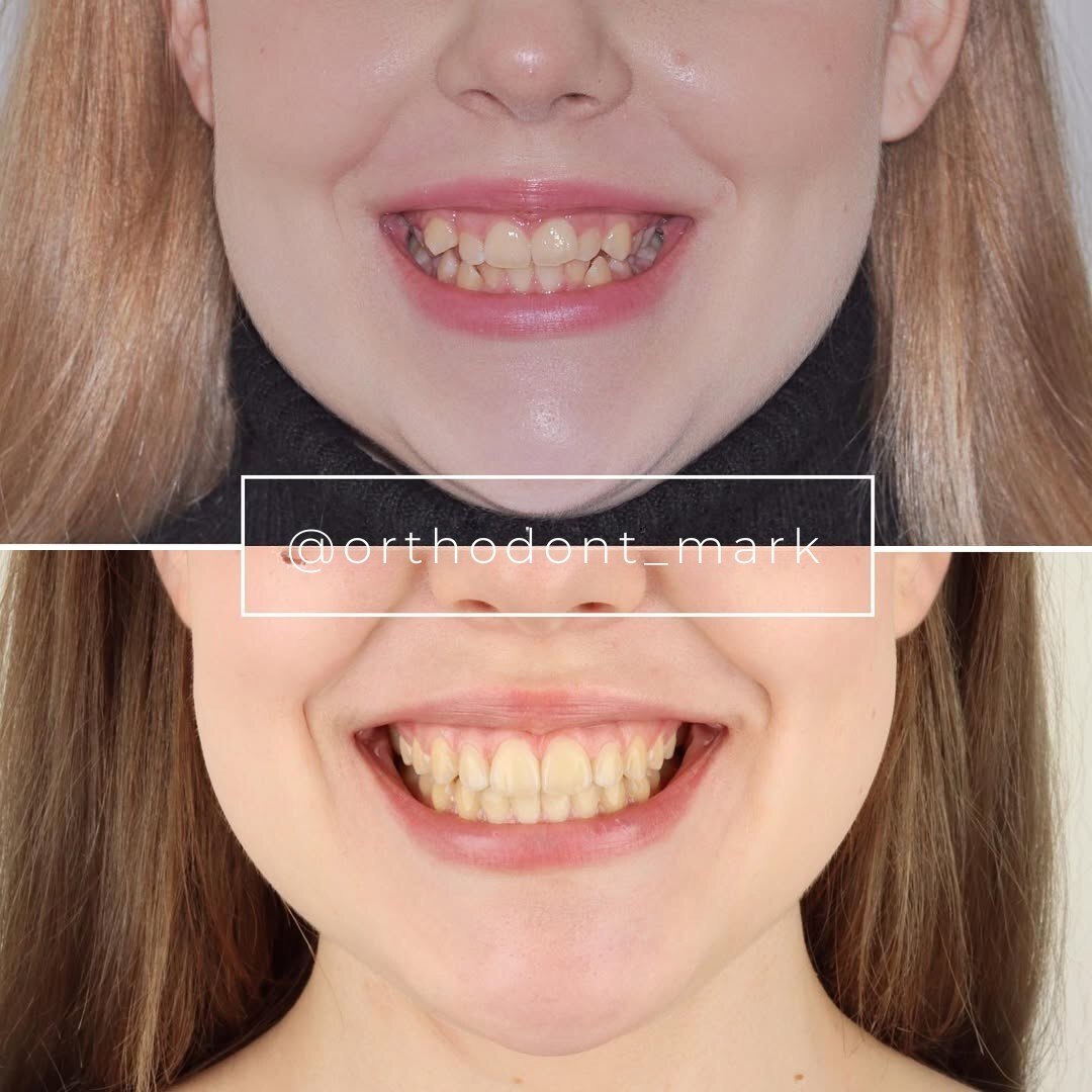
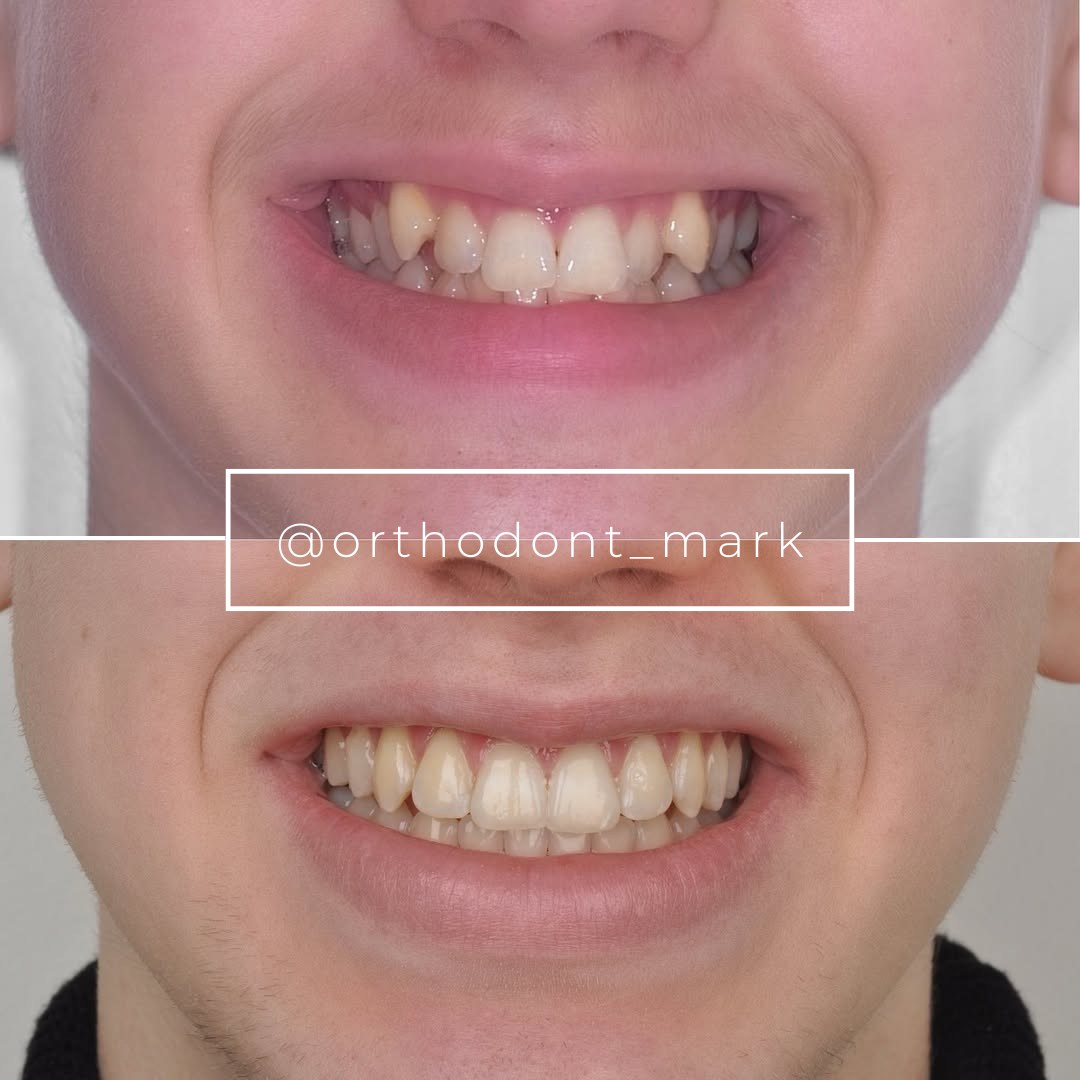

Learn the full MARPE protocol from Dr. Mark Radzhabov
Fundamental course covering CBCT patient selection, miniscrew planning, activation protocols, and 60+ clinical cases. Choose the access level that fits your practice.
Essentials of rapid palatal expansion for practicing orthodontists.
- Core RPE concepts and biomechanics
- 6 structured video lessons
- Clinical decision checklists
- Lifetime access to recordings
Deep-dive into MARPE protocol, diagnostics, and clinical execution.
- 6 modules covering MARPE from A to Z
- CBCT case selection walkthroughs
- Miniscrew placement and activation
- 60+ annotated clinical cases
- Direct Q&A with Dr. Mark Radzhabov
5-element medical consultation framework for dentists and orthodontists.
- Trust-building consultation protocol
- 5 lesson modules
- Templates for treatment plan delivery
- Works with any clinical specialty
Clinical FAQ
What cephalometric measurement indicates skeletal transverse deficiency in adults?
Maxillomandibular width analysis showing ≥7.5 mm discrepancy (maxillary width minus mandibular inter-molar width) is the threshold. Combine with palatal vault depth >10 mm and retroclined maxillary incisors for skeletal confirmation.
How do I differentiate skeletal transverse deficiency from dento-alveolar compensation?
Skeletal cases show high palatal vault, retroclined incisors, and stable mandibular inter-molar width with narrow maxilla. Dento-alveolar cases have normal vault depth, normally inclined incisors, and smaller inter-molar discrepancies. CBCT suture staging confirms.
What is the Angelieri staging system, and why is it used?
Angelieri classification grades midpalatal suture density (Stages A–D) on CBCT axial images. Stage A (patent) predicts 80–90% non-surgical expansion success. Stage D (fused) typically requires surgery. It predicts skeletal gain and relapse risk better than age alone.
What Hounsfield unit threshold indicates favorable expansion response?
Anterior palate cortical bone <800 HU with posterior <1000 HU suggests favorable expansion response. HU >1200 in posterior palate signals advanced ossification. Such patients carry higher relapse risk and may need surgical correction.
When should I refer for CBCT instead of relying on cephalometry alone?
CBCT is indicated when posterior crossbite is present, cephalometric inter-molar discrepancy is ≥6 mm, or adult age >35 years. It clarifies midpalatal suture maturity and palatal bone anatomy for surgical or MARPE planning.
How does midline shift in centric relation relate to transverse deficiency diagnosis?
Functional midline shift >2 mm from CO to CR correlates with significant skeletal constraint. Combined with posterior crossbite and cephalometric discrepancy, it signals skeletal transverse deficiency requiring imaging confirmation and non-trivial expansion planning.
What is the clinical significance of maxillary inter-molar width <31 mm?
Maxillary inter-molar width <31 mm in adults has high specificity for underlying skeletal transverse involvement. Widths <29 mm strongly suggest transverse maxillary deficiency requiring skeletal correction rather than dentoalveolar tipping.
How does palatal vault depth assessment improve transverse diagnosis?
High, narrow palate (vault depth >10 mm) combined with transverse constriction indicates long-standing skeletal constraint, not recent dental compensation. Normal vault depth with crossbite suggests dento-alveolar narrowing, not skeletal deficiency.
What role does cervical vertebral stage play in expansion prognosis?
Cervical vertebral maturation stage combined with midpalatal suture staging provides comprehensive skeletal maturity assessment. Early skeletal stages (CVMS 3–4) improve expansion stability. Advanced stages require careful suture-specific analysis via CBCT.
How do I counsel patients on relapse risk after diagnosing transverse maxillary deficiency?
Stage A/B patients: 8–12% relapse over 6 months. Emphasize retention protocol. Stage C patients: 12–18% relapse. Consider surgical correction. Stage D: >20% relapse. SARPE often preferred. Integrate suture stage and bone density into relapse prediction.
Early and accurate diagnosis of transverse maxillary discrepancy reshapes treatment planning and improves long-term stability. The integration of cephalometric transverse measurements, clinical posterior crossbite assessment, and midpalatal suture staging via CBCT creates a robust diagnostic framework that predicts both the feasibility and optimal timing of skeletal expansion. Dr. Mark Radzhabov's clinical protocols emphasize this multi-modal approach to avoid misdiagnosis. Review your posterior crossbite cases with these diagnostic criteria, or schedule a consultation to refine your approach to skeletal transverse expansion.

