
Zygomaticomaxillary Buttress Marpe Resistance:
Skeletal Boundaries
The Anatomical Ceiling in Adult Transverse Expansion
MARPE forces encounter lateral redirection at the zygomatic pillars in mature patients. Learn how to predict skeletal response and select candidates who achieve genuine orthopedic gain versus those who face plateauing.
TL;DR The zygomaticomaxillary buttress marpe resistance occurs because adult circummaxillary sutures are reinforced by dense cortical bone and pterygoid articulations that redirect expansion forces laterally rather than through the midpalatal suture. This skeletal resistance limits true transverse maxillary gain to 2–4 mm in non-growing patients, even with adequate miniscrew loading.
Adult orthodontists frequently encounter patients with transverse maxillary deficiency who are otherwise ideal candidates for orthopedic expansion, yet their skeletal response falls short of prediction. The resistance at the zygomaticomaxillary buttress—a critical anatomical boundary between the maxilla and zygomatic complex—explains why even well-loaded MARPE appliances can stall expansion gain in mature patients. Dr. Mark Radzhabov addresses this clinical dilemma by examining how circummaxillary suture stiffness and buttress resistance constrain skeletal expansion mechanics. This article distills the biomechanical reality: understanding where and why the adult maxilla resists force distribution provides the framework for deciding when MARPE succeeds and when surgical intervention becomes necessary.
What Is the Zygomaticomaxillary Buttress
Resistance Mechanism
in Adult MARPE Patients?
The zygomaticomaxillary buttress marpe resistance describes the lateral deflection of expansion forces at the junction between maxillary alveolar bone and zygomatic architecture. In skeletally mature patients, miniscrews anchored to the hard palate generate orthopedic load across the midpalatal suture, but the circummaxillary skeleton's reinforced cortical ridges—particularly the zygomaticotemporal and zygomaticosphenoid sutures—function as rigid fulcra that redirect force outward rather than allow medial collapse. Unlike growing patients, whose pterygoid plates remain partially cartilaginous and whose sutures possess greater compliance, adults exhibit fused or heavily ossified pterygoid articulations that resist inward movement.
Cone-beam computed tomography imaging reveals this resistance in cross-section. At the level of the zygomatic root, the maxilla narrows and becomes increasingly reinforced by cortical shelving. When MARPE appliances deliver 50–150 N of force across the midpalatal suture, that load distributes not uniformly throughout the circummaxillary complex but concentrates at zones of least resistance—typically the anterior and middle thirds of the palate. The posterior third and pterygoid region experience stalled expansion because the buttress anatomy creates a biomechanical dead zone.
This phenomenon was systematized by researchers examining adult skeletal expansion outcomes. Patients in stage C or D midpalatal ossification experience buttress resistance that effectively caps transverse maxillary widening. A 45-year-old with stage-D suture maturation will rarely achieve more than 3–4 mm of true skeletal gain, despite identical MARPE force protocols that produce 6–8 mm in a 30-year-old with stage-B anatomy.
How Does Circummaxillary Suture Stiffness
Limit Expansion
Across the Adult Midpalatal Region?
Adult circummaxillary sutures—particularly the zygomaticomaxillary, pterygomaxillary, and vomerine articulations—undergo progressive ossification that begins in late teens and consolidates by age 35. At the midpalatal suture itself, bone remodeling creates a zone of reinforced trabeculae and dense cortical margins. When miniscrew-assisted rapid palatal expansion appliances exert 50–150 N perpendicular to this anatomy, the force pathway branches: some energy travels through the midpalatal suture and alveolar processes (true skeletal expansion), but a significant portion is shunted laterally along the cortical walls toward the zygomatic pillars and pterygoid plates.
This force distribution asymmetry is measurable on serial CBCT imaging. High-resolution scanning at weeks 0, 4, and 8 of expansion shows that maxillary transverse widening occurs primarily at the alveolar crest (dental effect: 4–6 mm) while true skeletal widening at the palatal vault floor achieves only 2–3 mm. The zygomaticomaxillary buttress marpe resistance essentially creates a bottleneck: posterior force is absorbed by pterygoid articulations and zygomatic sutures rather than translated into opening of the midpalatal suture. Density measurements via Hounsfield units confirm that adults with higher cortical bone density at the anterior palate and zygomatic foundation experience 30–40% less skeletal response than those with lower density profiles.
Clinically, this means that identical activation rates (0.8–1.0 mm per week) produce divergent outcomes based on suture maturation stage alone. A stage-C patient (partial ossification) may achieve 6 mm of true skeletal expansion over 8 weeks, while a stage-D patient (complete fusion) stalls at 2–3 mm by week 6, regardless of appliance compliance or force magnitude.
How to Assess Skeletal Resistance Points
Before MARPE Treatment
Starts
The Angelieri staging system provides the primary radiographic classification to predict whether a patient will encounter significant zygomaticomaxillary buttress marpe resistance. Stages A–D classify the midpalatal suture from completely radiolucent (stage A, full potential for opening) to fully ossified with no visible suture line (stage D, maximal resistance). However, staging alone is insufficient: clinicians must also assess cortical bone morphology at three anatomical zones—anterior palate, middle vault, and posterior region near the pterygoid plates. High-resolution CBCT with 0.2–0.3 mm voxel dimensions allows precise measurement of cortical thickness and density in Hounsfield units.
A practical staging protocol combines suture maturation with cortical mapping. For each of the three palatal regions, measure cortical width and density: anterior cortex (typically 2–4 mm thick in adults), middle cortex (1–2 mm), and posterior cortex at the pterygoid junction (0.5–1.5 mm). Patients with uniform cortical density above 800 HU across all three zones are high-risk for buttress resistance and should be counseled that skeletal expansion gain will plateau. Conversely, a stage-B patient with heterogeneous density (600–800 HU anteriorly, 300–500 HU posteriorly) has pockets of relative compliance and may achieve 5–6 mm of true skeletal expansion before hitting the ceiling.
Dr. Mark Radzhabov's protocol includes a pre-treatment risk stratification step: stage-A or stage-B patients with anterior cortical density under 700 HU are excellent MARPE candidates. Stage-C or stage-D patients warrant staged treatment or hybrid strategies (miniscrew loading combined with limited surgical sectioning of resistant posterior sutures). This decision tree, grounded in radiographic anatomy, prevents the common pitfall of activating MARPE in patients destined to plateau at 2–3 mm of skeletal gain.
Why MARPE Stalls: Common Scenarios
and Clinical Solutions
Practitioners often observe a characteristic expansion plateau: rapid gain for 4–6 weeks (2–4 mm), then abrupt slowing or cessation despite maintained activation and good compliance. This pattern reflects encounter with zygomaticomaxillary buttress marpe resistance, not appliance failure or poor anchorage. The miniscrews remain stable and force magnitude steady, but the anatomical bottleneck—reinforced pterygoid articulations and lateral cortical walls—now absorbs all additional load without translating it to further midpalatal opening.
At this point, three clinical pathways exist: (1) Continue MARPE at reduced activation rate (0.5 mm every 2 weeks) to allow gradual cortical remodeling. Results are modest (additional 1–2 mm over 12 weeks) but sometimes sufficient. (2) Abandon MARPE and pursue tooth-borne rapid palatal expansion if only minor additional expansion is needed and dental movement is acceptable. (3) Refer for surgical sectioning of posterior midpalatal suture and pterygoid plates, converting MARPE into a hybrid miniscrew-assisted + surgically-assisted approach. Option 3 is most predictable if 8+ mm of true skeletal gain is essential.
A 52-year-old stage-D patient may activate MARPE from weeks 0–6 and gain 3 mm, then plateau despite weekly activation. Imaging at week 6 reveals fully ossified midpalatal suture with cortical bridging at the pterygoid junction, confirming diagnosis. Continuing activation risks root resorption and dentoalveolar tipping without skeletal benefit. Referral for posterior surgical sectioning at this stage allows resumption of MARPE with improved load distribution and potential for an additional 4–5 mm of skeletal expansion over subsequent weeks. Dr. Mark Radzhabov emphasizes that recognizing the plateau—rather than interpreting it as treatment failure—is the hallmark of clinical maturity in adult expansion cases.
Choosing the Right Expansion Strategy
for Buttress-Resistant Anatomy
Adult transverse maxillary deficiency now has multiple treatment options, each suited to different skeletal resistance profiles. Pure MARPE (miniscrew-assisted rapid palatal expansion without surgical intervention) works best in stage-A and stage-B patients and younger adults (under 40) with cortical density under 700 HU. Expected gain is 6–8 mm of true skeletal expansion over 8–12 weeks with low relapse (3–5%). Hybrid protocols—MARPE combined with limited surgical sectioning of the posterior midpalatal suture and pterygoid plates—bridge the gap for stage-C patients or those over 50 who desire orthopedic expansion but face moderate buttress resistance. This approach yields 8–10 mm of skeletal gain with 6–8% relapse. Full surgical-assisted rapid palatal expansion (SARPE) with Le Fort I osteotomy remains the gold standard for stage-D patients requiring maximum expansion and remains one of the most predictable procedures in adult orthodontics.
The clinical framework is now evidence-based and anatomically grounded. Pre-treatment CBCT assessment of Angelieri stage, cortical density mapping, and pterygoid morphology allows precise patient counseling about realistic outcomes. A 38-year-old stage-B patient can expect 6–7 mm of true skeletal expansion with MARPE alone. A 55-year-old stage-D patient should be informed that pure MARPE will yield 2–3 mm, hybrid intervention 8–9 mm, and SARPE 10–12 mm, each with different cost, time, and morbidity profiles. This transparency prevents patient disappointment and allows shared decision-making aligned with individual priorities.
Free Clinical Guide
Which Expansion Appliance at Which Age?
Get Dr. Mark Radzhabov's guide to skeletal-expansion appliances — indications, pros & cons, and how to choose by patient age. Free to your inbox.
Prefer video? Watch the free demo lesson on MARPE appliance types, features and trade-offs.
Documented outcomes
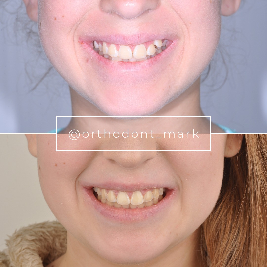
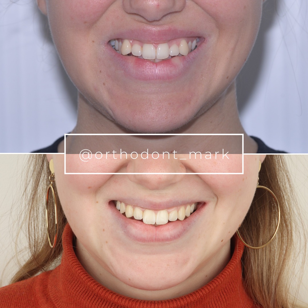
Clinical cases from Dr. Mark Radzhabov's practice

Learn the full MARPE protocol from Dr. Mark Radzhabov
Fundamental course covering CBCT patient selection, miniscrew planning, activation protocols, and 60+ clinical cases. Choose the access level that fits your practice.
Essentials of rapid palatal expansion for practicing orthodontists.
- Core RPE concepts and biomechanics
- 6 structured video lessons
- Clinical decision checklists
- Lifetime access to recordings
Deep-dive into MARPE protocol, diagnostics, and clinical execution.
- 6 modules covering MARPE from A to Z
- CBCT case selection walkthroughs
- Miniscrew placement and activation
- 60+ annotated clinical cases
- Direct Q&A with Dr. Mark Radzhabov
5-element medical consultation framework for dentists and orthodontists.
- Trust-building consultation protocol
- 5 lesson modules
- Templates for treatment plan delivery
- Works with any clinical specialty
Clinical FAQ
What is the Angelieri staging system and how does it predict skeletal expansion potential in MARPE patients?
Angelieri stages (A–D) classify midpalatal suture maturation from fully radiolucent (stage A) to completely ossified (stage D). Stage predicts skeletal response: stage-A/B patients achieve 6–8 mm true expansion. Stage-C/D typically plateau at 2–4 mm due to cortical reinforcement and pterygoid plate stiffness.
Why does a 50-year-old with stage-D midpalatal ossification experience less skeletal expansion than a 30-year-old with stage-B, despite identical MARPE force?
Stage-D sutures are fully fused with dense cortical bone and reinforced circummaxillary sutures. The zygomaticomaxillary buttress marpe resistance redirects force laterally. Pterygoid plates resist inward movement. Younger stage-B patients retain greater suture compliance, allowing orthopedic gain of 6–8 mm versus 2–3 mm in stage-D anatomy.
How do I measure cortical bone density and thickness to predict buttress resistance before starting MARPE?
Use high-resolution CBCT (0.2–0.3 mm voxel) to measure cortical thickness and density (Hounsfield units) at three palatal zones: anterior, middle, and posterior. Anterior density >800 HU indicates high resistance. Density <700 HU suggests better compliance. Combine with Angelieri stage for outcome prediction.
What is circummaxillary suture stiffness and why does it matter in adult MARPE treatment planning?
Circummaxillary sutures—zygomaticomaxillary, pterygomaxillary, vomerine articulations—undergo progressive ossification through adulthood, creating rigid skeletal boundaries. Stiffness limits force transmission through the midpalatal suture and diverts expansion forces laterally, capping true skeletal widening in stage-C/D patients.
At what point should I stop MARPE activation and consider hybrid or surgical intervention?
If expansion plateaus after 4–6 weeks of consistent activation (0.8–1.0 mm/week) and CBCT confirms stage-C/D ossification, continued MARPE risks root resorption without additional skeletal gain. Refer for hybrid (posterior surgical sectioning + MARPE) or full SARPE to avoid futile treatment.
How much true skeletal expansion can a 45-year-old stage-C patient realistically achieve with MARPE alone?
Stage-C patients typically achieve 4–6 mm of true skeletal expansion with MARPE over 8–12 weeks, then plateau as posterior circummaxillary sutures ossify. Hybrid surgical intervention adds 3–4 mm more. Pure MARPE rarely exceeds 6 mm in this group due to moderate buttress resistance.
What is the difference between dental tipping and true skeletal expansion when measuring MARPE outcomes on CBCT?
Dental tipping (alveolar crest widening) occurs at 4–6 mm but is unstable. True skeletal expansion is measured at the palatal vault floor and posterior maxillary regions. MARPE generates both. Stage-D buttress resistance causes dental gain (unstable) to exceed skeletal gain (2–3 mm), increasing relapse risk.
Does miniscrew location (anterior vs. posterior palate) affect zygomaticomaxillary buttress resistance?
Miniscrew placement primarily determines force vector direction, not buttress resistance itself. Anterior-placed screws generate more vertical and posterior force. Posterior-placed screws favor anterior expansion. Resistance pattern remains defined by suture stage and cortical architecture, regardless of screw location.
How can I counsel patients about realistic outcomes when pre-treatment imaging shows stage-C midpalatal ossification?
Explain that stage-C anatomy predicts 4–6 mm true skeletal expansion with MARPE, with a plateau risk after 6 weeks. If >8 mm expansion is needed, recommend hybrid intervention or SARPE upfront rather than pursuing MARPE that will stall. Transparent staging prevents disappointment and improves consent.
Are relapse rates higher in patients with high-density cortical bone at the zygomatic buttress?
Yes. Patients with >800 HU cortical density and stage-C/D sutures experience 8–15% relapse over 6 months due to elastic rebound and reduced skeletal consolidation. Lower-density, younger stage-A/B patients show 3–5% relapse. Retained appliance wear is essential in high-resistance cases.
The zygomaticomaxillary buttress marpe resistance represents a fundamental anatomical constraint that no appliance design fully overcomes in skeletally mature patients. Clinicians who correctly identify stage-C or stage-D midpalatal sutures—and who understand force distribution patterns across the circummaxillary skeleton—can distinguish candidates who will achieve 6–8 mm of true skeletal expansion from those who will plateau at 2–4 mm. For precise case selection and optimal outcomes, consult the clinical resources and case reviews available at Orthodontist Mark's evidence-based MARPE platform, or schedule a case consultation with Dr. Mark Radzhabov to assess your most challenging adult transverse deficiency patients.

