
First Week of RPE Activation:
Tissue Response,
Warning Signs & the Parent Briefing Script
A clinician-focused framework for monitoring palatal tissue, interpreting early osseous response, and structuring the post-activation conversation with families.
TL;DR The first week of RPE activation produces predictable soft-tissue and osseous responses, but suture maturation status critically shapes that response. Clinicians must distinguish transient discomfort from true warning signs—palatal blanching, periodontal recession, or absence of diastema—and brief parents on both before the patient leaves the chair.
Managing the first week of RPE activation well is what separates uneventful expansion from avoidable complications that erode patient trust and case outcomes. In adolescent patients, suture closure is already underway at ages that surprise many clinicians, making early tissue monitoring non-negotiable. Dr. Mark Radzhabov at ortodontmark.com has compiled this clinical guide to help practicing orthodontists navigate the post-activation tissue timeline, recognize genuine warning signs, and deliver a structured parent briefing that reduces panic calls and supports protocol adherence.
What Does the First Week of RPE Activation
Actually Tell You
Clinically?
The first week of RPE activation is the critical post-loading window during which clinicians document diastema formation, palatal tissue response, and early signs of suture opening or complication. The midline diastema — typically visible between the maxillary central incisors within the first two to three days of activation — is the most accessible clinical indicator that sutural forces are being transmitted skeletally rather than absorbed dentally. Absence of any diastema by day four warrants immediate reassessment of appliance fit, screw mechanics, and suture patency before continuing the activation schedule. Tissue blanching directly beneath the acrylic or metallic framework is an early pressure indicator that should be assessed at each activation visit. Persistent blanching beyond forty-eight hours, rather than transient post-activation pallor, signals excessive mucosal compression and requires appliance adjustment. Palatal tissue necrosis, periodontal recession, and failure of suture opening are all documented side effects following RME in skeletally mature patients, according to Omezli et al. (2020), making tissue surveillance in this age group particularly non-negotiable. The expansion vector also matters. Kinzinger et al. (2022) demonstrated that RME produces parallel expansion in children under 10 years old but shifts to a V-shaped opening pattern in adolescents aged 12 and older, meaning that the anterior diastema in an older patient will consistently appear wider than the posterior dental width gain. Clinicians interpreting the diastema in adolescent cases should account for this asymmetric geometry rather than using anterior gap width as a direct proxy for total skeletal gain. Documenting this distinction in the chart also supports the parent briefing: the gap looks dramatic precisely because the force is working.
Why Suture Maturation Changes What
You See
in the First Seven Days
Tissue response in the first week is not uniform across patients — it is directly shaped by the histological status of the midpalatal and accessory sutures at the time of loading. Govaerts et al. (2023) found that closed midpalatal sutures are present in 61% of 15-year-old females, meaning that a significant proportion of adolescent patients commonly perceived as still growing may already have limited sutural compliance at the time of appliance delivery. The clinical implication is that a 15-year-old female showing no diastema by day five is more likely to have a fused suture than a poorly fitted appliance. The transpalatal suture compounds this picture: Govaerts et al. (2023) reported 78–85% closure of the transpalatal suture from age 15 onward in females. Even when the midpalatal suture retains some mobility, resistance from the transpalatal suture limits posterior skeletal displacement, which can produce greater dental tipping relative to skeletal opening during the first activation week. This explains why early buccal root torque concerns are more pronounced in older adolescent cases. The pterygomaxillary suture adds a third constraint: Govaerts et al. (2023) documented 83–100% closure in females aged 13 to 17 years. In practical terms, this means that by mid-adolescence, all three principal suture systems involved in palatal expansion are at high risk of being fully or partially closed. A patient whose tissue response in the first week looks sluggish — minimal diastema, firm palatal tissue, no reported pressure sensation — may simply be encountering multi-sutural resistance rather than appliance failure. CBCT-based suture staging before appliance delivery, not after a failed first week, is the appropriate clinical response to this biology. For a deeper review of case selection criteria, see the MARPE activation protocol and skeletal expansion planning resource at ortodontmark.com.
Which Post-Activation Findings Require
Intervention
and Which Resolve on Their Own?
Distinguishing a self-limiting tissue response from an evolving complication is the core clinical skill of the first activation week. Most side effects following RME are temporary, and permanent complications are rare, as Omezli et al. (2020) confirm — but that reassurance only holds when clinicians actively triage rather than assuming all discomfort is benign. The relevant question is not whether a side effect is present, but whether it is progressing or resolving across successive days. Palatal mucosal ulceration under an acrylic pad that is present on day three but healing by day six falls within the expected range. A lesion that is enlarging or deepening on day seven is not. Similarly, mild gingival recession at the buccal surfaces of the anchor teeth during initial loading is a known risk, particularly in patients with thin periodontal biotypes, but should not be progressing after the first few activation turns. Periodontal recession is specifically listed among the documented complications of RME in skeletally mature patients by Omezli et al. (2020), and its early detection during this window directly affects the decision to continue at the planned activation rate. The most clinically serious first-week finding is evidence of dental rather than skeletal movement: buccal crown tipping without any measurable diastema, or rapid arch widening on the dental cast that exceeds what suture opening alone would produce. For patients managed with a conventional tooth-borne expander, this pattern suggests the force is being absorbed by the periodontal ligament rather than the suture. This is precisely the biomechanical context where reviewing rapid palatal expander management and tissue monitoring principles — and the distinction between tooth-borne and bone-borne force paths — becomes directly relevant to replanning the case.
How to Structure the Post-Activation
Parent Briefing
to Protect Protocol Adherence
A structured parent briefing delivered immediately after the first activation appointment is one of the most underutilized compliance tools in RPE management. Clinicians who frame the first week of RPE activation in concrete, observable terms — rather than generic reassurance — give parents a functional monitoring role and reduce the volume of anxious calls that disrupt clinic workflow. The briefing should name exactly what will happen, when, and what falls outside the expected range. Describe the diastema formation as a sign of success: parents who see the gap appearing between the upper front teeth and understand its mechanical meaning are less likely to panic and more likely to maintain the activation schedule. Explain that pressure, mild speech changes, and increased salivation during the first few days are transient responses to the new load on the palatal tissues. Explicitly name the three findings that warrant a same-day call: visible blanching of the gum that does not disappear within a day, a visible sore or white patch on the palate that is growing, and any appliance movement or detachment from an anchor tooth. The briefing should also address the age-specific biology honestly. In adolescent patients — particularly females in the mid-teen years — suture resistance can make the first-week response feel subtle or slow, and parents benefit from knowing that a measured, gradual response is preferable to rapid movement that exceeds the suture's capacity to remodel. This is not a conversation about failure. It is a conversation about biology. For practices building a systematic communication workflow around skeletal expansion, the structured MARPE training for your practice resource at ortodontmark.com offers protocol templates and consent frameworks developed from peer-reviewed imaging evidence.
Free Clinical Guide
Which Expansion Appliance at Which Age?
Get Dr. Mark Radzhabov's guide to skeletal-expansion appliances — indications, pros & cons, and how to choose by patient age. Free to your inbox.
Prefer video? Watch the free demo lesson on MARPE appliance types, features and trade-offs.
Documented outcomes
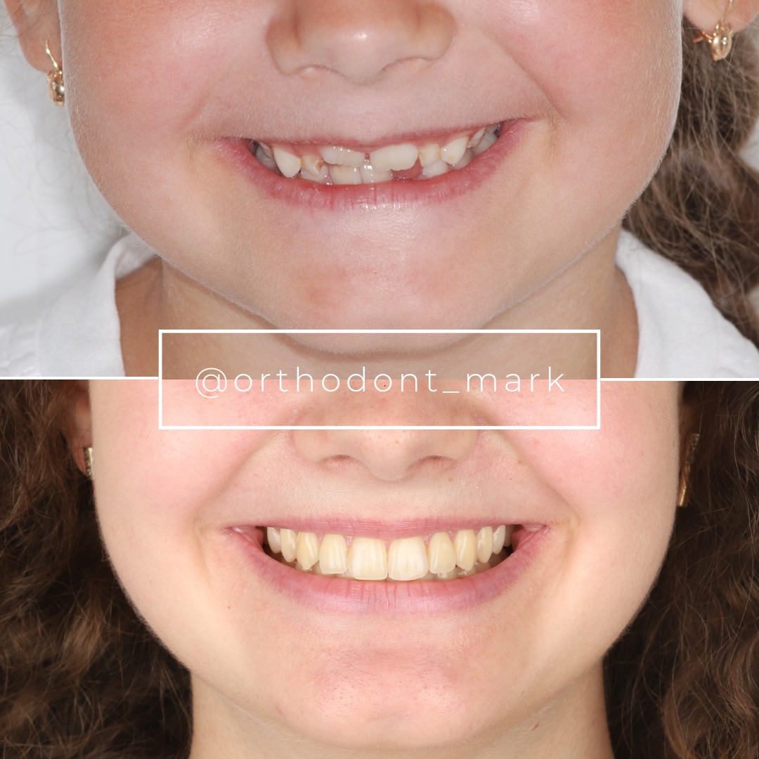
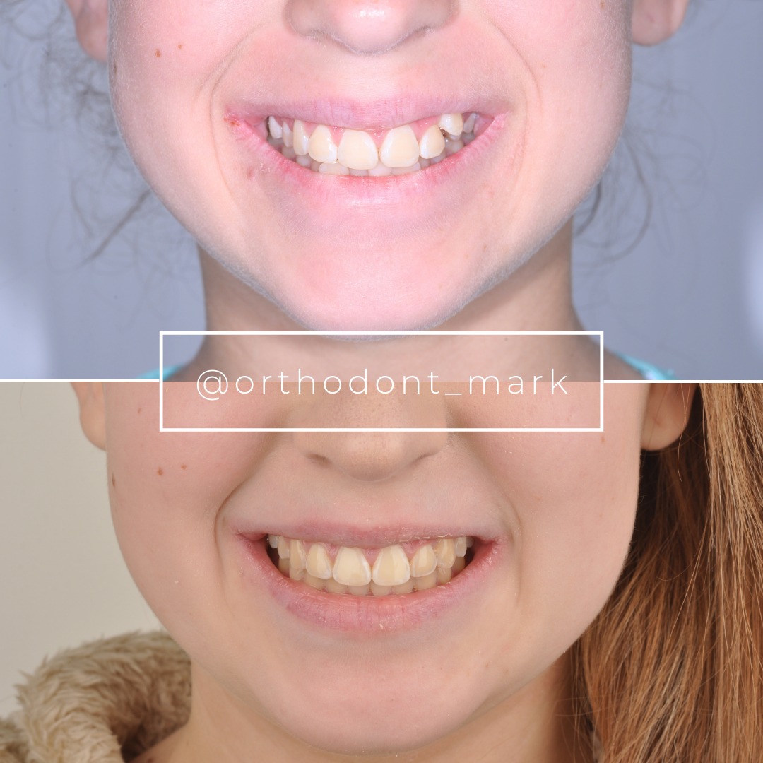
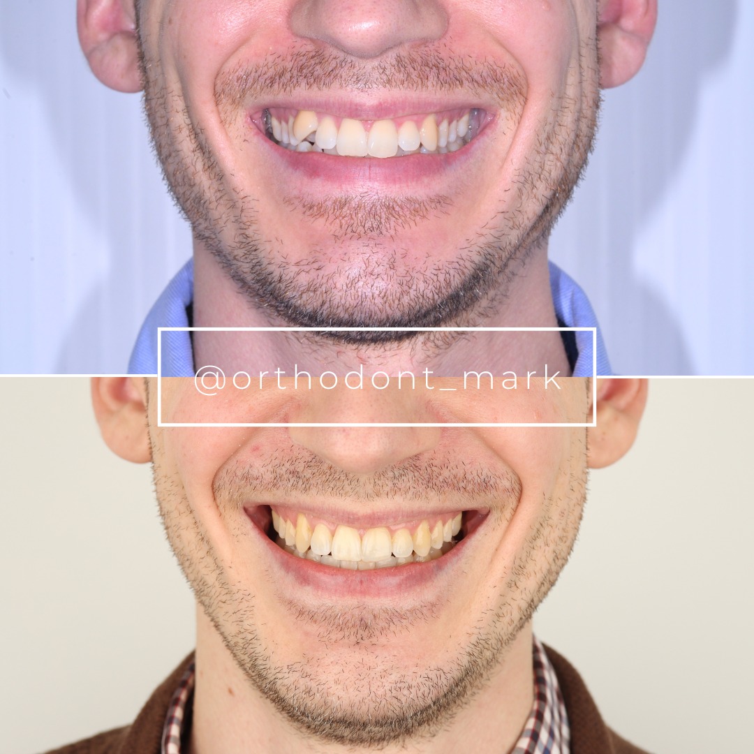
Clinical cases from Dr. Mark Radzhabov's practice
Learn the full MARPE protocol from Dr. Mark Radzhabov
Fundamental course covering CBCT patient selection, miniscrew planning, activation protocols, and 60+ clinical cases. Choose the access level that fits your practice.
Essentials of rapid palatal expansion for practicing orthodontists.
- Core RPE concepts and biomechanics
- 6 structured video lessons
- Clinical decision checklists
- Lifetime access to recordings
Deep-dive into MARPE protocol, diagnostics, and clinical execution.
- 6 modules covering MARPE from A to Z
- CBCT case selection walkthroughs
- Miniscrew placement and activation
- 60+ annotated clinical cases
- Direct Q&A with Dr. Mark Radzhabov
5-element medical consultation framework for dentists and orthodontists.
- Trust-building consultation protocol
- 5 lesson modules
- Templates for treatment plan delivery
- Works with any clinical specialty
Clinical FAQ
What is the clinical significance of the diastema during the first week of RPE activation?
Central incisor diastema formation within the first few days confirms that expansion forces are being transmitted to the midpalatal suture rather than absorbed dentally. Absent diastema by day four should prompt reassessment of suture patency and appliance seating before continuing the activation schedule.
How does RPE tissue response differ between patients under 10 and adolescents aged 12 and older?
Kinzinger et al. (2022) demonstrated parallel expansion in children under 10 years old and V-shaped expansion in adolescents aged 12 and older. In older patients, anterior diastema width overestimates total skeletal gain and greater buccal tipping of anchor teeth should be expected.
What percentage of 15-year-old females already have a closed midpalatal suture at RPE activation?
Govaerts et al. (2023) found closed midpalatal sutures in 61% of 15-year-old females. This means the majority of mid-adolescent female patients carry significant sutural resistance at the time of appliance delivery, regardless of chronological age.
Which post-activation RPE warning signs require same-day clinical intervention?
Persistent palatal blanching beyond 48 hours, an enlarging mucosal lesion, progressive gingival recession, and visible appliance movement at anchor teeth each require same-day evaluation. Omezli et al. (2020) list palatal tissue necrosis and periodontal recession as documented complications when these signs are missed.
How does transpalatal suture closure affect early expansion response in adolescents?
Govaerts et al. (2023) reported 78–85% transpalatal suture closure from age 15 onward in females. Resistance from this suture limits posterior skeletal displacement and shifts the force ratio toward dental tipping, making early-week buccal root torque concerns more pronounced in older adolescent cases.
Are permanent complications from RPE likely if early warning signs are missed?
Omezli et al. (2020) confirm that permanent complications from RME are rare and most side effects are temporary. However, that favorable outcome depends on active tissue surveillance — undetected progressive recession or palatal necrosis can result in lasting harm if not caught within the first week.
What does the V-shaped expansion pattern in adolescents mean for RPE activation monitoring?
Because Kinzinger et al. (2022) link V-shaped opening to patients aged 12 and older, the anterior diastema in these patients appears disproportionately wide relative to actual posterior skeletal gain. Clinicians should document cast measurements at the premolar and molar regions independently rather than relying solely on the anterior gap.
How should the post-activation parent briefing address suture resistance in mid-adolescent patients?
Parents should be told that a gradual or subtle first-week response in teenage patients can reflect multi-sutural resistance rather than appliance failure. Govaerts et al. (2023) document high closure rates across midpalatal, transpalatal, and pterygomaxillary sutures by mid-adolescence, supporting an honest discussion of biology over reassurance.
When during RPE treatment should CBCT suture staging be performed to avoid a failed first week?
CBCT-based suture staging should be completed before appliance delivery, not after a sluggish first-week response prompts retrospective investigation. Given that Govaerts et al. (2023) show substantial sutural closure from age 13 onward, pre-treatment imaging is the appropriate standard for any adolescent expansion case.
What is the clinical protocol for managing progressive gingival recession detected during the first week of RPE activation?
Pause or reduce the activation rate immediately and assess anchor tooth periodontal biotype. Periodontal recession is a documented complication of RME in skeletally mature patients per Omezli et al. (2020). Thin biotype cases warrant proactive monitoring from day one and may require transition to a bone-borne force path.
The first week of RPE activation is a diagnostic window, not just a waiting period — what the palatal tissue and the diastema tell you on day three and day seven directly informs whether to continue, pause, or refer for surgical evaluation. Permanent complications from RME are rare, as Omezli et al. (2020) confirm, but transient side effects are common enough that structured monitoring is essential. Dr. Mark Radzhabov invites colleagues to submit a case for review or enroll in structured MARPE training at ortodontmark.com to refine their activation protocols. Key sources: Omezli et al. (2020), BioMedicine, doi:10.37796/2211–8039.1007. Govaerts et al. (2023), Journal of Orofacial Orthopedics, doi:10.1007/s00056–023-00487-x. Kinzinger et al. (2022), doi:10.1007/s00056–022-00429-z.

