
Dental Case Presentation:
Problem-Consequences-Solution
A Framework for Clinicians
Build treatment plan acceptance by sequencing your clinical findings, imaging evidence, and appliance rationale in logical order—peer-reviewed and practice-tested.
TL;DR A dental case presentation built on a problem-consequences-solution structure gives clinicians a repeatable framework for communicating treatment rationale without pressure. Presenting the skeletal problem first, then its downstream effects, then the evidence-based solution—whether RPE, MARPE, or surgical expansion—sharpens case acceptance and aligns patient expectations with clinical reality.
A poorly structured dental case presentation is one of the most preventable causes of declined treatment in orthodontic practice. When clinicians jump straight to appliance selection without first establishing the clinical problem and its consequences, patients lack the diagnostic context needed to make an informed decision. Dr. Mark Radzhabov at ortodontmark.com teaches a problem-consequences-solution framework that sequences clinical findings, imaging interpretation, and treatment options in a logical order proven to reduce confusion and improve case acceptance. This article outlines that framework for orthodontists managing complex skeletal cases.
Why Does Presentation Sequence Determine
Case Acceptance?
A dental case presentation is a structured clinical communication sequence in which the clinician documents a diagnosed problem, explains its anatomical and functional consequences, and proposes a solution matched to the evidence before requesting a treatment decision. Most failed presentations collapse at the first step: the problem is never clearly named. Clinicians often move directly to appliance selection—RPE, MARPE, or surgery—before the patient understands what transverse maxillary deficiency is doing to their occlusion, airway, or alveolar bone. Establishing the problem first anchors every downstream conversation. The consequences step is the most underused part of the sequence. When a clinician explains that untreated posterior crossbite in a growing patient leads to asymmetric jaw loading and compensatory dental tipping, the treatment recommendation stops feeling arbitrary. Kinzinger et al. (2022) demonstrated that expansion patterns differ meaningfully by age group, with parallel expansion occurring in children under 10 years and a V-shaped pattern—anterior greater than posterior, inferior greater than superior—emerging by age 12. Showing a patient or parent why timing matters transforms the appliance choice from a clinical preference into a logical response to a documented finding. The solution stage should present at least two evidence-graded options with their clinical thresholds. For transverse skeletal deficiency, those options typically span tooth-borne rapid maxillary expansion, MARPE treatment planning and skeletal expansion protocols, and surgically assisted approaches. Framing the solution as a response to the imaging findings—suture maturity stage, bone density, arch width deficit—gives the clinician a defensible rationale and gives the patient a reason to proceed.
Which Imaging Criteria Support a Credible
Problem Statement
in Case Documentation?
Diagnostic imaging in case presentation serves two roles: it establishes the clinical problem objectively and it stratifies the patient into the correct solution pathway. For transverse maxillary deficiency, CBCT imaging allows the clinician to assess midpalatal suture maturation stage, basal bone width, and the bucco-palatal inclination of posterior teeth before any appliance is selected. Without this data, the problem statement rests on clinical impression rather than measurable anatomy. Force mechanics documented by Camporesi et al. (2013) illustrate why imaging-guided patient selection is non-negotiable when choosing between expansion systems. Hyrax and A2620 screws generated forces exceeding 20 kg in mechanical testing, while Palatal Split screws produced approximately 16 kg—Camporesi et al. (2013). Presenting these force magnitudes to a referring clinician, alongside the patient's suture density on CBCT, transforms a generic recommendation into a biomechanically grounded decision. The patient understands why a bone-borne device was selected over a tooth-borne alternative. Rapid maxillary expansion appliances generate forces ranging from 7.54 to 15.8 kg to overcome midpalatal suture resistance, according to Camporesi et al. (2013). Including this range in case documentation—alongside the patient's estimated suture resistance based on maturation stage—creates a direct link between the imaging finding and the mechanical rationale for the chosen appliance. That link is what distinguishes an evidence-based case presentation from a standard treatment proposal.
How Should Clinicians Sequence Appliance Options
by Skeletal Evidence
in the Solution Stage?
The solution stage of a dental case presentation must map directly to the problem documentation. For a patient with an open midpalatal suture on CBCT and a posterior crossbite of skeletal origin, tooth-borne rapid palatal expansion remains a first-line option—and the case file should state why. For a patient with a partially fused suture and insufficient alveolar bone for conventional band anchorage, the rationale for MARPE needs to be spelled out in the same language used to describe the problem: anatomy, imaging criteria, and force biology. Chun et al. (2022) reported that midpalatal suture separation occurred in 90% of RPE patients and 95% of MARPE patients immediately after identical expansion protocols—Chun et al. (2022). Presenting these separation rates alongside the patient's CBCT findings gives the treatment recommendation a probability basis rather than a preference basis. Clinicians who include this kind of referenced data in case documentation build a paper trail that supports both consent and outcome review. Internal resources on patient consultation skills and case presentation training can help structure how this data is communicated across the appointment. Standard RME screw mechanics offer another concrete reference point for the solution stage. Each full turn of a standard screw delivers 0.8 mm of expansion through 4 quarter-turns of 0.2 mm each, with a total screw length of 10 mm—Camporesi et al. (2013). Communicating this activation schedule as part of the treatment plan, alongside the target arch width gain, gives the patient a measurable timeline and gives the clinical record a documented protocol rather than a vague instruction.
What Documentation Gaps Undermine a Structured
Case Presentation
in Complex Expansion Cases?
The most common documentation gap in orthodontic case presentations is the absence of a consequences bridge. Clinicians present the imaging finding and jump immediately to appliance selection, leaving the patient without a clear understanding of what happens if they decline or delay treatment. For transverse skeletal deficiency, those consequences are anatomically specific: progressive alveolar bone loss over tipped posterior teeth, mandibular shift accommodating the crossbite, and reduced posterior occlusal contact area. Each of these should appear in the case file as a named finding, not as a verbal afterthought. A second pitfall is presenting the solution stage as a single option. When only one appliance is named—whether RPE or a MARPE device—the patient has no reference point for understanding why that choice was made. A two-option structure, in which a non-surgical pathway and a more intensive alternative are both presented with their respective imaging criteria and force mechanics, places the clinical decision in context. Appliance-specific data from Camporesi et al. (2013) supports this approach by providing measurable differences between device types that can be communicated without oversimplification. A third gap is the failure to document activation protocol in the treatment plan. Stating that the patient will 'turn the screw daily' is insufficient. A case file that records the specific screw length, turns per week, and target expansion in millimetres—referenced to published mechanical data—is both a clinical safeguard and a communication asset. Structured RPE training for early interceptive cases covers exactly this level of protocol documentation for tooth-borne expansion across paediatric and adolescent cohorts.
Free Clinical Guide
Which Expansion Appliance at Which Age?
Get Dr. Mark Radzhabov's guide to skeletal-expansion appliances — indications, pros & cons, and how to choose by patient age. Free to your inbox.
Prefer video? Watch the free demo lesson on MARPE appliance types, features and trade-offs.
Documented outcomes
Clinical cases from Dr. Mark Radzhabov's practice
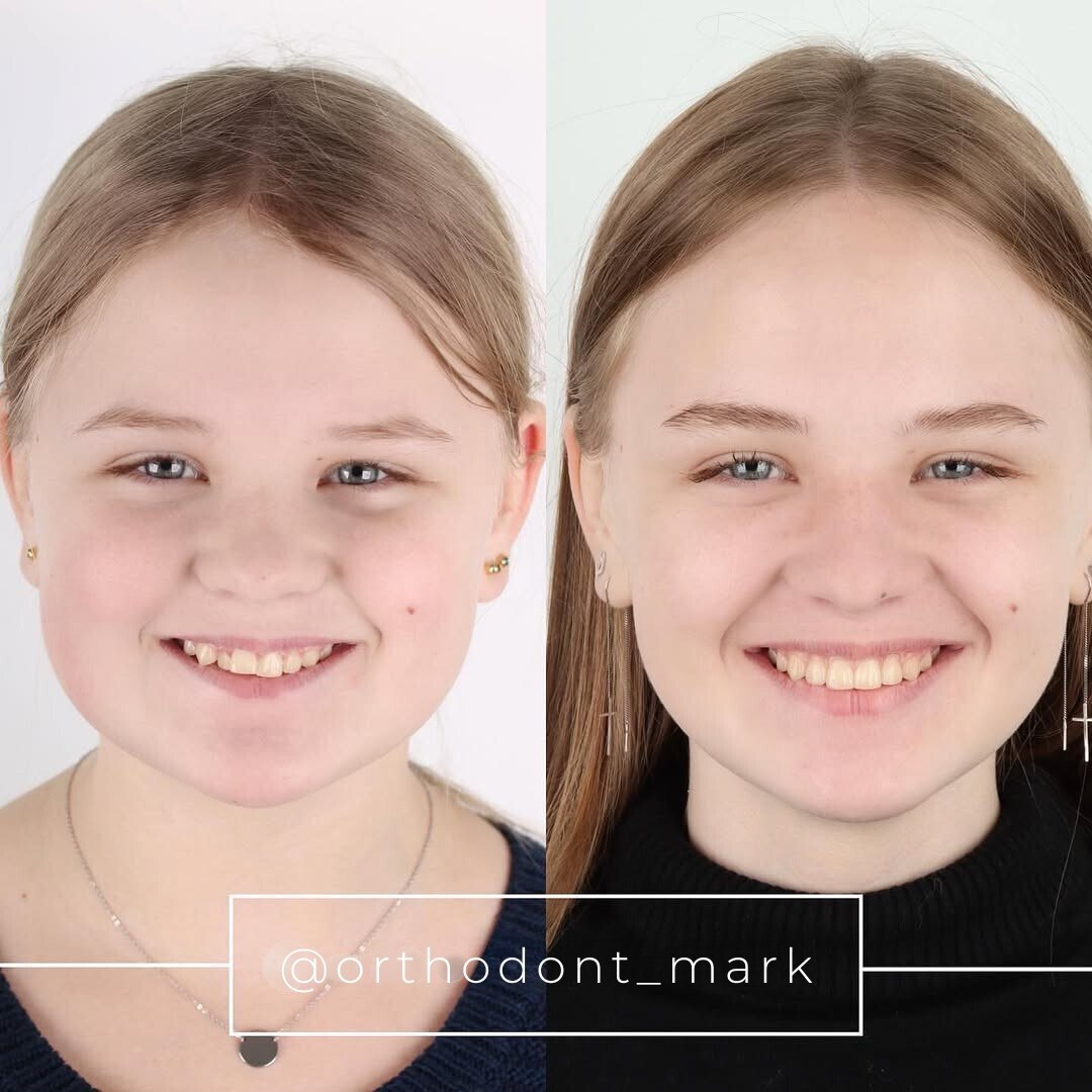

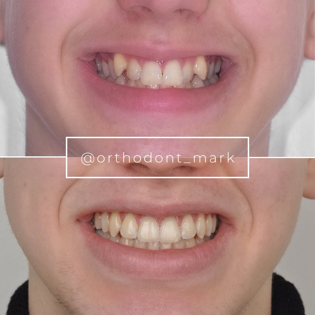
Learn the full MARPE protocol from Dr. Mark Radzhabov
Fundamental course covering CBCT patient selection, miniscrew planning, activation protocols, and 60+ clinical cases. Choose the access level that fits your practice.
Essentials of rapid palatal expansion for practicing orthodontists.
- Core RPE concepts and biomechanics
- 6 structured video lessons
- Clinical decision checklists
- Lifetime access to recordings
Deep-dive into MARPE protocol, diagnostics, and clinical execution.
- 6 modules covering MARPE from A to Z
- CBCT case selection walkthroughs
- Miniscrew placement and activation
- 60+ annotated clinical cases
- Direct Q&A with Dr. Mark Radzhabov
5-element medical consultation framework for dentists and orthodontists.
- Trust-building consultation protocol
- 5 lesson modules
- Templates for treatment plan delivery
- Works with any clinical specialty
Clinical FAQ
How should a dental case presentation differ for a skeletal expansion case versus a routine alignment case?
Skeletal cases require an additional imaging layer in the problem stage—CBCT suture assessment, arch width measurements, and bone density findings. The consequences must name specific anatomical risks, and the solution must distinguish between tooth-borne and bone-borne force pathways with referenced mechanical data.
Which imaging criteria should anchor the problem statement in an orthodontic case presentation?
Midpalatal suture maturation stage, basal bone width on CBCT, and posterior tooth inclination provide measurable anchors. Qualitative descriptions without imaging references weaken both the clinical record and the consent process.
How do I present RPE versus MARPE to a patient without making it sound like a sales pitch?
Frame both options around the imaging finding. Chun et al. (2022) reported midpalatal suture separation in 90% of RPE patients and 95% of MARPE patients—presenting these figures alongside the patient's suture stage keeps the conversation clinical rather than commercial.
What published data can I cite when documenting appliance force rationale in a treatment plan?
Camporesi et al. (2013) measured Hyrax and A2620 screws exceeding 20 kg and Palatal Split screws at approximately 16 kg. Rapid maxillary expansion appliances generate 7.54 to 15.8 kg to overcome suture resistance. These figures support written rationale for device selection.
How does age-stratified expansion pattern data improve treatment plan acceptance for paediatric cases?
Kinzinger et al. (2022) showed parallel expansion in children under 10 years and a V-shaped pattern at 12 and older. Presenting this progression helps parents understand why interceptive treatment at an earlier age produces a different—and more favourable—skeletal response.
What should be documented in the activation protocol section of an orthodontic treatment plan?
Record screw length, turns per activation, millimetres of expansion per turn, and total target width gain. Camporesi et al. (2013) confirmed standard RME screws measure 10 mm and deliver 0.8 mm per full turn through 4 quarter-turns of 0.2 mm each.
How do I structure a case presentation for a patient who has already declined expansion treatment once?
Return to the consequences stage first. Re-document the imaging findings, name the specific anatomical effects of inaction—occlusal asymmetry, alveolar bone strain—and present the updated solution with referenced force and separation data. A structured re-presentation differs meaningfully from a repeated recommendation.
What is the role of visual tools in explaining the problem-consequences-solution framework to a patient?
CBCT cross-sections, dental cast overlays, and annotated force diagrams translate abstract numbers into visible anatomy. Showing the suture density alongside device force thresholds from Camporesi et al. (2013) makes the appliance rationale immediately interpretable without clinical jargon.
How should a clinician document the consequences stage for a patient with untreated posterior crossbite?
Name specific findings: mandibular functional shift, asymmetric condylar loading, progressive dental tipping of anchorage teeth. Age-dependent expansion data from Kinzinger et al. (2022) supports explaining why the consequences compound differently in patients over 12 versus younger children.
Where can orthodontists access structured training on evidence-based case presentation and consultation skills?
Dr. Mark Radzhabov's consultation course at ortodontmark.com covers the problem-consequences-solution framework applied to skeletal expansion cases, including how to integrate imaging evidence and published mechanical data into a consent-ready treatment plan.
A well-sequenced dental case presentation is not a sales tool—it is a clinical communication standard that protects both the patient and the clinician. When the problem is defined with imaging evidence, the consequences are explained in anatomical terms, and the solution is matched to the documented skeletal finding, patients are far better positioned to consent meaningfully. Dr. Mark Radzhabov's structured approach to orthodontic case presentation, available through ortodontmark.com, gives practitioners a repeatable protocol they can apply from the first consultation to treatment plan sign-off. Key sources: Chun et al. (2022), BMC Oral Health, doi:https://doi.org/10.1186/s12903–022-02138-w. Camporesi et al. (2013), BioMedical Engineering OnLine, doi:http://www.biomedical-engineering-online.com/content/12/1/128.

