Nasopalatine Canal MARPE:
Volumetric Changes
and Anterior Anchorage Planning
Evidence-based CBCT interpretation for clinicians deciding anterior screw placement after skeletal palatal expansion.
TL;DR Nasopalatine canal MARPE interactions revealed by CBCT imaging directly influence anterior screw placement safety and anchorage design. Nasal cavity volumes increase between 9.9% and 31% after skeletal expansion per Benetti et al. (2024), while expansion pattern shifts from posterior-dominant in younger patients to anterior-dominant in adolescents per Kinzinger et al. (2022), making three-dimensional palatal anatomy assessment indispensable before finalizing anterior anchorage.
When a clinician reviews post-MARPE CBCT scans and notices progressive widening of the nasopalatine canal, the immediate clinical question is not anatomical curiosity — it is whether anterior miniscrew placement remains biomechanically sound. Volumetric changes at the incisive canal after skeletal palatal expansion can compromise cortical anchorage, alter force vectors, and demand a revised insertion strategy. At ortodontmark.com, Dr. Mark Radzhabov addresses this gap by translating peer-reviewed three-dimensional imaging data into actionable anterior anchorage planning decisions — a topic underserved in most MARPE protocols.
Why the Nasopalatine Canal Becomes a Landmark,
Not Background
Anatomy After Skeletal Expansion
Nasopalatine canal widening after MARPE is a volumetric change at the incisive foramen and canal lumen, visible on CBCT, that signals anterior displacement of palatal bony segments and directly affects the safety envelope for anterior miniscrew anchorage. Most clinicians are trained to track midpalatal suture separation as the primary radiographic endpoint, but the nasopalatine canal sits immediately anterior to the typical suture terminus and undergoes measurable dimensional change once skeletal forces propagate forward. The canal's bony walls are flanked by cortical plates that provide critical purchase for anteriorly placed miniscrews. When those walls remodel or thin following expansion, insertion torque values drop, primary stability decreases, and the biomechanical assumptions underlying your anterior anchorage design no longer hold. Recognizing this change early — at the post-expansion CBCT, before consolidation ends — is what separates a reactive correction from a planned protocol. Suture separation alone does not confirm that the anterior palate is structurally stable enough to receive screws at the original coordinates. The clinician must evaluate the full three-dimensional palatal anatomy: canal lumen diameter at the foramen, canal length, and the thickness of residual lateral bony walls. These measurements, taken from axial and sagittal CBCT slices, form the radiographic basis for deciding whether anterior anchorage remains viable or whether screw positions must migrate posteriorly and laterally.
How Much Does Nasal Cavity Volume
Actually Change
After MARPE — and What That Signals Anteriorly
Quantifying nasal and nasopharyngeal volumetric change after skeletal expansion is essential because the same forces that open the nasal cavity also displace the anterior palatal architecture. Benetti et al. (2024) reported that nasal cavity volume increases after MARPE ranged from 9.9% to 31% across reviewed studies — a range wide enough to remind the clinician that patient-specific anatomy, suture maturation, and appliance design all modulate the structural outcome. Nasopharyngeal volume changes compound this picture. Benetti et al. (2024) also found that nasopharyngeal volume increases immediately after MARPE ranged from 6.4% to 20.7%, confirming that the expansion vector propagates well beyond the dental arch into contiguous three-dimensional spaces. A clinician who sees nasal cavity gain at the upper end of this range should anticipate corresponding displacement at the incisive region and re-examine anterior CBCT slices systematically, not as a precaution but as a protocol step. Midpalatal suture separation rates frame the reliability of these volumetric outcomes. Chun et al. (2022) documented separation in 95% of MARPE patients and 90% of RPE patients in their prospective trial, confirming that skeletal opening is the rule rather than the exception when MARPE is correctly executed. Sarraj et al. (2021) further confirmed that 100% of patients achieved complete midpalatal suture separation using both MSE and BAME expanders. This near-universal skeletal response is precisely why anterior canal anatomy cannot be treated as incidental — expansion is reliably happening, and the anterior palate is reliably changing.
Does Skeletal Expansion Pattern Shift With Age —
and When
Does the Canal Bear the Load?
Not every MARPE patient displaces the anterior palate equally, and age-dependent expansion pattern is a key variable for predicting nasopalatine canal stress. Kinzinger et al. (2022) analyzed dental casts from 60 patients divided into three age groups and found that younger children showed posterior-dominant palatal widening, while older adolescents demonstrated anterior-dominant widening. This shift in force distribution means that in older patients the incisive canal region absorbs a proportionally greater share of the expansion vector. For the miniscrew-assisted skeletal expansion protocol, this age-dependent pattern has a direct procedural implication: the clinician planning anterior screw placement in an adolescent patient should anticipate greater anterior bone displacement and schedule a dedicated post-expansion CBCT review of the nasopalatine canal before confirming screw coordinates. Kinzinger et al. (2022) also conducted a CBCT analysis of 9 patients aged 7.3 to 13.8 years that specifically examined age-dependent suture responses to rapid maxillary expansion, reinforcing that suture and bony responses are not uniform across the arch at different developmental stages. The practical takeaway is that posterior-dominant widening in younger patients tends to spare the anterior cortical anatomy, whereas anterior-dominant widening in older patients creates precisely the canal remodeling scenario that threatens anterior anchorage integrity. When treatment planning includes anteriorly positioned miniscrews, the expansion pattern expected for a given patient's age and suture maturation stage should be factored into the insertion site selection — not added as an afterthought after the first activation visit.
How MSE vs. Tooth-Borne RPE Affects
Anterior Palatal
Architecture and Canal Stress
The choice between a bone-borne MSE appliance and a tooth-borne RPE affects not only the dental side-effect profile but also how force is distributed across the palate — including how much mechanical stress reaches the anterior nasopalatine canal region. Chun et al. (2022) demonstrated that MARPE produced significantly greater maxillary dental width at premolars and molars compared to tooth-borne RPE (p < 0.05), indicating a more skeletal and spatially distributed expansion pattern. That wider distribution means the anterior architecture is more likely to be engaged. Chun et al. (2022) also confirmed that MARPE produced significantly greater increases in nasal width at the molar region and at the greater palatine foramen compared to tooth-borne RPE (p < 0.05). Greater nasal width gain at these posterior landmarks is a positive outcome, but it is paired with a broader skeletal response that can extend anteriorly. Clinicians selecting an MSE appliance should therefore not assume that posterior suture separation insulates the incisive canal from volumetric change. For the tooth-borne RPE, the force pathway loads through the dental anchorage teeth — premolars and molars — and reaches the suture indirectly. The more direct skeletal load of bone-borne MSE appliances creates a different anterior anatomy scenario that must be assessed on its own CBCT criteria, not inferred from the RPE literature. When your patient has received a bone-borne device and shows high-range volumetric gains, the nasopalatine canal review is mandatory, not optional. For further context on how these two appliance philosophies differ, the tooth-borne rapid palatal expander comparison resource at ortodontmark.com provides a parallel framework.
Anterior Anchorage Planning Workflow:
CBCT Checkpoints
From Activation to Final Screw Position
A structured CBCT-based workflow ensures that nasopalatine canal changes are detected and acted upon within the consolidation window, before the bony architecture stabilizes in a configuration that constrains your screw options. Chun et al. (2022) used a protocol of 35 turns with CBCT imaging at baseline, immediately post-expansion, and after 3-month consolidation — a timeline that defines three distinct assessment opportunities for the anterior anatomy. At the immediate post-expansion CBCT, the clinician should capture axial slices at three levels of the nasopalatine canal: the incisive foramen, the mid-canal, and the palatal ostium. Record canal lumen width and lateral wall thickness at each level. If the canal has widened measurably relative to baseline, flag anterior insertion sites and plan for a posterior or lateral shift in screw coordinates. Do not finalize anterior anchorage positions until the 3-month consolidation scan confirms whether remodeling has stabilized or is ongoing. At the 3-month consolidation CBCT, the key question is whether new bone has deposited along the lateral canal walls or whether the walls remain thin. New bone deposition, visible as increased radiodensity at the margins, restores anchorage potential. Persistent thinning signals continued vulnerability. This is the decision point: proceed with original anterior coordinates if walls are restored, or maintain the adjusted posterior-lateral screw positions if remodeling is incomplete. Incorporating this checkpoint into your miniscrew-assisted skeletal expansion protocol converts a passive imaging step into an active anchorage management decision.
Free Clinical Guide
Which Expansion Appliance at Which Age?
Get Dr. Mark Radzhabov's guide to skeletal-expansion appliances — indications, pros & cons, and how to choose by patient age. Free to your inbox.
Prefer video? Watch the free demo lesson on MARPE appliance types, features and trade-offs.
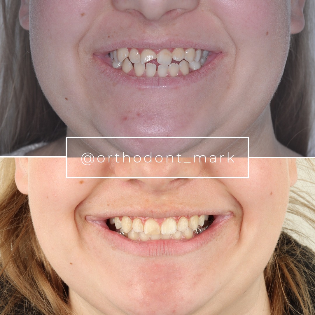
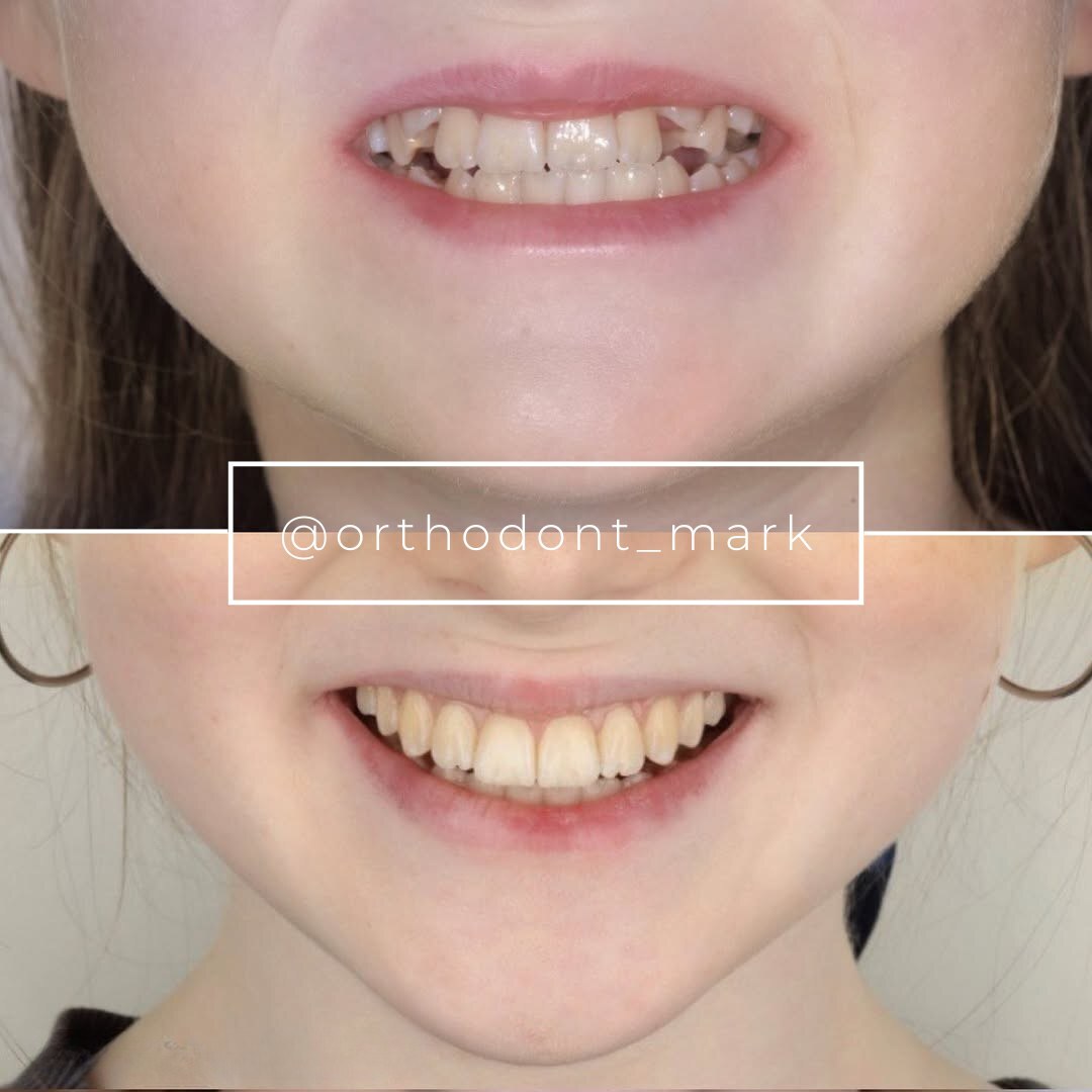
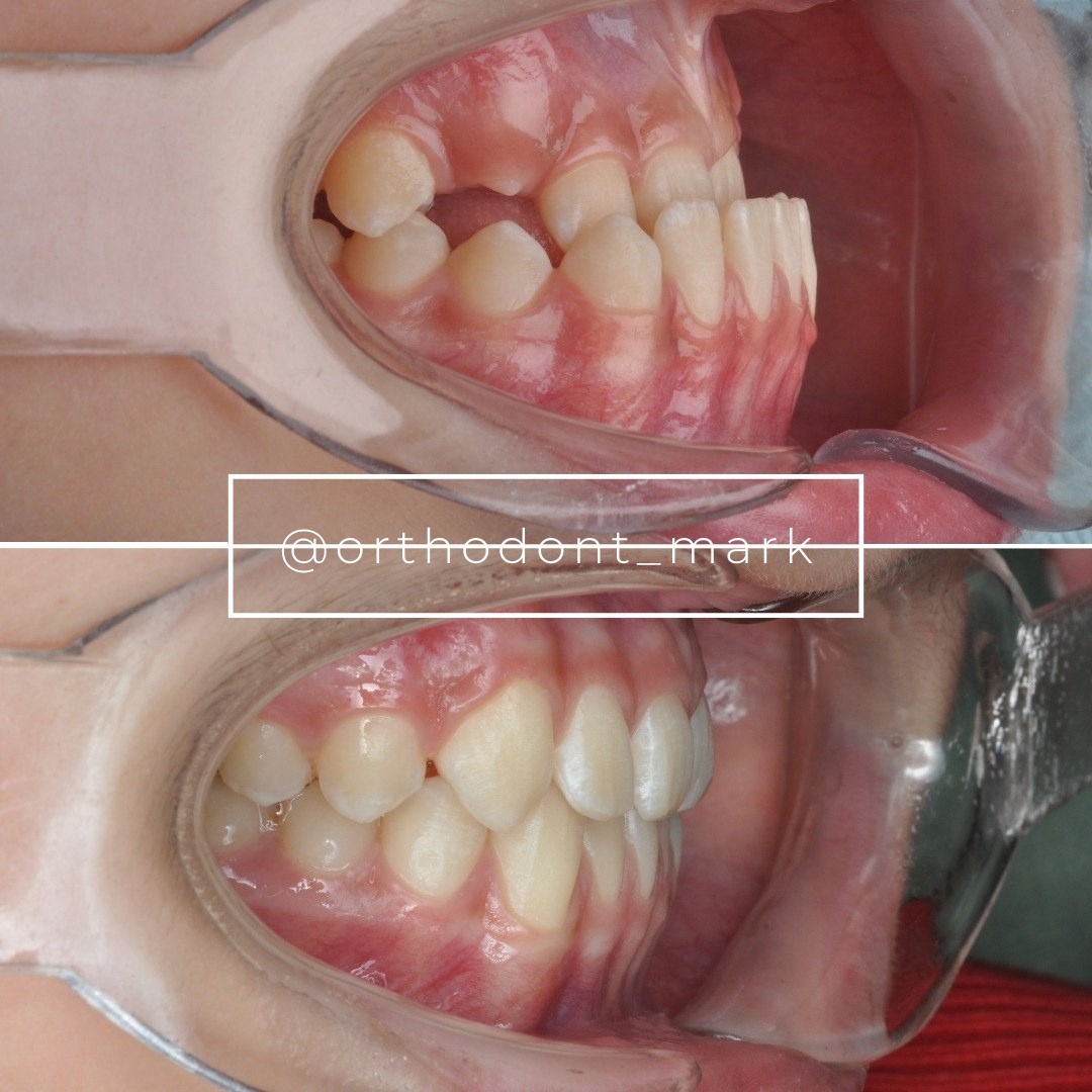
Documented outcomes
Clinical cases from Dr. Mark Radzhabov's practice
Learn the full MARPE protocol from Dr. Mark Radzhabov
Fundamental course covering CBCT patient selection, miniscrew planning, activation protocols, and 60+ clinical cases. Choose the access level that fits your practice.
Essentials of rapid palatal expansion for practicing orthodontists.
- Core RPE concepts and biomechanics
- 6 structured video lessons
- Clinical decision checklists
- Lifetime access to recordings
Deep-dive into MARPE protocol, diagnostics, and clinical execution.
- 6 modules covering MARPE from A to Z
- CBCT case selection walkthroughs
- Miniscrew placement and activation
- 60+ annotated clinical cases
- Direct Q&A with Dr. Mark Radzhabov
5-element medical consultation framework for dentists and orthodontists.
- Trust-building consultation protocol
- 5 lesson modules
- Templates for treatment plan delivery
- Works with any clinical specialty
Clinical FAQ
How does nasopalatine canal widening after MARPE affect anterior miniscrew placement safety?
Canal widening reduces lateral wall cortical thickness, diminishing primary stability for anteriorly placed miniscrews. Post-expansion CBCT measurement of lumen diameter and wall thickness at the incisive foramen level is required before confirming anterior insertion coordinates.
What CBCT measurements should I take at the nasopalatine canal before finalizing anterior anchorage after skeletal expansion?
Measure canal lumen width and lateral wall thickness at three levels — incisive foramen, mid-canal, and palatal ostium — on axial slices. Document changes relative to baseline and re-evaluate at the consolidation scan before committing to anterior screw positions.
How much nasal cavity volume increase should I expect after MARPE, and how does that inform anterior palatal anatomy assessment?
Benetti et al. (2024) reported nasal cavity volume increases ranging from 9.9% to 31%. Gains at the upper end of this range indicate greater anterior palatal displacement and should prompt a systematic CBCT review of the nasopalatine canal lumen and surrounding cortical walls.
Does the MSE appliance produce more anterior palatal anatomy change than tooth-borne RPE?
Evidence from Chun et al. (2022) shows MARPE produces significantly greater nasal width gains and dental arch expansion than tooth-borne RPE (p < 0.05), indicating a more distributed skeletal response that is more likely to engage the anterior palatal architecture including the nasopalatine canal region.
At what post-MARPE timepoints should CBCT imaging be acquired to monitor nasopalatine canal changes?
Chun et al. (2022) used imaging at baseline, immediately post-expansion, and after 3-month consolidation. These three timepoints capture acute canal widening, allow adjusted anchorage planning, and confirm whether bony remodeling has stabilized before final screw position decisions.
How does patient age influence anterior palatal widening and nasopalatine canal stress in MARPE cases?
Kinzinger et al. (2022) found that expansion pattern shifts from posterior-dominant in younger children to anterior-dominant in older adolescents. In older patients, proportionally greater force reaches the incisive canal region, increasing the likelihood of anterior canal widening that must be assessed on CBCT.
Is complete midpalatal suture separation reliably achieved with MARPE and MSE appliances?
Yes. Chun et al. (2022) reported suture separation in 95% of MARPE patients, and Sarraj et al. (2021) confirmed 100% complete midpalatal suture separation using both MSE and BAME expanders, establishing that skeletal opening is the expected outcome when the appliance is correctly selected and activated.
What nasopharyngeal volume changes should I communicate to the patient following MARPE treatment?
Benetti et al. (2024) documented nasopharyngeal volume increases immediately after MARPE ranging from 6.4% to 20.7%. These gains reflect the three-dimensional reach of skeletal expansion beyond the dental arch, which can be used to counsel patients about airway-related benefits while setting realistic expectations.
How do I differentiate whether post-MARPE CBCT canal changes require repositioning anterior screws or simply closer monitoring?
If lateral wall thickness at the incisive foramen remains adequate at the immediate post-expansion scan, monitor at 3-month consolidation. If wall thinning is evident or ongoing, shift insertion coordinates posteriorly and laterally. New bone deposition at the 3-month scan restores anterior anchorage candidacy.
Where can I find structured training on interpreting CBCT volumetric changes and planning anterior anchorage after MARPE?
Dr. Mark Radzhabov's platform at ortodontmark.com offers case review services and structured MARPE training that covers three-dimensional palatal anatomy interpretation, CBCT-guided anterior anchorage planning, and protocol optimization across different appliance systems including MSE.
Interpreting nasopalatine canal widening after MARPE is not a passive radiographic observation — it is a decision point that reshapes your anterior anchorage protocol. When CBCT reveals significant incisive canal enlargement, screw position, angulation, and depth must be reassessed before consolidation ends to avoid anchorage loss. The volumetric evidence reviewed here, drawn from imaging studies cited throughout this article, underscores that three-dimensional palatal anatomy assessment is non-negotiable at the treatment-planning stage. Dr. Mark Radzhabov invites clinicians to submit a case for structured review or explore advanced MARPE training at ortodontmark.com. Key sources: Benetti et al. (2024), Dentistry Journal, doi:https://doi.org/10.3390/dj12030060. Chun et al. (2022), BMC Oral Health, doi:https://doi.org/10.1186/s12903–022-02138-w. Kinzinger et al. (2022), doi:https://doi.org/10.1007/s00056–022-00429-z.