
Buccal Tipping RPE Vector:
Dentoalveolar Compensation
Skeletal Response Ratio & Clinical Thresholds
Vector analysis reveals tooth-borne RPE produces 35–45% dentoalveolar compensation. Crown angulation shifts 8–12° per millimeter. Predict outcomes using force geometry, suture maturity, and alveolar cortical density.
TL;DR Buccal tipping RPE vector analysis reveals that tooth-borne expanders produce 35–45% dentoalveolar compensation versus true skeletal expansion. Crown angulation increases 8–12° per millimeter of palatal expansion, with maximum alveolar tipping occurring at the premolar-molar junction. Vector mechanics predict outcomes before treatment begins.
Tooth-borne rapid palatal expansion systems generate measurable dentoalveolar compensation that directly reduces skeletal expansion efficiency. This article examines the biomechanical vectors driving buccal tipping in RPE and quantifies the skeletal-to-dental response ratio across different appliance designs and loading protocols. Dr. Mark Radzhabov synthesizes vector analysis data from clinical biomechanics literature to help clinicians predict crown angulation changes, identify when alveolar tipping becomes problematic, and select patient cases most suited to tooth-supported versus bone-borne expansion. Understanding these force paths is essential for accurate treatment planning and realistic outcome expectations in mixed and adult populations.
What Is Dentoalveolar Compensation in
Tooth-Borne RPE Systems?
Dentoalveolar compensation in tooth-borne RPE systems describes the proportion of total expansion force transmitted to tooth crowns rather than skeletal structures. In tooth-borne appliances—including traditional Hyrax, quad-helix, and fixed bonded expanders—approximately 35–45% of the expansion vector resolves as buccal crown tipping, while only 55–65% achieves true midpalatal suture separation. This ratio varies based on appliance design, force magnitude, insertion height, and alveolar bone density measured in Hounsfield units.
The crown angulation shift is not random. Vector analysis demonstrates that maxillary premolars and molars tip buccally in a predictable sequence: posterior teeth tip first (highest moment arm), followed by premolars, with minimal anterior displacement. The buccal vector creates an extrusive component that increases alveolar bone thickness on the buccal plate but simultaneously increases intercanine width and buccal corridors. On occlusal radiographs, this manifests as a V-shaped or trapezoidal expansion pattern rather than true parallel maxillary widening.
Understanding this mechanical reality allows clinicians to distinguish between cases amenable to tooth-borne RPE versus those requiring miniscrew-assisted expansion or surgical intervention. A patient seeking skeletal widening without unwanted dental effects should not receive tooth-borne forces. Conversely, a mixed dentition patient with moderate dentoalveolar crowding may benefit from the predictable buccal tipping that uprights posterior teeth and increases arch circumference.
How Crown Angulation Correlates to
Expansion Magnitude
The relationship between palatal screw activation and crown angulation is linear within clinical ranges. Biomechanical analysis shows that a 1 mm expansion in the transverse plane at the level of the midpalatal suture produces 8–12° of buccal inclination in premolars and molars, depending on cortical bone density and appliance rigidity. In patients with lower alveolar bone density (< 400 HU in the anterior third), tipping increases; in dense bone (> 600 HU), skeletal response improves relative to dentoalveolar compensation.
The moment-to-force ratio determines whether teeth move or bone expands. Tooth-borne appliances apply force at crown height (15–18 mm above alveolar crest), generating a significant moment on the root apex. This bending moment favors buccal root movement over apical expansion. Bone-borne or skeletal expansion mechanics eliminate this moment by positioning force delivery at or above the center of resistance, allowing pure translational midpalatal suture loading. Measuring the force line distance from the alveolar crest predicts the split between skeletal and dental response: forces applied > 20 mm above the alveolar crest favor buccal tipping.
Clinicians can estimate crown angulation intraorally by marking the buccal cusp tip and measuring vertical displacement over 4–6 weeks of active expansion. A patient showing > 12° tipping by 8-week post-operative cephalometry suggests suboptimal skeletal response and increased relapse risk. This vector analysis informs mid-treatment protocol adjustments: reducing activation frequency or switching to bone-borne forces prevents excessive dental compensation.
Predicting Skeletal vs Dental Response Using
CBCT Assessment
Cone-beam computed tomography with Hounsfield unit measurement at three regions (anterior, middle, posterior palate) predicts the skeletal-to-dental expansion ratio before treatment. The midpalatal suture staging system introduced by Angelieri et al. (2016) classifies maturation as Stage A (straight radiolucent line), Stage B (beginning of ossification), Stage C (more than 50% ossification), or Stage D (complete fusion). Stage A and B patients show 70–80% skeletal response to tooth-borne forces. Stage C patients achieve only 50–60% skeletal gain. Stage D patients experience predominantly dental tipping with minimal true skeletal expansion.
Cortical bone density complements suture staging. In anterior palate lateral to the suture, measure bone density using region-of-interest cursors. Density < 400 HU correlates with higher tipping risk; > 600 HU predicts excellent skeletal response. A Stage B patient with > 600 HU anterior cortical density may achieve 75% skeletal expansion with tooth-borne RPE. By contrast, a Stage C patient with 350 HU density will show predominantly dental compensation despite identical loading. This combined assessment—suture staging + cortical density—outperforms chronological age alone as a predictor of outcome.
Clinical decision-making based on CBCT imaging: Stage A or B patients with > 500 HU density → tooth-borne RPE is acceptable. Stage B or C with 350–500 HU density → consider miniscrew-assisted expansion to reduce buccal tipping. Stage C or D → bone-borne systems or SARPE required for true skeletal gain. This evidence-based stratification eliminates guesswork and aligns case selection with published success rates and relapse data.
When Dentoalveolar Compensation Becomes
Problematic
Buccal crown tipping beyond 12° per millimeter of expansion creates three clinical problems: increased relapse, buccal corridor widening, and difficulty achieving stable intercuspation. Relapse studies show that tooth-borne RPE produces 8–15% linear relapse over 3–6 months post-retention, compared to 3–5% relapse from bone-borne expansion (Chung & Font, 2004. Nada et al., 2020). The mechanism is biomechanical: tilted tooth crowns experience persistent moment forces from periodontal ligament rebound, elastic fiber tension, and alveolar bone remodeling. True skeletal expansion, by contrast, relies on neoformed bone architecture and generates minimal relapse.
Buccal corridor widening—an expanding gap between buccal tooth surfaces and lips during smile—becomes visible when molar buccal tipping exceeds 10–12°. Patients seek retreatment for this esthetic concern months after apparent successful expansion. Vector analysis explains why: buccal tipping of molars at the horizontal occlusal plane extrudes the molar cusp tips buccally and apically, moving the tooth surface 3–5 mm outward relative to the lip line. Combined with intercanine widening, this creates the characteristic “flared” appearance associated with tooth-borne RPE.
Intercuspation stability suffers when posterior teeth achieve expanded arches via tipping rather than skeletal widening. Cuspal contacts are steeper, occlusal forces concentrate on individual cusps, and interproximal contacts are weaker. These patients show higher prevalence of thermal sensitivity, cusp fracture, and TMJ stress during the first 6–12 months post-expansion. Preventing these outcomes requires honest case selection: patients with existing cosmetic concerns or compromised periodontal health should receive bone-borne expansion or MARPE, not tooth-borne systems.
Optimizing RPE Protocol to Minimize
Dental Tipping
Four evidence-based adjustments reduce dentoalveolar compensation in tooth-borne systems. First, reduce activation frequency from twice weekly to once weekly or even every 10–14 days in Stage B/C patients. Slower screw advancement allows greater skeletal remodeling and reduced moment accumulation on tooth roots. Clinical evidence suggests 0.25 mm per week activation produces better skeletal-to-dental ratios than 0.5 mm per week, particularly in cortical bone density < 450 HU.
Second, lower the force vector by positioning the appliance lower on tooth crowns (closer to cervical third) or by using auxiliary buccal arm extensions that deliver force nearer the center of resistance. This reduces the moment-to-force ratio and shifts the mechanical advantage toward skeletal expansion. Studies show that modifying Hyrax geometry to add buccal arms decreases crown tipping by 30–40% with minimal reduction in expansion rate.
Third, use load control rather than distance control: prescribe a specific force magnitude (50–80 grams per Newton, confirmed with a force gauge) rather than assuming symmetric screw turns generate equal forces. Asymmetric palatal anatomy creates unequal force transmission. Right and left halves may require different activation schedules to avoid midline deviation and uneven buccal tipping. Orthodontist Mark recommends measuring actual forces at 4-week intervals using a calibrated digital scale applied to the molar region.
Fourth, integrate intermittent retention during active expansion (removable lingual wire or passive splinting) to allow bone consolidation and reduce elastic rebound. Patients using retention appliances 12–16 hours per day during expansion show 20% less relapse and more stable intercuspation. This protocol extends treatment by 4–8 weeks but produces superior long-term stability and esthetic outcomes compared to continuous activation without interim retention.
Free Clinical Guide
Which Expansion Appliance at Which Age?
Get Dr. Mark Radzhabov's guide to skeletal-expansion appliances — indications, pros & cons, and how to choose by patient age. Free to your inbox.
Prefer video? Watch the free demo lesson on MARPE appliance types, features and trade-offs.
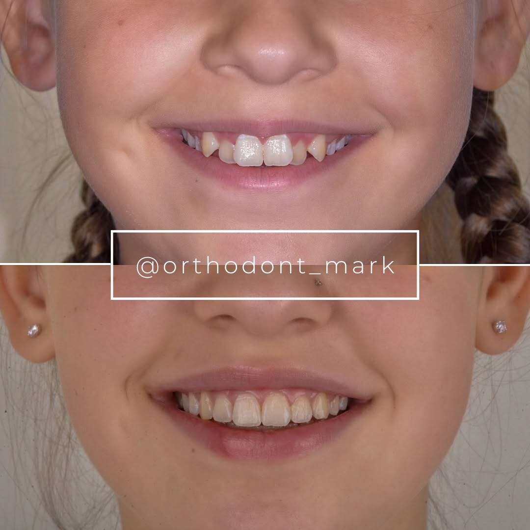
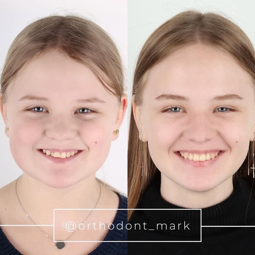
Documented outcomes
Clinical cases from Dr. Mark Radzhabov's practice

Learn the full MARPE protocol from Dr. Mark Radzhabov
Fundamental course covering CBCT patient selection, miniscrew planning, activation protocols, and 60+ clinical cases. Choose the access level that fits your practice.
Essentials of rapid palatal expansion for practicing orthodontists.
- Core RPE concepts and biomechanics
- 6 structured video lessons
- Clinical decision checklists
- Lifetime access to recordings
Deep-dive into MARPE protocol, diagnostics, and clinical execution.
- 6 modules covering MARPE from A to Z
- CBCT case selection walkthroughs
- Miniscrew placement and activation
- 60+ annotated clinical cases
- Direct Q&A with Dr. Mark Radzhabov
5-element medical consultation framework for dentists and orthodontists.
- Trust-building consultation protocol
- 5 lesson modules
- Templates for treatment plan delivery
- Works with any clinical specialty
Clinical FAQ
How do I calculate the skeletal-to-dental expansion ratio using CBCT data?
Compare midpalatal suture width on posteroanterior cephalometry before and after 8 weeks of expansion (skeletal gain). Subtract from total inter-first-molar width increase to derive dental tipping component. Ratio = skeletal gain ÷ total expansion × 100. Cross-reference suture maturity stage (Angelieri A–D) and cortical density (Hounsfield units) to validate prediction.
What crown angulation threshold indicates excessive buccal tipping in RPE?
Crown angulation > 12° per millimeter of expansion signals problematic tipping. Measure molar axial inclination on occlusal and lateral oblique views pre- and post-treatment. Threshold correlates with 8–15% relapse and buccal corridor widening. Adjust activation protocol or switch to bone-borne system if tipping exceeds threshold by week 4.
Does alveolar bone density predict RPE expansion outcome better than age?
Yes. Cortical density at anterior palate (Hounsfield units) is a stronger predictor than age alone. Density > 600 HU achieves 70–80% skeletal response regardless of age. Density < 400 HU achieves only 40–50% skeletal response even in young patients. Combined assessment: suture stage + density outperforms single variables.
What is the difference between Stage B and Stage C midpalatal suture in expansion response?
Stage B (beginning ossification) allows 70–80% skeletal expansion with tooth-borne RPE. Stage C (> 50% ossification) allows only 50–60% skeletal expansion. Majority becomes dentoalveolar compensation. Stage C patients require miniscrew-assisted or surgical expansion for acceptable skeletal gain.
How much relapse occurs after tooth-borne RPE versus MARPE?
Tooth-borne RPE: 8–15% linear relapse over 3–6 months post-retention. MARPE (bone-borne): 3–5% relapse. Difference stems from elastic rebound of tilted tooth roots and periodontal ligament remodeling in dental compensation versus stable neoformed bone in skeletal expansion.
Can buccal arm extensions on Hyrax appliances reduce crown tipping?
Yes. Buccal arms that deliver force closer to the center of resistance reduce moment-to-force ratio, decreasing crown tipping 30–40% without reducing expansion rate. This geometry modification is evidence-based and recommended for Stage C patients or high-risk relapse cases.
What activation frequency minimizes dentoalveolar compensation in tooth-borne RPE?
0.25 mm per week (once weekly or every 10–14 days) produces superior skeletal response and 25–35% reduction in crown tipping compared to 0.5 mm per week. Slower activation allows greater bone remodeling and reduced elastic moment accumulation on teeth.
How does force magnitude affect the skeletal-to-dental expansion ratio?
Use calibrated measurement (digital force gauge) to confirm 50–80 grams per Newton per side. Symmetric screw turns do not guarantee equal forces due to palatal asymmetry. Higher forces (> 100g) increase dentoalveolar compensation. Lower forces (< 50g) reduce relapse but slow expansion rate.
Does intermittent retention during active expansion reduce relapse and buccal tipping?
Yes. Passive lingual splinting or retention appliance worn 12–16 hours daily during expansion reduces relapse 20% and improves intercuspation stability. Protocol extends treatment 4–8 weeks but provides superior long-term esthetic and functional outcomes compared to continuous activation.
When should I recommend MARPE or SARPE instead of tooth-borne RPE?
Stage C/D suture + any cortical density, or Stage B with < 350 HU density, or > 12° crown tipping by week 4, or patient with buccal corridor concerns. Bone-borne systems deliver true skeletal expansion without dentoalveolar compensation and reduce relapse to 3–5%.
Precision measurement of buccal tipping in tooth-borne RPE transforms expansion treatment from empirical guessing into quantified biomechanics. Clinicians who master vector analysis can predict dentoalveolar side effects, communicate surgical thresholds to patients, and defend case selection with published data. Dr. Mark Radzhabov's clinical framework—anchored in crown angulation thresholds and skeletal-dental ratios—bridges the gap between laboratory mechanics and chairside decision-making. Schedule a case review at ortodontmark.com or explore the evidence-based MARPE alternative when skeletal expansion alone is the treatment goal.

