
MARPE Appointment Sequencing:
Bonding, Activation
and Monitoring — A Published-Protocol Framework
Evidence-based chairside workflow covering staff roles, screw-force benchmarks, and CBCT timing for skeletal expansion cases.
TL;DR MARPE appointment sequencing structures each clinical visit around bonding, activation, and consolidation checkpoints drawn from published expansion protocols. Imaging at baseline and after a defined number of turns tracks suture response, while force-monitoring data from in vitro screw studies inform safe activation thresholds. Precise visit scheduling reduces dental tipping and improves skeletal outcomes.
Getting MARPE appointment sequencing right is the difference between a controlled skeletal split and an avoidable complication — yet published chairside workflows are rarely consolidated into a single clinical reference. On ortodontmark.com, Dr. Mark Radzhabov synthesizes bonding criteria, activation schedules, and consolidation checkpoints drawn directly from peer-reviewed imaging and mechanical studies, giving practicing orthodontists a decision-ready protocol they can adapt from the first insertion visit through retention. This article covers staff coordination, screw-force benchmarks, and the CBCT timing that peer-reviewed trials use to verify suture separation.
Why Does Appointment Structure Drive
Suture Separation
Outcomes in MARPE Cases?
MARPE appointment sequencing is the organized series of chairside visits — covering miniscrew-assisted appliance bonding, controlled screw activation, mid-expansion imaging, and consolidation monitoring — that guides a palatal expansion case from insertion to stable retention. Without a published-protocol framework, activation decisions become ad hoc and imaging is delayed until complications surface rather than used proactively to verify suture response. The bonding visit sets the mechanical foundation. Miniscrew position relative to the median palatal suture, appliance seating across the first molars and premolars, and passive fit verification all occur here before a single activation turn is delivered. Staff roles at this appointment include isolation management, torque confirmation on each miniscrew, and photographic documentation of screw head orientation for future turn-count reference. Imaging timing follows a structured logic grounded in clinical trials. Chun et al. (2022) performed CBCT at baseline, immediately after 35 turns of expansion, and after a 3-month consolidation period, providing a three-point radiographic record that captures initial suture response, end-of-activation width, and post-consolidation stability. Adopting these same imaging checkpoints in your own activation and monitoring schedule aligns chairside decisions with the evidence rather than with arbitrary convention. The consolidation visit is frequently under-resourced in practice. Retaining the appliance passively while new bone fills the expanded suture demands the same level of clinical attention as activation — staff must document that no inadvertent turns have occurred and that the patient reports no nocturnal appliance movement.
What Screw-Force Data Should
Guide Your Activation
Schedule at Each Visit?
Activation visit design must account for the force each appliance generates per quarter-turn, because palatal tissue tolerance differs markedly from the force ranges that in vitro testing documents. Camporesi et al. (2013) evaluated screw forces at 1, 5, 10, 15, and 20 quarter-turns using a 5 kN load cell, mapping the non-linear force accumulation that occurs as the screw progresses through its range of motion. This data directly informs how many turns are safe per visit before soft-tissue or bony resistance warrants a clinical pause. Camporesi et al. (2013) found that the Hyrax and A2620 screws generated over 20 kg of force, while the Palatal Split screw generated approximately 16 kg. Knowing which appliance is bonded changes the activation increment clinically appropriate for each visit. For context, RME appliances typically produce expansion forces in the range of 7.54 to 15.8 kg according to Camporesi et al. (2013) — a range that clarifies why MARPE-specific screws demand more conservative per-visit turn protocols than classical RME benchmarks might suggest. Chairside staff should log cumulative turn count at every activation visit and flag any patient-reported sharp palatal pain that could indicate force accumulation beyond tissue tolerance. A turn-count log maintained alongside the activation monitoring record gives the supervising orthodontist an immediate, auditable dataset for each appointment.
How Should Monitoring Appointments Track
Anchor-Tooth Displacement
Differently for MARPE Versus RPE Cases?
One of the clearest clinical arguments for structured MARPE appointment sequencing — rather than reactive check-ins — is the differential effect on anchor-tooth position. Chun et al. (2022) demonstrated that MARPE produced less buccal displacement of anchor teeth compared to RPE during both expansion and consolidation phases. That advantage is only realized if the monitoring visits are frequent enough to detect early tipping before it accelerates. At each activation and monitoring appointment, the clinician should assess the buccal inclination of the first molars against baseline dental cast records or intraoral scans. Deviation from the pre-treatment axial inclination is the earliest chairside signal that the appliance is transmitting too much force through the dental anchorage rather than directly to bone. Bone-borne force transmission — the defining feature of well-placed miniscrews — should minimize this, but soft-tissue torquing or miniscrew micro-mobility can shift the load pathway toward dental anchorage at any visit. For reference on age-related expansion geometry, Kinzinger et al. (2022) showed that palatal expansion after RME is parallel in children under 10 years but becomes V-shaped in adolescents aged 12 and older — a finding that explains why monitoring the posterior versus anterior width gain at sequential visits yields different patterns depending on the patient's skeletal maturity. This informs where clinical photography and caliper measurements are taken at each chairside check.
Does Patient Age Change Which
Expansion Checkpoints
Your Visits Should Prioritize?
The visit schedule cannot be age-agnostic. Kinzinger et al. (2022) evaluated nine patients aged 7.3 to 13.8 years with CBCT measurements of the median palatal suture, documenting that suture morphology and expansion geometry differ substantially across this developmental window. For the clinician designing a MARPE appointment sequencing template, that variability means the imaging and measurement protocol must specify which anatomical landmarks are being tracked — posterior nasal spine width, anterior intermolar distance, or midpalatal suture density — because each landmark responds differently depending on the patient's maturation stage. Kinzinger et al. (2022) also stratified a dental cast analysis of 60 patients into three age groups of 20 patients each: under 10 years, 10 to under 12 years, and 12 years and older, demonstrating that the clinical measurement approach shifts as skeletal maturity advances. In the youngest group, parallel expansion means symmetric width gain is detectable at all measurement points simultaneously. In the oldest group, the V-shaped pattern concentrates visible gain anteriorly first — so a monitoring visit that only checks posterior intermolar width may underestimate total expansion progress. For the MARPE appointment sequencing framework at ortodontmark.com, this translates into age-stratified measurement sheets: anterior and posterior intermolar widths recorded at every visit for adolescent and adult cases, with the CBCT midplane documented at the defined turn-count threshold. Staff training on landmark identification ensures measurement consistency across operators throughout the monitoring phase.
Which Imaging Criterion Confirms
Successful Suture Opening
at the Mid-Expansion Visit?
The mid-expansion imaging visit is the clinical pivot point: it either confirms suture separation and validates the activation protocol, or it reveals insufficient opening and triggers a protocol adjustment. Chun et al. (2022) reported midpalatal suture separation in 90% of RPE patients and 95% of MARPE patients, establishing the separation rates that published protocols are designed to achieve. If your mid-expansion CBCT — taken at the defined turn-count threshold rather than on a calendar schedule — does not show a clear radiolucent gap along the midpalatal suture, the visit sequence and activation rate need immediate review. The imaging criterion itself must be pre-specified. The midpalatal suture appears as a dark radiolucent line on axial CBCT slices. Separation is confirmed when that line widens uniformly from the anterior palatal spine toward the posterior nasal spine without unilateral hinging or asymmetric opening. Asymmetric opening detected at the mid-expansion visit is an actionable finding: it may indicate unequal miniscrew engagement, asymmetric suture resistance, or differential bone density across the two palatal halves. For a complete skeletal expansion MARPE protocol review, clinicians can explore the MARPE activation and monitoring protocol resources at ortodontmark.com, where the imaging checkpoints described above are mapped to specific appliance systems and patient profiles. Standardizing the imaging trigger — turns rather than weeks — keeps the visit schedule tied to mechanical reality rather than a fixed calendar.
How Should Chairside Staff Roles
Be Assigned
Across Bonding, Activation, and Consolidation Visits?
A well-designed expansion visit schedule fails in practice if staff assignments are ambiguous. The bonding appointment requires the highest clinical density: the orthodontist confirms miniscrew position and passive appliance fit, while the chairside assistant manages isolation, documents screw head orientation, and prepares the bonding sequence. No activation occurs at this visit — separating bonding from first activation eliminates the confound of post-bonding tissue tenderness masking early appliance-tolerance problems. Activation visits are high-frequency and brief. The assistant verifies the cumulative turn count from the log, confirms the patient reports no sharp palatal pain between visits, and records buccal inclination visually before the orthodontist delivers that session's turns. This two-person checkpoint — assistant review followed by clinician activation — builds an auditable record that supports both clinical decision-making and, where required, documentation for insurance or medicolegal purposes. Consolidation monitoring visits shift the staff role toward measurement and imaging coordination. The assistant takes standardized intraoral photographs, records anterior and posterior intermolar widths from intraoral scan overlays or calipers, and schedules the CBCT if the visit coincides with the defined turn-count imaging threshold. Orthodontist Mark's clinical framework for the skeletal expansion monitoring phase assigns the clinician's time to imaging interpretation and appliance-removal decision-making rather than data collection, freeing bandwidth in a busy schedule while maintaining protocol fidelity.
Free Clinical Guide
Which Expansion Appliance at Which Age?
Get Dr. Mark Radzhabov's guide to skeletal-expansion appliances — indications, pros & cons, and how to choose by patient age. Free to your inbox.
Prefer video? Watch the free demo lesson on MARPE appliance types, features and trade-offs.
Documented outcomes
Clinical cases from Dr. Mark Radzhabov's practice
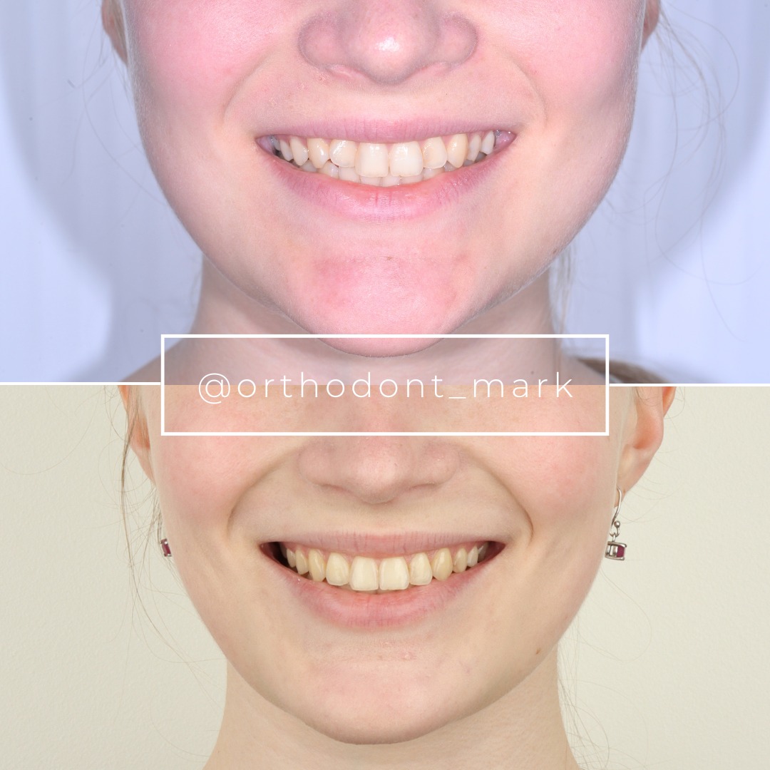
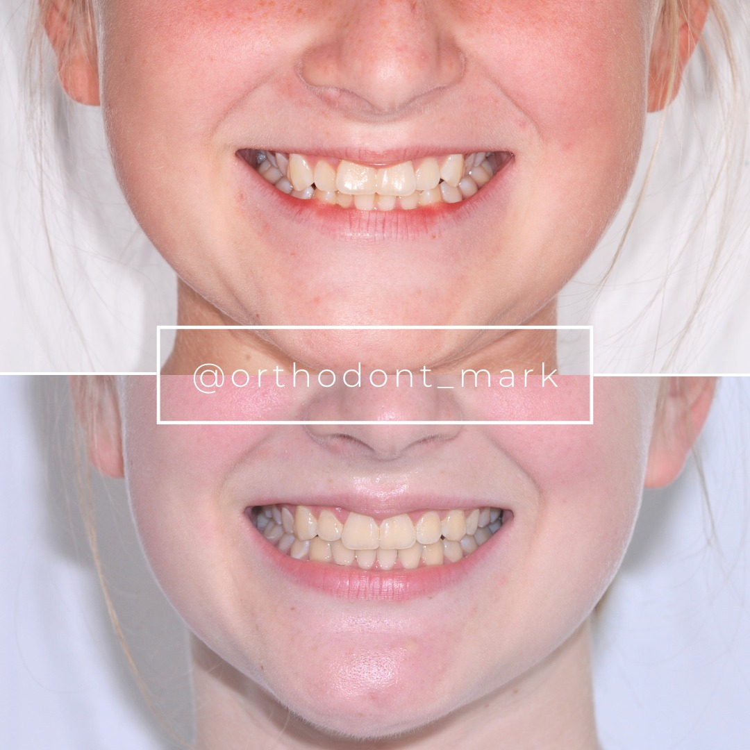

Learn the full MARPE protocol from Dr. Mark Radzhabov
Fundamental course covering CBCT patient selection, miniscrew planning, activation protocols, and 60+ clinical cases. Choose the access level that fits your practice.
Essentials of rapid palatal expansion for practicing orthodontists.
- Core RPE concepts and biomechanics
- 6 structured video lessons
- Clinical decision checklists
- Lifetime access to recordings
Deep-dive into MARPE protocol, diagnostics, and clinical execution.
- 6 modules covering MARPE from A to Z
- CBCT case selection walkthroughs
- Miniscrew placement and activation
- 60+ annotated clinical cases
- Direct Q&A with Dr. Mark Radzhabov
5-element medical consultation framework for dentists and orthodontists.
- Trust-building consultation protocol
- 5 lesson modules
- Templates for treatment plan delivery
- Works with any clinical specialty
Clinical FAQ
What is the evidence-based imaging checkpoint for a MARPE activation sequence?
Chun et al. (2022) performed CBCT at baseline, after 35 turns of expansion, and after a 3-month consolidation period. Using the turn count rather than elapsed weeks as the imaging trigger aligns the monitoring visit with the mechanical state of the appliance rather than an arbitrary calendar date.
How does screw selection affect per-visit activation increments in a MARPE protocol?
Camporesi et al. (2013) found that Hyrax and A2620 screws exceeded 20 kg of force at maximum activation, while the Palatal Split screw reached approximately 16 kg. Higher-force devices warrant more conservative per-visit turn counts to stay within tissue tolerance thresholds.
Should the bonding visit and first activation visit be separated in an expansion visit schedule?
Separating bonding from first activation is sound protocol practice. Post-bonding tissue tenderness can mask early appliance-tolerance problems, and verifying passive fit before loading the appliance ensures the miniscrews are fully engaged before suture forces are introduced.
How does expansion geometry differ between younger and older patients when monitoring width gain?
Kinzinger et al. (2022) showed that expansion is parallel in children under 10 years but becomes V-shaped in those aged 12 and older. Monitoring visits for older patients should record anterior and posterior intermolar widths separately to avoid underestimating total expansion progress.
What suture separation rate should a structured MARPE appointment sequence be designed to achieve?
Chun et al. (2022) reported suture separation in 95% of MARPE patients and 90% of RPE patients. These published figures represent the outcome benchmark against which a well-structured activation and monitoring schedule should be evaluated at the mid-expansion imaging visit.
Which chairside staff roles are most critical during the consolidation monitoring phase?
The assistant should record standardized intermolar widths and intraoral photographs at each consolidation visit, freeing the orthodontist to focus on imaging interpretation and the appliance-removal decision. Consistent staff-led measurement reduces inter-appointment variability across a long consolidation window.
What force range do conventional RME appliances produce compared to MARPE screws?
Camporesi et al. (2013) measured typical RME forces between 7.54 and 15.8 kg, while MARPE-specific screws such as the Hyrax exceeded 20 kg. This gap means activation protocols developed for conventional RME cannot be directly transferred to MARPE cases without adjustment.
How should anchor-tooth buccal displacement be tracked across MARPE monitoring appointments?
Chun et al. (2022) demonstrated that MARPE produces less buccal displacement of anchor teeth than RPE. Monitoring visits should compare molar axial inclination against baseline intraoral scans at every activation appointment to detect early tipping before it becomes clinically significant.
What is the clinical rationale for a turn-count log maintained across every activation visit?
A cumulative turn-count log gives the clinician an auditable activation record, ensures the imaging threshold is triggered at the correct mechanical state rather than by calendar, and provides documentation for consent, insurance, and medicolegal purposes throughout the expansion visit schedule.
How does the V-shaped expansion pattern in adolescents change the staff measurement protocol at chairside?
Because V-shaped expansion concentrates anterior width gain first in patients aged 12 and older per Kinzinger et al. (2022), the measurement sheet must specify both anterior and posterior intermolar widths as separate data points — a single posterior caliper reading underrepresents true expansion progress in this age group.
Structuring MARPE visits around evidence-based checkpoints — bonding verification, turn-by-turn force awareness, mid-expansion imaging, and a defined consolidation window — removes guesswork from a mechanically demanding procedure. Clinicians who standardize these appointments report fewer dental side effects and clearer end-point decisions. Dr. Mark Radzhabov recommends reviewing your current activation and monitoring schedule against published trial protocols before your next case. A structured case review or enrollment in a focused MARPE training program can accelerate that alignment. Key sources: Chun et al. (2022), BMC Oral Health, doi:10.1186/s12903–022-02138-w. Camporesi et al. (2013), BioMedical Engineering OnLine. Kinzinger et al. (2022), doi:10.1007/s00056–022-00429-z.