MARPE Chairside Workflow:
Appointment Sequencing
from Bonding Through Retention Handoff
An evidence-informed visit structure that protects skeletal gains, limits dental side effects, and gives orthodontists a clear decision gate at every stage.
TL;DR The MARPE chairside workflow structures each appointment from initial bonding through final retention handoff to protect skeletal outcomes and minimize dental side effects. Chun et al. (2022) confirmed that MARPE produced less buccal displacement of anchor teeth than RPE, validating a bone-borne force path. A disciplined visit sequence—bonding, activation, monitoring, consolidation, retention—is the clinical foundation for reproducible results.
A poorly sequenced MARPE case rarely fails at the screw. It fails at the appointment level—missed consolidation imaging, premature appliance removal, or inconsistent activation records that leave the clinician without a clear clinical picture at each decision point. The MARPE chairside workflow, from bonding day through retention handoff, deserves the same systematic attention as appliance selection itself. At ortodontmark.com, Dr. Mark Radzhabov outlines an appointment-by-appointment protocol grounded in peer-reviewed mechanics and imaging data, giving orthodontists a decision-ready framework they can apply starting with their next case.
What Happens at the Bonding Appointment That Determines
Everything After?
The MARPE chairside workflow is the appointment-by-appointment clinical sequence—spanning bonding, activation, monitoring, consolidation, and retention handoff—that governs how a miniscrew-assisted palatal expander is delivered, loaded, and transitioned to passive retention. Bonding day is not purely a placement visit. It is the moment when the clinician confirms that mechanical prerequisites are met before a single activation turn is made. Miniscrew seating depth and torque feedback at insertion are the primary quality checks at this appointment. Primary stability requires bicortical purchase through the palatal mucosa into cortical bone. Any detectable micromotion at the screw head under finger pressure indicates inadequate purchase and warrants reinsertion before the patient leaves the chair. The appliance frame should be assessed for full band seating on the anchor teeth, verifying that no rocking or gapping exists at the molar bands—an unsupported band concentrates lateral force on the tooth root rather than transmitting it to the midpalatal suture. A baseline CBCT taken prior to bonding provides the anatomical reference against which suture separation will later be measured. The imaging plane should capture the anterior, middle, and posterior thirds of the midpalatal suture so that post-expansion scans can document the characteristic shift from parallel to triangular orientation that Kinzinger et al. (2022) described with transverse force application. Confirming this baseline before the patient leaves ensures that any post-activation ambiguity is resolvable by direct image comparison rather than clinical estimation alone.
How Should the Activation Schedule Be Structured
to Drive Skeletal Rather Than Dental Movement?
The mechanical output of the chosen expansion screw directly shapes the activation schedule. Camporesi et al. (2013) tested 10 mm expansion screws on an Instron machine with a 5 kN load cell, measuring forces at 1, 5, 10, 15, and 20 quarter-turns, and found that Hyrax and A2620 devices generated forces exceeding 20 kg while Palatal Split screws produced approximately 16 kg. Knowing that the tested screw range delivers 16–20 kg of force (Camporesi et al., 2013) allows the clinician to match activation rate to the suture's expected resistance rather than applying a generic turn-per-day protocol regardless of appliance type. In the prospective trial by Chun et al. (2022), both the RPE and MARPE groups received identical expansion of 35 turns, with CBCT imaging performed at baseline, immediately post-expansion, and after 3 months of consolidation. That standardized 35-turn protocol provides a documented benchmark for total activation volume, though individual suture resistance, patient age, and CBCT-confirmed suture staging should inform whether the clinician decelerates activation if the patient reports asymmetric palatal pressure or the appliance frame shows unilateral binding. Critically, Chun et al. (2022) demonstrated that MARPE produced significantly less buccal displacement of anchor teeth compared to RPE across the expansion and consolidation periods—a direct consequence of routing force through the miniscrews rather than the periodontal ligament. Preserving this advantage requires that the activation schedule be managed consistently: irregular patient-administered turns at home introduce force spikes that partially defeat the bone-borne path. Clinicians using the MARPE skeletal expansion protocol at ortodontmark.com recommend in-office activation for at least the early turns so that the force envelope remains controlled.
Which Mid-Treatment Findings Should Trigger a
Protocol Adjustment
Before Expansion Is Complete?
Monitoring appointments during active expansion serve as clinical decision gates, not routine check-ins. At each visit, the examiner should verify miniscrew stability by attempting gentle mesiodistal torque on each screw head—any micro-rotation or soft-tissue ingrowth around the collar signals impending loss of primary stability and should prompt a CBCT to evaluate bone contact before continuing activation. Nasal response is an underused chairside indicator. Chun et al. (2022) reported that MARPE produced greater increases in nasal width at the molar region and greater palatine foramen width compared to RPE, confirming that nasal airway widening is an expected structural correlate of successful midpalatal separation. The absence of any patient-reported nasal sensation or occlusal diastema formation after sufficient turns may indicate that the suture is not separating and warrants a mid-course occlusal radiograph or CBCT before further activation. Molar occlusion and canine overjet should be recorded at each monitoring visit against the bonding-day cast. Because MARPE produced less buccal displacement of anchor teeth than RPE in the Chun et al. (2022) trial, progressive buccal flare noted at a monitoring visit is a red flag that the bone-borne force path has been compromised—possibly by loose bands redistributing load to the periodontal ligament. Corrective action at this stage, rather than at the post-expansion CBCT, protects the skeletal result and avoids the compensatory dental movements that increase retention complexity.
When Is the Midpalatal Suture Stable Enough to
Transition Out of Active Retention?
The consolidation phase begins when the target expansion turns are completed and the clinician locks the screw to prevent reversal. Its purpose is mineralization of the mid-sutural connective tissue that filled the separation gap during active expansion. Chun et al. (2022) used a 3-month consolidation window with a post-consolidation CBCT as the endpoint measurement, providing a documented timeframe against which clinical practice can be benchmarked. The post-consolidation CBCT should be read with the same regional approach used at baseline: anterior, middle, and posterior suture thirds are evaluated independently because mineralization does not proceed uniformly. Kinzinger et al. (2022) showed that the suture's geometric response to transverse force is non-uniform, with changes in width varying across the cranial-caudal and anterior-posterior axes. A suture third that shows persistent radiolucency at the 3-month mark is not necessarily a failure, but it does justify extending passive retention before appliance removal to avoid elastic recoil. Patiently running the full consolidation interval also protects against the increased relapse risk that accompanies premature removal. A tooth-borne RPE comparison arm in Chun et al. (2022) recorded suture separation in 90% of RPE patients, affirming that both appliance types can open the suture—but the post-consolidation skeletal stability depends less on the appliance type than on the adequacy of the consolidation window before the expander is deactivated.
How Should the Retention Handoff Be Structured to
Protect Long-Term Skeletal Width?
Retention handoff is a distinct clinical appointment with its own checklist, not simply the visit at which the MARPE appliance is removed. Before removal, the clinician should confirm via the most recent CBCT that all three suture thirds show adequate mineralization, that miniscrew sites are free of periapical pathology, and that the transverse dental relationship is consistent with the measured skeletal gain. Miniscrew removal follows appliance debond and requires the same sterile field discipline as insertion. The palatal mucosa at each screw site should be probed for depth and healing quality before removal. A site showing granulation tissue or suppuration warrants debridement and a short antibiotic course rather than simple screw extraction. Post-removal occlusal records—digital scan or stone cast—provide the legally and clinically essential baseline against which any future transverse relapse is measured. For review of how miniscrew-assisted expansion compares to a tooth-borne rapid palatal expander in long-term retention planning, the tooth-borne RPE comparison data from Chun et al. (2022) provides a useful reference: MARPE's reduced buccal displacement of anchor teeth throughout the expansion period translates into a retention phase that begins from a cleaner dental position, with less compensatory root torquing required before fixed appliance mechanics resume. Linking the retention appliance design—whether a transpalatal arch, bonded retainer, or Hawley-type device—to the post-expansion CBCT findings rather than defaulting to a generic protocol is the final step in a disciplined MARPE chairside workflow.
Free Clinical Guide
Which Expansion Appliance at Which Age?
Get Dr. Mark Radzhabov's guide to skeletal-expansion appliances — indications, pros & cons, and how to choose by patient age. Free to your inbox.
Prefer video? Watch the free demo lesson on MARPE appliance types, features and trade-offs.
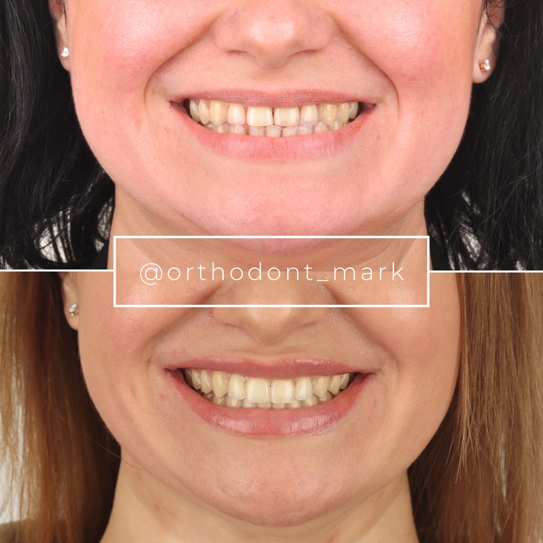
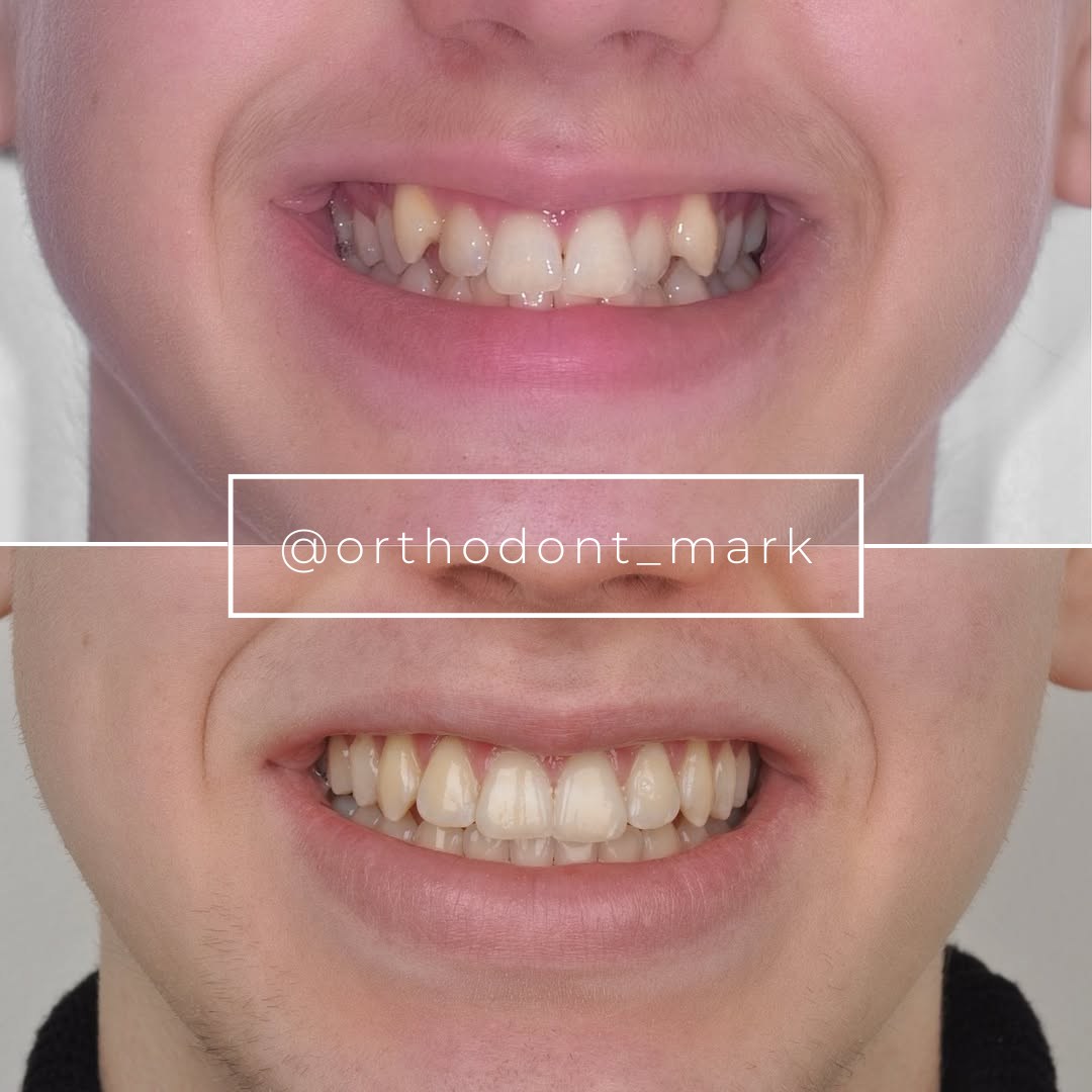
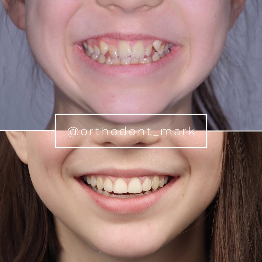
Documented outcomes
Clinical cases from Dr. Mark Radzhabov's practice
Learn the full MARPE protocol from Dr. Mark Radzhabov
Fundamental course covering CBCT patient selection, miniscrew planning, activation protocols, and 60+ clinical cases. Choose the access level that fits your practice.
Essentials of rapid palatal expansion for practicing orthodontists.
- Core RPE concepts and biomechanics
- 6 structured video lessons
- Clinical decision checklists
- Lifetime access to recordings
Deep-dive into MARPE protocol, diagnostics, and clinical execution.
- 6 modules covering MARPE from A to Z
- CBCT case selection walkthroughs
- Miniscrew placement and activation
- 60+ annotated clinical cases
- Direct Q&A with Dr. Mark Radzhabov
5-element medical consultation framework for dentists and orthodontists.
- Trust-building consultation protocol
- 5 lesson modules
- Templates for treatment plan delivery
- Works with any clinical specialty
Clinical FAQ
How many activation turns are used in a standardized MARPE expansion protocol?
Chun et al. (2022) used 35 turns as the standardized total across both the RPE and MARPE groups in their prospective trial, providing a documented benchmark for total activation volume in a controlled clinical setting.
How long should the consolidation phase last before MARPE appliance removal?
Chun et al. (2022) used a 3-month consolidation period with a post-consolidation CBCT as the endpoint. Regional sutural mineralization assessed across anterior, middle, and posterior thirds should guide whether this window needs extending before debond.
What force range do common expansion screws generate during MARPE activation?
Camporesi et al. (2013) measured Hyrax and A2620 screws exceeding 20 kg and Palatal Split screws producing approximately 16 kg during mechanical testing of 10 mm screws at sequential activation levels using an Instron machine.
Does MARPE cause less buccal tipping of anchor teeth than RPE during expansion?
Yes. Chun et al. (2022) demonstrated that MARPE produced significantly less buccal displacement of anchor teeth than RPE across both the expansion and consolidation periods, confirming the dental-side advantage of a bone-borne force path.
What CBCT changes confirm successful midpalatal suture separation after MARPE?
Kinzinger et al. (2022) described the suture shifting from parallel to triangular orientation with transverse force, with decreased width in cranial and dorsal directions. Absence of this geometric change at the post-expansion scan warrants clinical reassessment before consolidation proceeds.
How does MARPE affect nasal width compared to tooth-borne RPE?
Chun et al. (2022) found that MARPE produced greater increases in nasal width at the molar region and greater palatine foramen width than RPE. Monitoring nasal response during active expansion serves as a useful chairside indicator of ongoing sutural separation.
What miniscrew stability check should be performed at each monitoring appointment?
Apply gentle mesiodistal torque to each screw head with a finger. Any detectable micromotion or soft-tissue ingrowth around the screw collar indicates compromised primary stability and should prompt a CBCT before further activation turns are delivered.
When in the MARPE chairside workflow should the expansion screw be locked?
The screw should be locked immediately when the target activation turns are completed. Physical lock or bonded composite block prevents inadvertent reversal during the consolidation phase and should be verified at every subsequent consolidation-period visit.
How should post-consolidation CBCT findings guide retention appliance selection after MARPE?
Each sutural third—anterior, middle, posterior—should be evaluated independently for mineralization. Persistent radiolucency in any region justifies a longer passive retention period. The retention appliance design should correspond to CBCT findings rather than default to a generic protocol.
What records should be obtained at the MARPE retention handoff appointment?
A digital scan or stone cast taken on the debond visit establishes the transverse baseline for long-term relapse monitoring. Combined with the post-consolidation CBCT, these records form the medicolegal and clinical reference for all future transverse assessments.
A reproducible MARPE chairside workflow is not a luxury—it is the mechanism by which bone-borne skeletal expansion consistently outperforms dental tipping protocols in adults. Each appointment gate, from bonding-day torque verification through three-month consolidation CBCT, transforms a technically demanding procedure into a manageable clinical sequence. Orthodontists who want to audit their own visit structure or plan their first case are encouraged to review the full protocol resources at ortodontmark.com or book a direct case consultation with Dr. Mark Radzhabov to stress-test their sequencing decisions before chair time. Key sources: Chun et al. (2022), BMC Oral Health, doi:https://doi.org/10.1186/s12903–022-02138-w. Camporesi et al. (2013), BioMedical Engineering OnLine, doi:http://www.biomedical-engineering-online.com/content/12/1/128.