
MARPE Protocols in Narrow High-Arched Palates:
Appliance Fit
and Screw Trajectory Evidence
A CBCT-grounded review of how steep palatal vault morphology alters miniscrew angulation, band seating, and suture separation outcomes for practicing orthodontists.
TL;DR MARPE protocols in patients with narrow high-arched palates require appliance adaptation to steep palatal vault morphology and adjusted miniscrew angulation. Holm et al. (2016) quantified palatal bone thickness at paramedian sites, and Chun et al. (2022) documented expansion with CBCT at baseline and after consolidation. Suture separation success varies markedly by anatomical and biological factors.
When a patient presents with a narrow high-arched palate, the standard MARPE appliance geometry that performs reliably in a flatter arch becomes a clinical liability: band seating, screw trajectory, and miniscrew purchase angle all shift. Dr. Mark Radzhabov at ortodontmark.com examines how documented MARPE protocols account for these anatomical realities, drawing on CBCT bone-thickness data and prospective trial imaging to help clinicians refine appliance fit, choose insertion vectors, and anticipate biological variation before placing a single miniscrew.
How Does Palatal Vault Geometry Dictate
Miniscrew
Insertion Strategy in Deep-Arch Cases?
MARPE (miniscrew-assisted rapid palatal expansion) is a bone-borne skeletal expansion technique in which titanium miniscrews anchored directly in the palatal midline transmit orthopedic force across the midpalatal suture without relying on tooth roots for anchorage. In a steep, narrow palate, the lateral walls rise at a sharper angle than in a broad, flat arch, which shifts the optimal insertion vector away from perpendicular and demands a more oblique approach to achieve adequate cortical engagement without perforating the nasal floor. CBCT mapping of palatal bone stock is the prerequisite that makes this decision defensible rather than intuitive. Holm et al. (2016) measured palatal bone thickness at paramedian distances of 2, 4, 6, and 8 mm on both left and right sides, establishing a site-specific reference that clinicians can overlay on their own pre-treatment scans. The practical implication is that the thickest available bone — typically in the anterior palate lateral to the raphe — is not uniformly distributed, so a single insertion depth or angle cannot be extrapolated from one patient to another. Sex differences in palatal bone stock add another layer of complexity. Holm et al. (2016) reported that male patients demonstrated 1.2 mm greater average palatal bone thickness than female patients, a finding with direct consequence for miniscrew selection and thread engagement length. In patients with a narrow high-arched palate and thinner cortical plates — a profile more common in female patients — the clinician must reduce insertion torque targets and consider shorter miniscrews to avoid fenestration, rather than applying a protocol calibrated for denser bone.
What CBCT Imaging Schedule Best Captures
Suture Separation
Progress in MARPE Cases?
Deciding when to image during an active MARPE case is not arbitrary — the scan timing must align with the expansion mechanics to distinguish true skeletal opening from dental tipping or appliance distortion. Chun et al. (2022) performed CBCT imaging at three defined timepoints: baseline, immediately after 35 turns of identical expansion, and again following a 3-month consolidation period. This protocol creates an objective before-and-after record of suture morphology change rather than relying on clinical indicators alone. The 35-turn standardization used by Chun et al. (2022) is clinically meaningful because it eliminates interoperator variability in activation amount, making the imaging data directly comparable across patients in a prospective randomized clinical trial. For orthodontists adapting this framework to their own workflow, the key principle is that expansion magnitude must be fixed before the post-expansion scan, otherwise the CBCT captures an arbitrary mid-treatment state. The 3-month consolidation window documented by Chun et al. (2022) is long enough for early mineralization of the sutural gap to begin while still allowing the clinician to assess relapse risk before appliance removal. In narrow high-arched palate cases, where the vault geometry concentrates stress at a smaller cross-sectional suture area, confirming radiographic fill before debonding is especially important. A CBCT showing a persistent dark cleft at the midpalatal suture after consolidation indicates incomplete ossification and argues for extending the retention phase.
Which Patient Factors Predict Suture Opening
Failure
Before You Bond the MARPE?
Even a well-fitted appliance with accurate miniscrew angulation cannot guarantee midpalatal suture separation when the underlying biology is unfavorable. Jeon et al. (2022) reported an overall MARPE suture separation success rate of 79.53% in a retrospective cohort study, and that aggregate figure conceals substantial subgroup divergence. Understanding which patients sit at the lower end of that distribution is the selection problem every MARPE clinician faces before treatment consent. Jeon et al. (2022) found that female patients achieved suture separation in 94.17% of cases, while male patients reached only 61.05%, a difference large enough to change the pre-treatment conversation entirely. Given that Holm et al. (2016) independently documented 1.2 mm greater average palatal bone thickness in males, the lower separation rate in male patients likely reflects both denser interdigitation of the midpalatal suture and heavier cortical resistance — not simply a force-delivery problem solvable by increasing activation frequency. For the clinician planning a miniscrew-assisted palatal expansion in a male adult with a narrow high-arched palate, the Jeon et al. (2022) data create a clear counseling threshold: the probability of non-separation is substantially elevated relative to a female patient of comparable skeletal maturity. This does not automatically indicate surgical referral, but it demands an explicit informed-consent discussion and a pre-defined decision point — typically assessed by CBCT at mid-expansion — at which failure to achieve radiographic suture opening redirects the treatment plan. Appliance design, screw trajectory optimization, and CBCT-guided insertion all improve the conditions for success without altering the underlying suture biology.
How Should Appliance Fit Be Modified
for Deep Palatal
Anatomy in Skeletal Expansion Cases?
A tooth-borne rapid palatal expander transfers expansion forces primarily through the periodontal ligament, which in a narrow high-arched palate creates buccal crown tipping before meaningful sutural separation. The bone-borne force path of a MARPE appliance eliminates this pathway, but only if the miniscrews are placed where cortical bone is adequate — and in a steep vault, that zone is narrower in the transverse dimension than CBCT scouts often suggest at first glance. Band adaptation for the maxillary premolars and molars in a high-arch case must account for the steeper buccolingual angulation of the posterior teeth. Standard bands seated to a flat palate template will rock occlusally on the lingual cusp shelf, creating a gap that compromises load transfer and risks cement failure under the cyclic forces of daily activation. Clinicians should verify band seating with bite registration material before final cementation and confirm that the expander body lies parallel to the palatal plane across its full anterior-to-posterior span. The screw housing position within the appliance also changes in high-vault anatomy: the inferior surface of the expansion screw sits further from the palatal mucosa than in a flat arch, which increases the moment arm through which activation force passes before reaching the miniscrews. This geometry does not negate efficacy, but it does mean that the connecting arms between the screw body and the miniscrew heads must be rigid enough to prevent flex during activation — a fabrication detail that distinguishes a protocol built for palatal morphology variations from a generic laboratory prescription.
Free Clinical Guide
Which Expansion Appliance at Which Age?
Get Dr. Mark Radzhabov's guide to skeletal-expansion appliances — indications, pros & cons, and how to choose by patient age. Free to your inbox.
Prefer video? Watch the free demo lesson on MARPE appliance types, features and trade-offs.
Documented outcomes
Clinical cases from Dr. Mark Radzhabov's practice
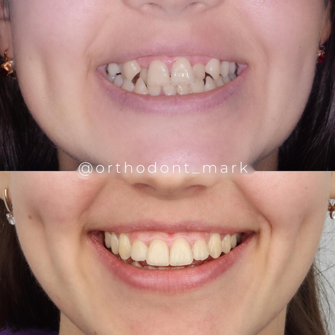
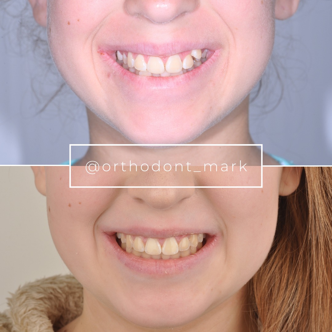
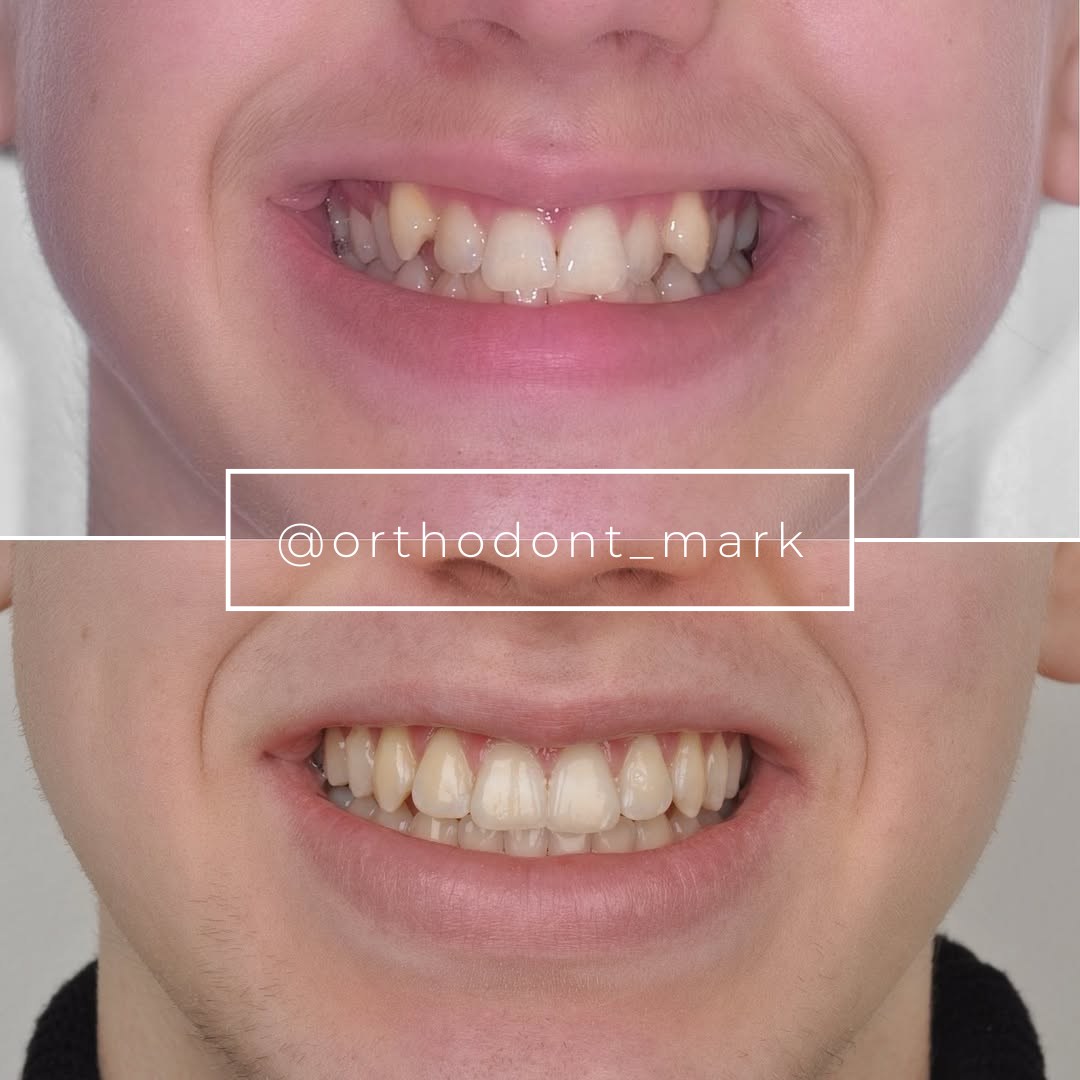
Learn the full MARPE protocol from Dr. Mark Radzhabov
Fundamental course covering CBCT patient selection, miniscrew planning, activation protocols, and 60+ clinical cases. Choose the access level that fits your practice.
Essentials of rapid palatal expansion for practicing orthodontists.
- Core RPE concepts and biomechanics
- 6 structured video lessons
- Clinical decision checklists
- Lifetime access to recordings
Deep-dive into MARPE protocol, diagnostics, and clinical execution.
- 6 modules covering MARPE from A to Z
- CBCT case selection walkthroughs
- Miniscrew placement and activation
- 60+ annotated clinical cases
- Direct Q&A with Dr. Mark Radzhabov
5-element medical consultation framework for dentists and orthodontists.
- Trust-building consultation protocol
- 5 lesson modules
- Templates for treatment plan delivery
- Works with any clinical specialty
Clinical FAQ
How does palatal vault height affect miniscrew angulation in MARPE appliance placement?
A steep vault shifts the safe insertion vector away from perpendicular. The clinician must angle miniscrews to stay within cortical bone while avoiding nasal floor proximity. CBCT measurement of paramedian bone thickness at multiple sites guides this angulation decision before any surgical step.
What does CBCT palatal bone thickness mapping show about optimal miniscrew insertion sites?
Holm et al. (2016) measured thickness at 2, 4, 6, and 8 mm paramedian distances, revealing site-specific variation. The anterior palate lateral to the raphe typically offers the greatest cortical depth. This map prevents placement into zones with insufficient bone for primary stability.
Does sex influence miniscrew-assisted palatal expansion suture separation outcomes?
Yes. Jeon et al. (2022) reported separation in 94.17% of female patients versus 61.05% of male patients. Holm et al. (2016) independently found 1.2 mm greater average palatal bone thickness in males, suggesting denser suture interdigitation contributes to the lower male separation rate.
What is the overall MARPE suture separation success rate across mixed-sex adult populations?
Jeon et al. (2022) reported an overall rate of 79.53% in their retrospective cohort study. This aggregate figure masks significant sex-based divergence, so using it as the sole counseling figure without subgroup stratification underestimates risk for male patients.
How should the CBCT imaging schedule be structured for a standardized MARPE protocol?
Chun et al. (2022) used three timepoints: baseline, immediately after 35 turns of identical expansion, and after a 3-month consolidation period. Fixing activation amount before the post-expansion scan is essential for comparing suture response across patients.
Why does a narrow high-arched palate create band-seating problems with standard MARPE appliances?
Steep vault geometry increases the buccolingual tilt of posterior teeth, causing standard bands to rock on the lingual cusp shelf. This creates a seating gap that compromises load transfer and risks early cement failure during daily activation. Verify full band contact with bite registration material before final cementation.
How is the bone-borne force path in MARPE different from tooth-borne RPE in deep-arch patients?
A tooth-borne rapid palatal expander transmits force through the periodontal ligament, producing buccal crown tipping before sutural separation — particularly pronounced in steep vaults. MARPE delivers force directly to palatal bone via miniscrews, bypassing dental tipping while requiring accurate cortical insertion.
When should a mid-expansion CBCT be scheduled to detect MARPE non-separation early?
A decision-point scan after a defined activation period — comparable to the post-35-turn imaging used by Chun et al. (2022) — allows radiographic confirmation of suture opening before continuing to load. Absence of a visible sutural cleft at this checkpoint should trigger surgical consultation.
How does palatal bone thickness data from CBCT change miniscrew length selection in deep-vault cases?
Holm et al. (2016) showed sex- and site-specific thickness differences up to 1.2 mm on average. Thinner bone in female patients or at specific paramedian distances requires shorter miniscrews with reduced insertion torque targets to prevent fenestration into the nasal floor.
What laboratory specifications reduce arm flex in MARPE appliances designed for high-arch anatomy?
Greater distance between the expander screw body and palatal mucosa increases the moment arm during each activation turn, amplifying flex forces on connecting arms. Rigid arm construction must be explicitly specified in the laboratory prescription for deep-vault cases rather than relying on standard appliance geometry.
Palatal morphology is not a footnote in MARPE planning — it is the variable that determines whether your miniscrews anchor productively or torque against thin cortical bone. Matching appliance geometry and screw angulation to the specific vault profile of each patient is the first clinical decision, not the last. Dr. Mark Radzhabov encourages colleagues to audit their current CBCT interpretation workflow and compare it against the imaging criteria reviewed here. Structured MARPE training is available for orthodontists who want a systematic framework for these cases. Key sources: Holm et al., 2016, Angle Orthodontist, doi:10.2319/091515–622.1. Chun et al., 2022, BMC Oral Health, doi:10.1186/s12903–022-02138-w. Jeon et al., 2022, Clinical Oral Investigations, doi:10.1007/s00784–021-04281–0.