
Transverse Vertical Discrepancy Diagnosis:
Dual-Plane
Maxillary Problems at the Planning Stage
Integrate suture maturation staging, multi-suture fusion data, and vertical landmark assessment before selecting MARPE, RPE, or surgical referral.
TL;DR Transverse vertical discrepancy diagnosis requires evaluating midpalatal suture maturation, pterygomaxillary and zygomaticomaxillary fusion, and vertical maxillary relationships before selecting an expansion modality. Suture staging via cone-beam CT is the most reliable case-selection filter. MARPE achieves higher skeletal suture separation rates than RPE, making modality choice pivotal at the planning stage.
A patient presenting with both a narrow maxilla and excessive vertical facial dimension forces a treatment-planning decision that neither a single arch wire nor a standard expander can resolve alone. Transverse vertical discrepancy diagnosis sits at the intersection of suture biology, three-dimensional imaging, and biomechanical sequencing — and errors made here compound throughout treatment. At ortodontmark.com, Dr. Mark Radzhabov addresses this dual-plane diagnostic challenge by integrating cone-beam CT suture staging with multi-suture fusion assessment, giving clinicians a reproducible framework for selecting between MARPE, conventional RPE, and surgical referral.
Why Does a Combined Transverse and Vertical Problem
Change
the Entire Diagnostic Sequence?
A dual-plane maxillary discrepancy is a simultaneous transverse deficiency and vertical excess of the maxilla that requires coordinated diagnostic imaging and sequenced mechanical correction rather than single-axis treatment. Most planning errors arise when clinicians address one plane while ignoring the other, allowing the untreated dimension to worsen during active mechanics. Recognizing the combined problem early is what separates a predictable outcome from a series of reactive adjustments. The vertical component of a maxillary dimensional analysis governs several downstream decisions: whether an intrusive mechanics vector is needed alongside expansion, whether the palatal vault morphology will assist or resist suture opening, and whether orthognathic surgery must eventually manage the vertical dimension independently. Identifying excessive lower anterior face height, steep mandibular plane angle, and hyperdivergent skeletal pattern on the lateral cephalogram flags the vertical axis as clinically significant before any expander is placed. For the transverse plane, cone-beam CT suture staging provides information that a conventional periapical or panoramic film cannot. The midpalatal suture, pterygomaxillary suture, and zygomaticomaxillary suture each fuse on independent timelines, and fusion in any one of them alters the biomechanical response to palatal expansion. Reviewing all three in a single CBCT volume — rather than inferring biology from chronological age alone — is the diagnostic standard that underpins modern three-dimensional maxillary diagnosis.
Which Sutures Actually Determine Whether
Expansion
Is Still Viable Without Surgery?
Suture fusion is neither uniform across anatomical sites nor predictable from a birth certificate. Govaerts et al. (2023) reported that pterygomaxillary suture closure ranges from 83% to 100% in females aged 13 to 17 years — a spread wide enough to mean that two patients at the same chronological age can have radically different biomechanical substrates for palatal expansion. The midpalatal suture presents an equally striking picture. Govaerts et al. (2023) found that at age 15 years, 61% of females showed closed midpalatal sutures in maturation stages D and E. If the majority of 15-year-old female patients already show advanced suture fusion, deferring CBCT imaging until the mid-teens means the diagnostic window for non-surgical expansion has likely narrowed considerably. Govaerts et al. (2023) placed the recommended age cutoff for surgical rather than orthodontic rapid palatal expansion at 15.1 years according to orthodontists and 14.8 years according to surgeons. The zygomaticomaxillary suture adds a third dimension to this complexity. Govaerts et al. (2023) documented zygomaticomaxillary suture closure in 32% to 47% of females aged 15 to 17 years. Partial fusion at this lateral buttress limits the lateral skeletal response even when the midpalatal suture remains amenable to opening, making multi-suture assessment indispensable for accurate skeletal maxillary expansion planning.
When Does the Suture Stage Justify MARPE
Over
Conventional RPE in the Dual-Plane Case?
Once suture staging identifies a patient who remains a candidate for non-surgical expansion, the choice between MARPE and conventional RPE directly affects how much of the applied force translates into true skeletal widening. Chun et al. (2022) reported that midpalatal suture separation occurred in 90% of RPE patients and 95% of MARPE patients following identical amounts of expansion — a difference that becomes clinically significant when the dual-plane case demands maximal skeletal response with minimal dental tipping. Chun et al. (2022) also found that MARPE produced greater skeletal expansion of nasal width and palatine foramen dimensions compared to RPE during both the expansion and consolidation phases. In a patient with vertical maxillary excess, unwanted dentoalveolar tipping from a tooth-borne expander can exacerbate the open-bite tendency. The bone-borne force pathway of MARPE skeletal expansion reduces this risk by directing force more orthopedically through the palatal vault rather than through the buccal root surfaces. For clinicians building the diagnostic sequencing framework for combined discrepancy planning, these separation data inform a clear decision threshold: where suture staging suggests partial but not complete fusion, MARPE's higher suture separation rate makes it the preferred starting point before considering surgical assistance. When multi-suture fusion is extensive, the imaging evidence — not the clinical intuition — should prompt early surgical consultation rather than a trial of non-surgical mechanics. Reviewing the evidence base for MARPE skeletal expansion protocol decisions is a practical step before finalizing any dual-plane treatment plan.
How Should Cervical Vertebral Maturation
Inform
Vertical Maxillary Assessment in Non-Growing Patients?
Skeletal growth status shapes the biomechanical context for both transverse and vertical correction. Manabe et al. (2022) allocated 80 patients to each cervical vertebral maturation stage group, totalling 400 patients, to establish a reproducible growth-staging reference. This scale allows clinicians to objectively position a patient's remaining growth potential on a continuum rather than relying on chronological age as a proxy — a meaningful distinction when vertical excess and transverse deficiency co-exist and growth vectors are still active. In the hyperdivergent patient with residual growth, the direction of continued mandibular growth can either worsen or partially self-correct the vertical component of the discrepancy. Cervical vertebral staging identifies whether the patient is pre-peak, at-peak, or post-peak — information that determines the urgency of vertical control mechanics and the timing of palatal expansion relative to growth deceleration. A post-peak patient with combined vertical maxillary excess and transverse deficiency requires a fundamentally different treatment sequence than a pre-peak patient with identical cephalometric measurements. Integrating cervical vertebral maturation data with multi-suture CBCT staging produces a two-axis diagnostic map: one axis for skeletal growth status, one for suture fusion biology. When both axes point toward limited remaining plasticity, the surgical pathway becomes difficult to avoid regardless of the patient's chronological age. Documenting this dual assessment systematically — rather than leaving it to clinical impression — is what makes the diagnostic sequencing process auditable and reproducible across a practice.
What Is the Practical Diagnostic Sequence for a
Combined
Transverse and Vertical Maxillary Problem?
The diagnostic sequence for a dual-plane maxillary problem begins with lateral cephalometric analysis to quantify the vertical component: mandibular plane angle, lower anterior face height, and palatal plane inclination establish whether vertical maxillary excess is contributing to the facial pattern. These measurements set the biomechanical context before any transverse imaging is reviewed, because the vertical status determines which expansion mechanics are tolerable and which carry unacceptable risk of open-bite progression. The second step is cone-beam CT acquisition with volumetric review of all three relevant sutures — midpalatal, pterygomaxillary, and zygomaticomaxillary — assessed against an established maturation staging system. Reviewing rapid palatal expander mechanics alongside MARPE data at this stage helps clarify whether tooth-borne or bone-borne force distribution is more appropriate for the specific suture biology encountered. The surgical-referral age thresholds reported by Govaerts et al. (2023) — 15.1 years for orthodontists and 14.8 years for surgeons — serve as calibration benchmarks during this review. The third step is growth staging via cervical vertebral maturation, using the standardized framework validated across 400 patients by Manabe et al. (2022). Combining this with suture staging produces a complete pre-treatment biological profile: growth trajectory on one axis, skeletal plasticity on the other. Only after all three layers of diagnostic information are documented should appliance selection and sequencing be finalized. For clinicians building or refining this workflow, exploring structured MARPE training resources provides both the imaging interpretation skills and the biomechanical knowledge needed to manage these cases confidently.
Free Clinical Guide
Which Expansion Appliance at Which Age?
Get Dr. Mark Radzhabov's guide to skeletal-expansion appliances — indications, pros & cons, and how to choose by patient age. Free to your inbox.
Prefer video? Watch the free demo lesson on MARPE appliance types, features and trade-offs.
Documented outcomes
Clinical cases from Dr. Mark Radzhabov's practice

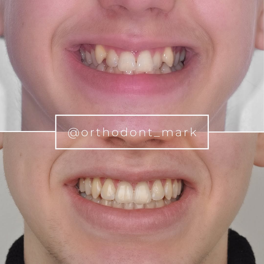
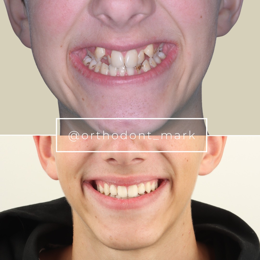
Learn the full MARPE protocol from Dr. Mark Radzhabov
Fundamental course covering CBCT patient selection, miniscrew planning, activation protocols, and 60+ clinical cases. Choose the access level that fits your practice.
Essentials of rapid palatal expansion for practicing orthodontists.
- Core RPE concepts and biomechanics
- 6 structured video lessons
- Clinical decision checklists
- Lifetime access to recordings
Deep-dive into MARPE protocol, diagnostics, and clinical execution.
- 6 modules covering MARPE from A to Z
- CBCT case selection walkthroughs
- Miniscrew placement and activation
- 60+ annotated clinical cases
- Direct Q&A with Dr. Mark Radzhabov
5-element medical consultation framework for dentists and orthodontists.
- Trust-building consultation protocol
- 5 lesson modules
- Templates for treatment plan delivery
- Works with any clinical specialty
Clinical FAQ
How does transverse vertical discrepancy diagnosis differ from evaluating each plane in isolation?
Evaluating both planes together reveals interactions invisible in single-axis assessment. Vertical excess alters which expansion mechanics are safe — tooth-borne force in a hyperdivergent patient risks worsening open bite — while suture fusion status determines whether non-surgical transverse correction is biologically achievable at all.
At what age does midpalatal suture fusion make non-surgical expansion unreliable in female patients?
Govaerts et al. (2023) found that 61% of females at age 15 already showed closed midpalatal sutures in advanced maturation stages, and recommended surgical referral from age 14.8 to 15.1 years depending on specialty. Suture staging by CBCT remains more informative than age alone.
How does MARPE compare to RPE for achieving skeletal suture separation?
Chun et al. (2022) reported midpalatal suture separation in 95% of MARPE patients and 90% of RPE patients following identical expansion amounts. MARPE also produced greater nasal width and palatine foramen skeletal expansion during both expansion and consolidation, making it preferable when maximal skeletal response is required.
Why must pterygomaxillary and zygomaticomaxillary sutures be assessed separately from the midpalatal suture?
Each suture follows an independent fusion timeline. Govaerts et al. (2023) found pterygomaxillary closure in 83% to 100% of females aged 13 to 17, while zygomaticomaxillary closure was present in 32% to 47% of females aged 15 to 17. Fusion at lateral buttresses limits skeletal response even when the midpalatal suture remains open.
How does cervical vertebral maturation staging contribute to dual-plane maxillary treatment planning?
Manabe et al. (2022) validated CVM staging across 400 patients, enabling objective growth-trajectory mapping. In hyperdivergent patients, knowing whether mandibular growth is pre-peak or post-peak determines the urgency of vertical control mechanics and the timing of expansion relative to growth deceleration.
What cephalometric landmarks should confirm vertical maxillary excess before CBCT suture review?
Assess mandibular plane angle, lower anterior face height, and palatal plane inclination on the lateral cephalogram. Steep mandibular plane and increased lower face height define a hyperdivergent pattern and indicate that tooth-borne palatal expansion carries greater risk of vertical dimension worsening.
Does MARPE produce different skeletal effects than RPE during the consolidation phase?
Yes. Chun et al. (2022) found MARPE produced greater skeletal expansion of nasal width and palatine foramen dimensions compared to RPE during both active expansion and the consolidation phase, suggesting the bone-borne force pathway maintains broader skeletal effects beyond the active activation period.
How should a clinician decide between MARPE and surgical referral in a borderline suture fusion case?
Document midpalatal, pterygomaxillary, and zygomaticomaxillary fusion status on CBCT, then map against the referral thresholds from Govaerts et al. (2023). When multiple sutures show advanced closure, the imaging evidence — not clinical intuition — should prompt surgical consultation rather than trialing non-surgical mechanics.
What sample size supported the cervical vertebral maturation staging reference used for growth assessment?
Manabe et al. (2022) allocated 80 patients to each cervical vertebral maturation stage group, totalling 400 patients. This standardized distribution provides a reproducible reference baseline for positioning individual patients along the growth continuum in clinical practice.
How can orthodontic practices standardize dual-plane diagnostic sequencing across clinicians?
Establishing a written three-step protocol — lateral cephalometric vertical analysis, multi-suture CBCT staging, and cervical vertebral maturation assessment — before any appliance selection creates an auditable, reproducible workflow. Dr. Mark Radzhabov at ortodontmark.com offers structured training resources to help practices implement this framework consistently.
A combined transverse and vertical maxillary problem rewards clinicians who invest in thorough diagnostic imaging before committing to an appliance: suture maturation stage, pterygomaxillary fusion status, and vertical landmarks must align before activation begins. When the dual-plane assessment points toward MARPE, the evidence supports superior skeletal separation over conventional RPE. Dr. Mark Radzhabov offers structured resources at ortodontmark.com for clinicians ready to refine their diagnostic sequencing and expand their case-selection confidence — a valuable next step for any practice managing complex skeletal discrepancies. Key sources: Chun et al. (2022), BMC Oral Health, doi:10.1186/s12903–022-02138-w. Govaerts et al. (2023), Journal of Orofacial Orthopedics, doi:10.1007/s00056–023-00487-x. Manabe et al. (2022), PLOS ONE, doi:10.1371/journal.pone.0265272.