
Unilateral vs Bilateral Crossbite Diagnosis
Etiology
at the First Exam: Expansion Decision Framework
Separate functional shift from skeletal deficiency before choosing between bone-borne and tooth-borne expansion. A single exam finding changes the entire treatment arc.
TL;DR Unilateral vs bilateral crossbite diagnosis at the first exam determines whether expansion targets a true skeletal transverse deficiency or a functional mandibular shift. A bilateral skeletal presentation calls for bone-borne expansion. A unilateral shift demands functional correction first. Missing this distinction leads to overcorrection, relapse, or unnecessary surgical referral.
Distinguishing a true bilateral skeletal crossbite from a functionally driven unilateral vs bilateral crossbite diagnosis error is among the most consequential calls an orthodontist makes at the first exam. Misclassifying a functional shift as a fixed skeletal asymmetry directs treatment toward expansion protocols that solve the wrong problem — and misclassifying a bilateral skeletal deficiency as a simple dental tipping issue leads to inadequate force mechanics and predictable relapse. At ortodontmark.com, Dr. Mark Radzhabov has structured this clinical guide around the examination findings, imaging criteria, and TMD history that reliably separate these two presentations before a single activation turn is made.
Why Crossbite Laterality Assessment Changes
Everything
About Appliance Selection
Posterior crossbite etiology diagnosis is the clinical and radiographic process of identifying whether a crossbite arises from a true skeletal transverse maxillary deficiency, a dental compensation, or a functional mandibular shift — a distinction that directly governs appliance choice and expansion mechanics. Two patients can present with an identical occlusal photograph and require completely different interventions depending on what the mandible does when guided to centric relation. In maximum intercuspation, a unilateral crossbite can reflect a bilaterally constricted maxilla whose midline coincides with the mandibular midline in centric relation — a bilateral skeletal problem masked by a habitual lateral slide. Alternatively, the maxillary arch may be genuinely asymmetric with dental tipping confined to one quadrant. These three presentations — bilateral skeletal, unilateral skeletal, and unilateral dental-functional — demand separate mechanical strategies, and confusing them is one of the most avoidable sources of post-treatment relapse. The first exam must therefore include both a dental and a postural component: occlusal contacts in maximum intercuspation, midline coincidence in centric relation, and an assessment of whether a lateral slide is present and repeatable. Midline deviation that disappears on guided closure confirms a functional component and shifts the immediate priority toward eliminating the shift before committing to any fixed expansion appliance. For bone-borne skeletal expansion planning, the bilateral skeletal presentation is the primary indication.
When the Patient's Pain History Alters
Crossbite
Treatment Sequencing
A thorough orofacial history changes the clinical framing of a crossbite presentation. Patients referred to orofacial pain clinics carry a measurable burden of overlapping symptoms, and a crossbite with a functional shift component may be embedded in that same symptom cluster. Broberg et al. (2017) found that among patients with anterior bite opening, 62% reported tiredness or orofacial pain, 41% headache, 24% TMJ clicking, and 41% sensitive or tender teeth — a symptom profile that, when present alongside a unilateral crossbite and lateral slide, signals that the mandibular position may be pain-driven rather than purely structural. The sequencing implication is direct: initiating transverse expansion in a patient whose mandibular shift is maintained by a pain-avoidance posture risks stabilizing the wrong jaw position. Broberg et al. (2017) also documented that 66% of patients reported prior TMD symptoms before onset of the bite change, suggesting that the joint environment often precedes rather than follows the occlusal finding. Screening for TMD history before recording crossbite laterality is therefore a diagnostic prerequisite, not an add-on. Furthermore, Broberg et al. (2017) reported that two-thirds of patients with bite changes reported parafunction or bruxism — a cofactor that loads the posterior dentition asymmetrically and can mimic or exacerbate a dental crossbite on one side. When parafunction is confirmed, the apparent crossbite depth on mounted models may exceed the true skeletal discrepancy, leading to overestimation of the required expansion magnitude.
How to Conduct a Crossbite Etiology Assessment
Step by Step
at the Initial Examination
The unilateral vs bilateral crossbite diagnosis workflow begins with the dental midline in two jaw positions: maximum intercuspation and centric relation. Record the midline offset in both positions. If the offset is zero or reversed in centric relation, a functional mandibular shift is present and the crossbite laterality in maximum intercuspation is not a reliable representation of the skeletal discrepancy. This single observation recalibrates every downstream decision. Next, palpate the lateral pterygoid region and the TMJ poles bilaterally during opening and closing. A repeatable click or capsular tenderness on the contralateral side to the crossbite is consistent with a habitual shift protecting that joint. Broberg et al. (2017) excluded patients with TMJ arthrosis, systemic rheumatic disease, neuromuscular disease, jaw fractures, and prior orthodontic treatment to isolate idiopathic bite changes — a methodological choice that underlines how powerfully comorbidities confound the occlusal picture in general clinical populations. For arch width measurement, Fes et al. (2022) showed that Pont's Index and CBCT-derived arch width analysis produced no statistically significant difference (p=0.85 by McNemar's test), meaning that a well-executed dental cast measurement remains diagnostically equivalent to CBCT for transverse arch width alone. CBCT adds value specifically for suture maturation staging and bony cortical thickness assessment — not for confirming whether a width discrepancy exists. Order the scan to guide expansion mechanics, not to detect the crossbite itself. After laterality is confirmed and the functional component is excluded or quantified, classify the presentation: bilateral skeletal (expand with a rapid palatal expander for dental crossbite correction or a bone-borne device), unilateral skeletal (asymmetric mechanics or unilateral surgical assist), or unilateral functional (interceptive occlusal guidance, then reassess).
Matching Expansion Protocol to Confirmed
Crossbite
Etiology and Skeletal Status
Once the etiology is confirmed as bilateral skeletal, the expansion decision pivots to suture maturation and patient age. In growing patients, tooth-borne expansion applies force through the periodontal ligament to the alveolar process and indirectly to the midpalatal suture — effective when sutural resistance is low and dental anchorage is adequate. In skeletally mature patients with a confirmed bilateral skeletal crossbite, bone-borne force vectors applied directly to the palatal cortex offer superior skeletal displacement with reduced dental tipping. For clinicians building or refining this protocol, submit a case for clinical consultation at ortodontmark.com to review imaging and expansion sequencing. A bilateral crossbite with a confirmed functional shift component treated with symmetric bilateral expansion risks fixing the mandible in its shifted position — a scenario that creates an iatrogenic asymmetric skeletal result. The treatment sequence must resolve the shift first, reconfirm arch width needs in centric relation, and then activate expansion to address only the verified transverse deficit. Among referred patients with bite changes, Broberg et al. (2017) found that 52% presented with occlusal problems alone and 48% with combined occlusal and pain presentations — a near-even split that reminds clinicians that pain-free patients still carry a meaningful probability of underlying functional or joint-mediated contributions to their crossbite. A negative pain history does not rule out a functional shift. Centric relation assessment remains mandatory regardless of symptom status.
Pitfalls That Compromise Crossbite
Differential
Diagnosis and Expansion Outcomes
The most consistent diagnostic error is recording crossbite laterality only in maximum intercuspation without a centric relation check. This omission converts a bilateral skeletal problem into an apparent unilateral one when the mandible habitually shifts to achieve best-fit posterior contact — or inverts a genuine unilateral asymmetry into a bilateral appearance when shift direction coincides with the narrow side. Neither situation is rare, and neither is recoverable without repeating the records. A second pitfall is treating the crossbite in isolation from the TMD and parafunction history. Broberg et al. (2017) excluded patients with rheumatic disease, neuromuscular disease, and prior orthodontic treatment to isolate idiopathic presentations — in clinical practice, these comorbidities are present and must be actively screened because each one can alter jaw position, occlusal loading, and apparent arch width in ways that mimic or amplify a structural crossbite. An unchecked bruxism habit, for instance, depresses posterior vertical dimension asymmetrically and narrows the apparent transverse space on the working side. Finally, over-relying on CBCT to confirm crossbite presence — rather than using it selectively for suture staging and bone density assessment — adds radiation exposure and cost without changing the crossbite diagnosis. Fes et al. (2022) demonstrated that Pont's Index and CBCT-derived measurements produced statistically equivalent arch width results (McNemar's test p=0.85). Reserve the scan for the expansion mechanics decision, not the etiology decision. Crossbite laterality is a clinical diagnosis. CBCT is a biomechanical planning tool.
Free Clinical Guide
Which Expansion Appliance at Which Age?
Get Dr. Mark Radzhabov's guide to skeletal-expansion appliances — indications, pros & cons, and how to choose by patient age. Free to your inbox.
Prefer video? Watch the free demo lesson on MARPE appliance types, features and trade-offs.
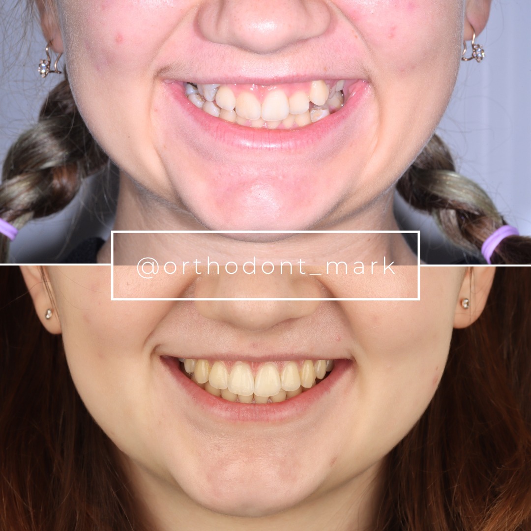
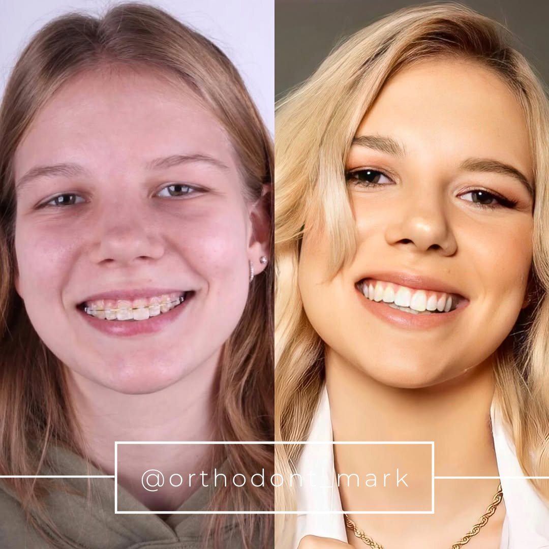
Documented outcomes
Clinical cases from Dr. Mark Radzhabov's practice

Learn the full MARPE protocol from Dr. Mark Radzhabov
Fundamental course covering CBCT patient selection, miniscrew planning, activation protocols, and 60+ clinical cases. Choose the access level that fits your practice.
Essentials of rapid palatal expansion for practicing orthodontists.
- Core RPE concepts and biomechanics
- 6 structured video lessons
- Clinical decision checklists
- Lifetime access to recordings
Deep-dive into MARPE protocol, diagnostics, and clinical execution.
- 6 modules covering MARPE from A to Z
- CBCT case selection walkthroughs
- Miniscrew placement and activation
- 60+ annotated clinical cases
- Direct Q&A with Dr. Mark Radzhabov
5-element medical consultation framework for dentists and orthodontists.
- Trust-building consultation protocol
- 5 lesson modules
- Templates for treatment plan delivery
- Works with any clinical specialty
Clinical FAQ
How do I distinguish a bilateral skeletal crossbite from a unilateral functional shift at the first exam?
Guide the mandible to centric relation and record the dental midline. If the crossbite becomes bilateral or the midline coincides in CR, a functional shift was present in maximum intercuspation. A persistent unilateral crossbite in CR indicates a true unilateral skeletal or dental etiology.
When should I perform a centric relation check during posterior crossbite etiology diagnosis?
At every first exam where a unilateral crossbite is observed. Habitual lateral mandibular displacement to achieve best-fit posterior contacts is common and renders maximum-intercuspation occlusal photographs unreliable for classifying crossbite laterality and expansion indication.
Does a pain-free patient still need a TMD screen before crossbite expansion planning?
Yes. Broberg et al. (2017) found that 52% of patients with bite-position changes presented with occlusal problems alone and no pain complaint. A negative pain history does not exclude a functional mandibular shift. Centric relation assessment remains mandatory regardless of symptom status.
How does parafunction or bruxism affect posterior crossbite etiology assessment?
Parafunction loads the posterior dentition asymmetrically and can depress vertical dimension on the working side, making the transverse discrepancy appear greater than the underlying skeletal deficit. Broberg et al. (2017) found two-thirds of patients with bite changes reported parafunction, warranting screening before arch width measurement.
Is CBCT necessary to confirm the presence of a posterior crossbite or determine its laterality?
No. Fes et al. (2022) showed no statistically significant difference between Pont's Index and CBCT for transverse arch width assessment (p=0.85). CBCT is indicated for midpalatal suture maturation staging and bone density planning — not for confirming the crossbite diagnosis itself.
What clinical examination findings specifically support a functional shift diagnosis in unilateral crossbite cases?
A dental midline discrepancy in maximum intercuspation that reduces or disappears in centric relation. A repeatable lateral slide path on closing. And the absence of a skeletal or dental asymmetry on clinical and radiographic records all support a functional shift rather than a fixed structural etiology.
When is MARPE expansion protocol indicated for a bilateral posterior crossbite versus a tooth-borne RPE?
Bone-borne MARPE is preferred in skeletally mature patients with a confirmed bilateral skeletal transverse deficiency and advanced midpalatal suture maturation on CBCT. Tooth-borne RPE remains appropriate in growing patients with low sutural resistance where dental anchorage is adequate and the etiology is bilateral skeletal.
How does prior TMD history alter the crossbite expansion treatment sequence?
Broberg et al. (2017) documented that 66% of patients with bite changes had prior TMD symptoms before onset — suggesting joint pathology can precede and drive the occlusal shift. Expansion into an unresolved TMD-driven posture risks stabilizing the mandible in an asymmetric, symptom-avoiding position.
What is the diagnostic risk of recording crossbite laterality only in maximum intercuspation?
Habitual mandibular shifts convert a bilateral skeletal deficiency into an apparent unilateral crossbite in maximum intercuspation. Acting on that apparent laterality leads to asymmetric expansion of a symmetric problem — an error that produces iatrogenic dental and skeletal asymmetries and elevated relapse risk.
How should I integrate Pont's Index into a posterior crossbite differential diagnosis workflow?
Use Pont's Index from dental casts to quantify transverse arch width discrepancy during the etiology workup. Fes et al. (2022) confirmed it performs equivalently to CBCT measurement (p=0.85). Proceed to CBCT only when suture maturation staging or palatal bone density assessment is needed to choose between expansion modalities.
Accurate crossbite laterality assessment at the first exam is not a preliminary step — it is the clinical decision that determines every downstream protocol choice, from appliance selection to CBCT acquisition to surgical threshold. A functional shift caught early resolves with guidance mechanics. A bilateral skeletal deficiency in a mature patient requires bone-borne force delivered across the midpalatal suture. Before committing to either path, review the TMD history, the midline discrepancy in centric relation, and the suture maturation stage on CBCT. Dr. Mark Radzhabov offers structured case review and mentorship for clinicians refining this diagnostic workflow — begin with a consultation at ortodontmark.com. Key sources: Broberg et al. (2017), The Open Dentistry Journal, doi:10.2174/1874210601711010628. Fes et al. (2022), Dentistry Journal, doi:10.3390/dj10020023.

