
RPE Candidate Transverse Assessment:
Skeletal vs Dental
A Clinical Decision Framework
CBCT suture geometry, cast analysis, and appliance force data — applied together to confirm true transverse deficiency before RPE case selection.
TL;DR Accurate RPE candidate transverse assessment requires separating true skeletal constriction from dentoalveolar compensation before appliance selection. Suture opening geometry shifts from parallel to V-shaped as patients age, altering force distribution and treatment goals. Appliance forces, suture histology, and cast analysis together guide case selection more reliably than arch width alone.
Determining whether a patient presenting with posterior crossbite reflects genuine maxillary skeletal constriction or compensatory dental tipping remains one of the most consequential decisions in orthodontic treatment planning. An incorrect diagnosis leads either to under-treatment — leaving a structural discrepancy unresolved — or to inappropriate expansion that moves teeth without meaningfully widening the basal arch. At ortodontmark.com, Dr. Mark Radzhabov integrates CBCT-derived suture geometry, dental cast analysis, and appliance biomechanics to build a reliable clinical decision framework. This guide consolidates that evidence so clinicians can perform a rigorous RPE candidate transverse assessment before committing to appliance selection.
Why Posterior Crossbite Does Not Equal
Skeletal Constriction
in Every RPE Candidate
Transverse deficiency diagnosis in RPE candidate assessment is the process of determining whether a patient's posterior arch narrowing originates from skeletal constriction of the maxillary base, dentoalveolar tipping of the posterior teeth, or a combination of both — a distinction that directly dictates appliance type and expansion vector. A posterior crossbite in an eight-year-old and the same crossbite in a thirteen-year-old carry different structural implications, and treating them identically produces very different results at the end of retention. The midpalatal suture is the primary anatomical variable in this assessment. A 2022 CBCT and dental cast analysis confirmed that expansion geometry is not uniform across age groups: patients under 10 years of age show parallel suture opening during RPE, while those aged 12 and older develop a V-shaped pattern in which anterior opening exceeds posterior opening. This anterior-dominant vector is not a complication — it is a predictable biomechanical consequence of progressive suture interdigitation — but it changes how much true basal expansion is achievable at a given skeletal age. Dental compensation complicates the picture further. When posterior teeth have tipped buccally in response to a narrow palate, arch width measurements at the cusp tips overestimate the true skeletal transverse dimension. A clinician relying solely on intermolar width risks selecting an RPE when the deficiency is predominantly dentoalveolar, or conversely, underestimating skeletal severity when severe tipping masks a wider functional arch. CBCT cross-sections through the alveolar base — not the dental crowns — are required to separate these contributions cleanly before committing to a rapid palatal expander protocol.
What Suture Biopsies Reveal About
Tissue Response
and Timing Windows
The suture is not a passive hinge — it is a metabolically active connective tissue zone whose histological state determines whether applied force produces bone displacement or simply compresses fibrous tissue. Caprioglio et al. (2017) obtained midpalatal suture biopsies from 3 growing patients with a mean age of 8.3 years, including two treated with RME and one untreated control, providing direct histological evidence of the tissue-level response to expansion force in the growing palate. The histological timeline matters for clinical decision-making. Caprioglio et al. (2017) showed that by 30 days after RME, newly formed bone trabeculae with perpendicular orientation to the suture axis were already detectable. This rapid ossification window has a direct implication for retention protocol: if new bone is forming within 30 days, active retention must bridge the immature mineralization phase or relapse force from the stretched palatal mucosa will act on incompletely calcified trabeculae. Bud et al. (2023) identified palatal mucosal hyperplasia as a frequently encountered local complication of RPE, attributable to mechanical trauma from the expander body. This finding is not merely a comfort issue — chronic mucosal inflammation at the appliance contact zone can mask the early tissue signs of adequate suture separation and confound clinical monitoring. Incorporating this knowledge into the RPE candidate transverse assessment means planning appliance design and activation rate to minimize soft-tissue impingement from the first activation visit.
How Screw Design and Force Output Shape
Skeletal vs Dental
Expansion Outcomes
Choosing between a bone-borne and a tooth-borne palatal expander cannot be reduced to age alone — it requires understanding what force each appliance delivers and how that force is transmitted across the transverse suture. Camporesi et al. (2013) conducted in vitro mechanical testing of three RPE screw designs and found that Hyrax and A2620 screws generated forces exceeding 20 kg, while Palatal Split screws generated approximately 16 kg. These magnitudes are not interchangeable. A clinician selecting a lower-force screw in a patient with borderline skeletal-versus-dental presentation is making a different biomechanical bet than one selecting a high-force design. Activation increment is equally precise. Camporesi et al. (2013) documented that standard RPE screws are 10 mm in total size, with each full turn delivering 0.8 mm of expansion across 4 quarter-turn activations of 0.2 mm each. This granularity matters when managing the V-shaped expansion pattern seen in older patients: more frequent, smaller activations may distribute force more evenly across the anterior-posterior palatal gradient than single large increments. The broader force range documented by Camporesi et al. (2013) — spanning 7.54 to 15.8 kilograms across device designs — underscores why screw selection must be deliberate rather than habitual. In a patient with predominantly dentoalveolar compensation and a patent suture, the higher end of this range risks excessive tipping of the anchor teeth before meaningful skeletal displacement occurs. Pairing screw selection with CBCT-confirmed suture status is the only reliable way to match force to biology within a skeletal expansion protocol.
Building the RPE Candidate Assessment
Decision Sequence
From Imaging to Appliance
A reproducible decision sequence protects against confirmation bias and ensures that appliance selection follows the evidence rather than clinical habit. The sequence begins with CBCT acquisition centered on the midpalatal suture, reviewed in axial, coronal, and sagittal planes. The axial view confirms suture patency and its anterior-to-posterior continuity. The coronal view quantifies alveolar base width independent of crown angulation. And the sagittal reconstructions reveal whether the suture shows the open radiolucent appearance of an immature structure or the interdigitated opacity of a maturing one. Dental cast analysis runs parallel to imaging, not as a substitute. Intermolar width measured at the cusp tips is compared with a second measurement at the gingival margin to estimate the magnitude of buccal tipping. A large discrepancy between crown-level and cervical-level width indicates substantial dentoalveolar compensation and shifts the primary treatment objective toward torque correction before or concurrent with expansion. Cases where the two measurements converge suggest that the arch narrowing is predominantly skeletal in origin and that RPE force will be transmitted more directly to the palatal base. Once imaging and cast data are reconciled, appliance selection follows logically. A young patient with a patent, parallel-opening suture and minimal dental tipping is a straightforward candidate for a conventional tooth-borne expander within the force range documented by Camporesi et al. (2013). An older patient showing a V-shaped pattern and significant dentoalveolar compensation warrants closer consideration of bone-borne anchorage to reduce dental side effects and concentrate force at the basal level. Connecting this assessment to a case review consultation before the first activation visit is the most direct way to avoid post-treatment transverse relapse.
Free Clinical Guide
Which Expansion Appliance at Which Age?
Get Dr. Mark Radzhabov's guide to skeletal-expansion appliances — indications, pros & cons, and how to choose by patient age. Free to your inbox.
Prefer video? Watch the free demo lesson on MARPE appliance types, features and trade-offs.
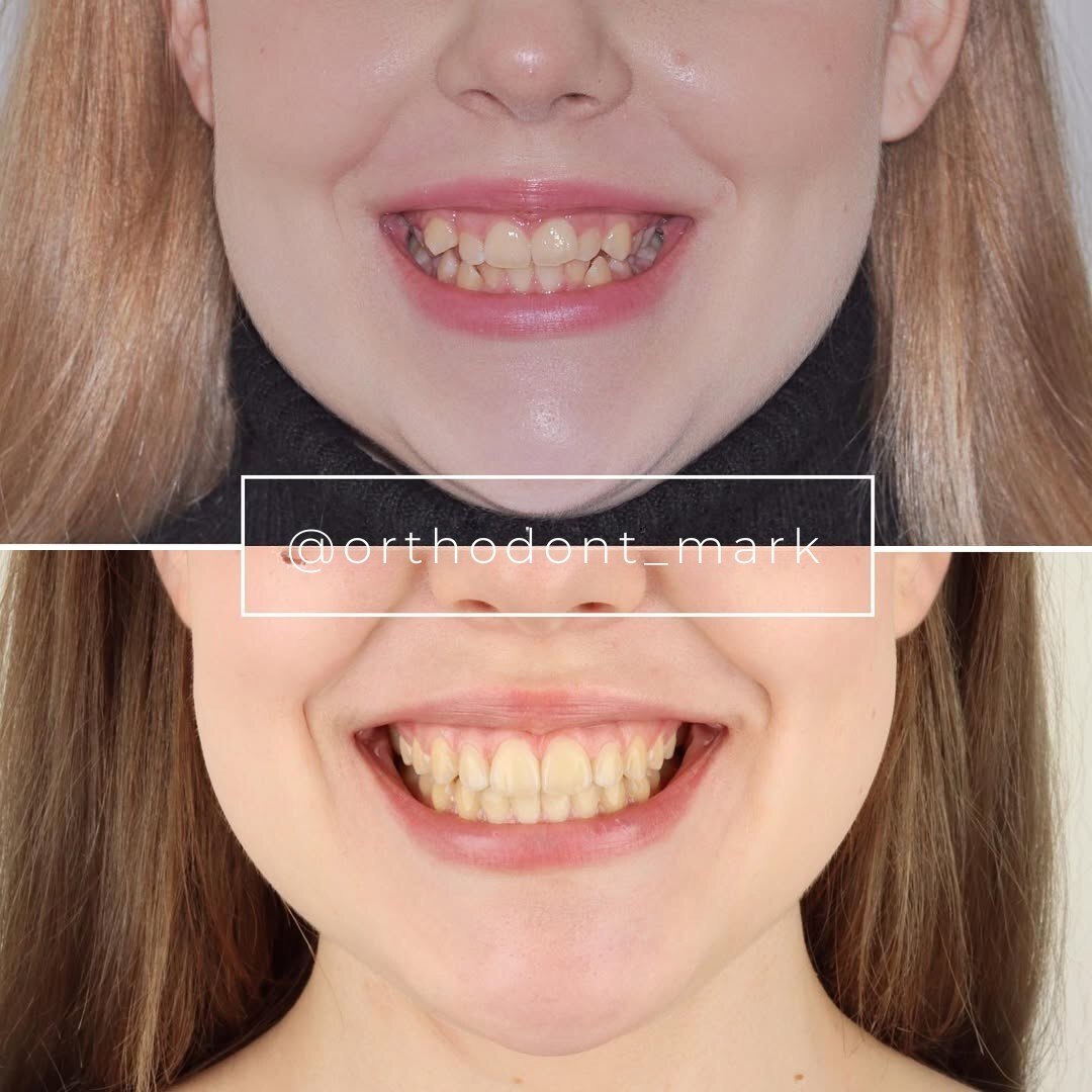
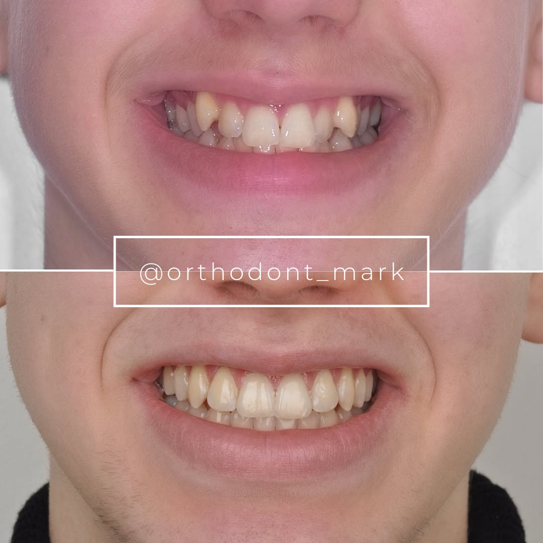
Documented outcomes
Clinical cases from Dr. Mark Radzhabov's practice

Learn the full MARPE protocol from Dr. Mark Radzhabov
Fundamental course covering CBCT patient selection, miniscrew planning, activation protocols, and 60+ clinical cases. Choose the access level that fits your practice.
Essentials of rapid palatal expansion for practicing orthodontists.
- Core RPE concepts and biomechanics
- 6 structured video lessons
- Clinical decision checklists
- Lifetime access to recordings
Deep-dive into MARPE protocol, diagnostics, and clinical execution.
- 6 modules covering MARPE from A to Z
- CBCT case selection walkthroughs
- Miniscrew placement and activation
- 60+ annotated clinical cases
- Direct Q&A with Dr. Mark Radzhabov
5-element medical consultation framework for dentists and orthodontists.
- Trust-building consultation protocol
- 5 lesson modules
- Templates for treatment plan delivery
- Works with any clinical specialty
Clinical FAQ
How do I distinguish true skeletal transverse deficiency from dentoalveolar compensation on CBCT?
Measure alveolar base width on coronal CBCT slices rather than at crown level. A narrow base with upright posterior teeth confirms skeletal origin. Normal base width with buccally tipped crowns points to dentoalveolar compensation. Cast analysis of cusp-tip versus cervical intermolar width reinforces the CBCT finding.
At what age does RPE suture opening shift from parallel to V-shaped?
A 2022 CBCT and dental cast analysis found that patients under 10 years show parallel expansion, while those aged 12 and older develop a V-shaped opening pattern with greater anterior than posterior separation. This geometry change alters force distribution and retention planning.
What force do standard RPE screws generate, and does screw design matter for case selection?
Camporesi et al. (2013) showed RPE devices generate 7.54 to 15.8 kg depending on design. Hyrax and A2620 screws exceeded 20 kg while Palatal Split screws produced approximately 16 kg. Higher-force screws increase dental side effects in predominantly dentoalveolar cases — screw selection should follow the diagnosis.
How quickly does new bone form after RME, and why does it matter for retention timing?
Caprioglio et al. (2017) documented perpendicular bone trabeculae within 30 days of RME in growing patients. Retention must begin while this new bone is still immature. Premature appliance removal before mineralization is complete increases relapse risk from palatal soft-tissue recoil.
What is palatal mucosal hyperplasia, and how does it affect RPE monitoring?
Bud et al. (2023) identified palatal mucosal hyperplasia as a frequent local complication caused by mechanical trauma from the expander body. Soft-tissue overgrowth can obscure suture opening signs on clinical inspection, making CBCT verification more important than visual monitoring alone.
How does each activation increment translate to palatal opening in a standard RPE screw?
Camporesi et al. (2013) documented that each full turn of a standard 10 mm RPE screw delivers 0.8 mm of expansion across 4 quarter-turn activations of 0.2 mm each. This precision is relevant when titrating force in older patients with a V-shaped suture opening pattern.
When should I choose a bone-borne over a tooth-borne palatal expander for transverse deficiency?
Bone-borne anchorage is preferred when cast analysis reveals significant dentoalveolar compensation — large crown-to-cervical width discrepancy — combined with CBCT evidence of adequate alveolar bone for miniscrew insertion. Tooth-borne designs are appropriate when tipping is minimal and suture geometry is patent and parallel.
How do I use dental cast analysis alongside CBCT to confirm RPE candidate suitability?
Measure intermolar width at cusp tips and again at the cervical margin on articulated casts. Compare the two figures: a large gap signals buccal tipping and dentoalveolar compensation. Reconcile with CBCT alveolar base width to determine the skeletal versus dental contribution before appliance selection.
Does a posterior crossbite in a 12-year-old always indicate a need for skeletal RPE expansion?
Not necessarily. A 2022 CBCT and dental cast analysis showed that V-shaped suture opening develops in patients aged 12 and older, meaning posterior expansion is reduced relative to anterior. CBCT alveolar base measurement and cast tipping analysis must confirm genuine skeletal narrowing before selecting RPE over camouflage mechanics.
What histological evidence supports early intervention in growing RPE candidates?
Caprioglio et al. (2017) biopsied the midpalatal suture of 3 growing patients with a mean age of 8.3 years and found active bone remodeling in response to RME force, with oriented trabeculae evident at 30 days. This confirms that the growing suture responds structurally to expansion and supports earlier rather than delayed intervention.
Distinguishing true transverse deficiency from dental compensation is not a single-measurement decision — it depends on imaging, cast analysis, appliance biomechanics, and suture histology working together. A parallel suture opening pattern in a young patient, a V-shaped vector in an adolescent, and the tissue response documented at 30 days all carry direct protocol implications. Clinicians who want a structured framework for applying this evidence to individual cases are welcome to book a case review or explore the structured MARPE training available through Dr. Mark Radzhabov at ortodontmark.com. Key sources: a 2022 CBCT and dental cast analysis, J Orofac Orthop, doi:https://doi.org/10.1007/s00056–022-00429-z. Caprioglio et al. (2017), Int. J. Mol. Sci., doi:10.3390/ijms18030615. Camporesi et al. (2013), BioMedical Engineering OnLine, doi:http://www.biomedical-engineering-online.com/content/12/1/128.

