
Gingival Biotype Before Palatal Expansion
Recession Prediction Tool
Baseline soft tissue screening for skeletal widening
Pre-expansion periodontal screening identifies thin-phenotype patients at risk for buccal recession. Learn the clinical protocol for transgingival measurement and risk stratification.
TL;DR Gingival biotype before palatal expansion is a critical predictor of buccal soft tissue recession risk during skeletal expansion. Thin phenotypes (≤0.5 mm) show 3–4× higher recession incidence than thick biotypes. Baseline buccal gingival assessment using transgingival ultrasound or clinical probing determines whether to modify loading protocols or select alternative treatment.
Skeletal expansion via miniscrew-assisted rapid palatal expansion (MARPE) achieves predictable transverse gain in non-growing patients, yet soft tissue response remains underestimated in treatment planning. Gingival biotype before palatal expansion fundamentally determines whether buccal bone loss and recession will accompany skeletal widening. Dr. Mark Radzhabov and the clinical community increasingly recognize that pre-expansion periodontal screening—specifically measurement of gingival thickness at the premolar-molar region—distinguishes recession-prone from stable cases. This guide reviews the evidence and protocol for baseline gingival assessment, enabling clinicians to counsel patients on risk and optimize loading mechanics before beginning expansion.
Why Gingival Biotype Matters Before MARPE
soft tissue
Understanding baseline phenotype
Gingival biotype is the phenotypic thickness and architecture of marginal gingiva, measured as keratinized tissue width and sulcus depth, which predicts soft tissue response to orthopedic and orthodontic force. Clinical observation and emerging ultrasound studies show that thin biotypes (keratinized tissue <0.5 mm and sulcus depth >3 mm) recede 3–4 times more frequently than thick phenotypes during transverse expansion. The mechanism is biomechanical: thin gingiva cannot distribute buccal bone loads evenly. Cortical plate dehiscence develops in the anterior-middle third of the palate, where expansion velocity peaks. A 35-year-old with thin recession-prone gingival biotype undergoing MARPE may experience 1–2 mm of buccal soft tissue loss within 12 months, whereas a thick-biotype peer shows minimal recession.
Baseline assessment before loading the miniscrew appliance is therefore not cosmetic surveillance but risk stratification. Patients who decline soft tissue measurement or have no baseline record face post-treatment surprise: what was labeled 'stable bone levels' at treatment start may mask unrecognized recession. Radiographic imaging (cone-beam CT) does not capture soft tissue phenotype. Only direct measurement or transgingival ultrasound reveals the true architecture. Orthodontist Mark emphasizes that skipping this step is equivalent to prescribing heavy force without assessing bone density—the outcome is unpredictable.
The clinical question is straightforward: does this patient have sufficient gingival thickness to tolerate expansion-induced buccal bone resorption? If not, loading protocol and force magnitude must be modified, or alternative treatment (surgical assistance or staged protocols) becomes necessary. Documentation at baseline also protects the practice: clear records of pre-treatment phenotype and informed consent separate unavoidable soft tissue change from iatrogenic complication.
How to Measure Buccal Soft Tissue Thickness
baseline evaluation
Transgingival ultrasound and probing methods
Transgingival ultrasound (probe frequency 20 MHz) is the gold standard for non-invasive gingival thickness measurement. Clinicians place the ultrasound tip perpendicular to the gingival margin at the buccal plate of maxillary first premolars and molars, recording thickness from the epithelial surface to the alveolar crest. Readings of ≤0.5 mm denote thin biotype; 0.75–1.0 mm represents moderate thickness; >1.0 mm indicates thick, stable phenotype. The measurement takes 90 seconds per side and costs minimal practice overhead. Studies show intraobserver and interobserver reliability (ICC >0.87) when performed by trained staff, making it suitable for routine baseline screening.
Clinical probing—while less precise—remains accessible and valid. Gently insert a periodontal probe to the sulcus base without tissue penetration. Note sulcus depth (normal 1–3 mm. Deepened sulci >3 mm suggest thin biotype). Simultaneously assess keratinized tissue width: measure from the gingival margin to the mucogingival junction using a stent or caliper. Width <2 mm at premolars and <3 mm at molars correlates with thin phenotype and recession risk. Record both sulcus depth and keratinized width at baseline—the combination is more predictive than either measurement alone.
Documentation form should include site-specific readings (mesial, mid, distal of each buccal quadrant), photograph of the gingival margin, and a risk classification (thin/moderate/thick). This baseline becomes the anchor for post-treatment comparison at 12 and 24 months. Patients with mixed biotypes—thin in the anterior, thick in the posterior—require zone-specific load adjustments. Expansion velocity may need reduction anterior to the transition zone. Digital imaging (standardized photograph at 90° angle) also captures baseline tissue contour for objective assessment of post-treatment gingival margin shift.
Adjusting Expansion Strategy for Recession-Prone Cases
load management
Thin biotype modification protocols
Once thin gingival biotype is confirmed at baseline—particularly keratinized tissue <2 mm or sulcus depth >3.5 mm—the MARPE loading protocol must be individualized. Standard expansion velocity in non-thin phenotypes targets 0.8–1.0 mm per week. Thin-biotype patients should be capped at 0.4–0.6 mm weekly activation. Slower velocity allows buccal cortical plate time to remodel rather than fracture, reducing dehiscence risk. A 45-year-old with thin biotype in the anterior region may require extended treatment duration (12–14 months for 8 mm gain versus 8–10 months for standard protocol) but exits treatment with stable soft tissue. Conversely, pushing standard force on thin tissue risks 1.5–2 mm of permanent gingival recession by month 9.
Clinicians should also consider force vector and miniscrew insertion site. Miniscrews placed in the hard palate (vs. alveolar process) distribute loads more posteriorly, reducing anterior buccal plate stress in thin-biotype cases. Asymmetric activation—slightly higher force on the thick-phenotype side, reduced force on the thin side—is a pragmatic middle-ground strategy when mixed biotypes are present. Communication with the patient is essential: explain that slower expansion protects soft tissue, set realistic timeline expectations, and document the modified protocol in informed consent.
Adjunctive soft tissue management (connective tissue graft or free gingival graft before expansion if keratinized width <1 mm) is rarely necessary but should be offered to high-risk patients. Post-treatment retraction or gingival contouring can address minor margin shift, though prevention via load modulation remains superior to correction.
What Clinicians Overlook: Mixed Biotypes and Age Effects
predictive factors
Beyond phenotype: Systemic and anatomical modifiers
Gingival biotype is not uniform across the dental arch. Many patients present with thin anterior-buccal zones and thick posterior zones. Clinical screening that measures only one site misses zone-specific risk. A 52-year-old with 0.4 mm thickness at premolars but 1.2 mm at molars faces anterior recession but posterior stability. Load asymmetry and staged expansion (anterior region last, after posterior is complete) can mitigate this. Ultrasound scanning should cover at least three buccal sites per quadrant to build a risk map.
Age and bone density interact with gingival phenotype. Patients over 50 show slightly higher recession susceptibility even with thick biotypes, attributed to reduced bone turnover and slower osseous adaptation. A 58-year-old with 0.7 mm thickness (moderate-thin) may behave like a 40-year-old with 0.5 mm. Age correction of ±1 year's physiologic response is clinical wisdom. Systemic factors—smoking, diabetes, bisphosphonate use—amplify recession risk. Thin biotype + smoking becomes a contraindication for standard MARPE without surgical alternative.
Periodontally compromised patients (pre-existing bone loss, pockets >4 mm) represent a separate category. Thin biotype + existing periodontitis = absolute contraindication for rapid expansion. These cases require referral for periodontal consult and possible staged approach (perio stabilization first, then conservative skeletal expansion under modified load). Orthodontist Mark's screening protocol includes a single-question periodontal screen:
Recording Baseline Assessment and Informed Consent
risk disclosure
Procedure notes and patient education
Documentation of baseline gingival thickness must be explicit and quantified—not merely “normal” or “good biotype.” Chart entries should include: (1) measurement method (ultrasound 20 MHz / clinical probe), (2) thickness readings at specific sites (e.g.,
Free Clinical Guide
Which Expansion Appliance at Which Age?
Get Dr. Mark Radzhabov's guide to skeletal-expansion appliances — indications, pros & cons, and how to choose by patient age. Free to your inbox.
Prefer video? Watch the free demo lesson on MARPE appliance types, features and trade-offs.
Documented outcomes
Clinical cases from Dr. Mark Radzhabov's practice
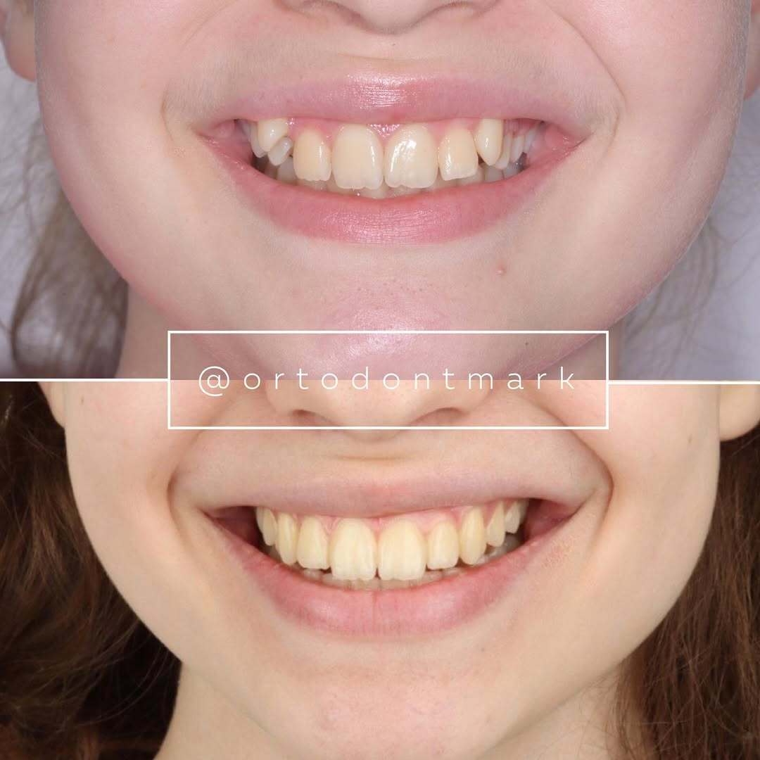
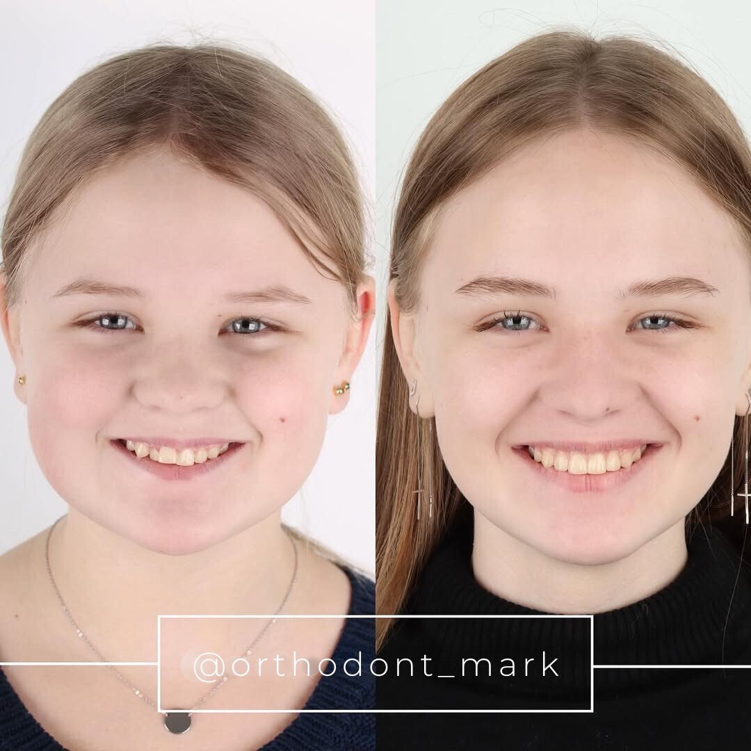

Learn the full MARPE protocol from Dr. Mark Radzhabov
Fundamental course covering CBCT patient selection, miniscrew planning, activation protocols, and 60+ clinical cases. Choose the access level that fits your practice.
Essentials of rapid palatal expansion for practicing orthodontists.
- Core RPE concepts and biomechanics
- 6 structured video lessons
- Clinical decision checklists
- Lifetime access to recordings
Deep-dive into MARPE protocol, diagnostics, and clinical execution.
- 6 modules covering MARPE from A to Z
- CBCT case selection walkthroughs
- Miniscrew placement and activation
- 60+ annotated clinical cases
- Direct Q&A with Dr. Mark Radzhabov
5-element medical consultation framework for dentists and orthodontists.
- Trust-building consultation protocol
- 5 lesson modules
- Templates for treatment plan delivery
- Works with any clinical specialty
Clinical FAQ
How is gingival thickness measured before skeletal expansion?
Transgingival ultrasound (20 MHz probe perpendicular to gingival margin) is the gold standard. Clinical probing of sulcus depth and keratinized width provides accessible alternatives. Both methods achieve ICC >0.87 when performed by trained staff.
What gingival thickness threshold predicts recession during MARPE?
Keratinized tissue ≤0.5 mm and/or sulcus depth >3 mm indicate thin biotype with 3–4× higher recession incidence. Thickness >1.0 mm confers stable phenotype. Moderate (0.75–1.0 mm) cases require individualized load monitoring.
Should expansion protocol change if baseline buccal soft tissue is thin?
Yes. Reduce activation to 0.4–0.6 mm/week (vs. standard 0.8–1.0 mm/week), extend treatment duration 2–4 months, and consider asymmetric load or posterior miniscrew placement to spare anterior buccal plate.
Can thin gingival biotype be a contraindication for MARPE?
Thin biotype alone is not absolute contraindication, but combined with smoking, diabetes, or pre-existing periodontitis it becomes high-risk. Consult periodontics first. Consider surgical expansion or conservative protocol modification.
How does age interact with gingival biotype in expansion outcomes?
Patients >50 years show 2–3× higher recession risk even with thick biotypes, due to reduced bone turnover. Treat patients >50 with thin phenotype as physiologically 1–2 mm thinner. Apply modified load protocols accordingly.
What is the clinical difference between thin and thick gingival phenotypes?
Thin phenotype: keratinized width <2 mm, sulcus >3 mm, compromised load distribution. Thick phenotype: keratinized width >3 mm, shallow sulcus, stable under expansion force. Mixed biotypes require zone-specific assessment.
Should patients with thin gingival biotype avoid palatal expansion?
No. Slower expansion velocity (0.4–0.6 mm/week), extended timeline, and careful load management make MARPE feasible. Document baseline phenotype and obtain informed consent explicitly disclosing soft tissue risk.
How often should soft tissue thickness be re-assessed during MARPE?
Baseline measurement before loading, then at 6 and 12 months post-activation. Serial ultrasound readings identify early recession trends and guide load adjustment or protocol modification to minimize further soft tissue loss.
Can connective tissue grafting improve outcomes in thin-biotype expansion cases?
Grafting before expansion (if keratinized width <1 mm) improves soft tissue volume but adds surgical time and cost. Prevention via slower activation and modified load is preferred. Post-treatment grafting corrects minor recession if needed.
What documentation protects the practice when soft tissue change occurs?
Record baseline ultrasound readings (specific mm thickness), photographs, sulcus depth, keratinized width, risk classification, and explicit informed consent disclosing soft tissue risk. Serial follow-up measurements objective validate expected vs. excessive recession.
Documenting baseline gingival biotype before expansion is no longer optional. It is foundational to informed consent and protocol selection. A simple transgingival ultrasound measurement or clinical probe reading at baseline provides the single most useful predictor of whether a patient will experience buccal bone loss during skeletal expansion. Clinicians who integrate pre-expansion periodontal screening into their case workup—as Dr. Mark Radzhabov demonstrates in his clinical education resources—achieve superior soft tissue outcomes and avoid post-treatment recession regret. Enroll in our advanced MARPE consultation module or submit your case for review on ortodontmark.com to refine your baseline assessment strategy.

