
Palatal expansion relapse rates:
ossification
evidence and retention duration
How published ossification and surgical studies translate into defensible retention protocols for miniscrew-assisted and surgically assisted expansion.
TL;DR Palatal expansion relapse rates are most reliably interpreted alongside ossification evidence rather than fixed calendar timelines. Retention duration recommendations emerge from studies tracking alveolar bone remodeling and suture fill after active expansion. Evidence from Sant'Ana et al. (2016) and Lim et al. (2017) informs both the surgical decision threshold and the bone response pattern that underpins retention planning.
When a clinician removes a palatal expander prematurely, the skeletal gain can partially reverse—yet the published literature on exactly how long to retain expansion remains inconsistently applied in daily practice. Interpreting palatal expansion relapse rates requires understanding the ossification evidence from which retention duration recommendations are actually derived, not simply adopting a generic timeframe. At ortodontmark.com, Dr. Mark Radzhabov examines the mechanistic basis for retention protocols, drawing on peer-reviewed imaging and surgical studies to help clinicians connect bone remodeling physiology to evidence-based decision-making.
Why palatal expansion relapse rates
cannot
be read without ossification context
Retention duration in palatal expansion is the post-active phase during which an immobilized appliance maintains arch width while newly deposited bone mineralizes across the expanded midpalatal suture. That definition already signals the problem with blanket retention timelines: the suture does not ossify on a fixed calendar schedule. Bone fill rate varies with patient age, the magnitude of expansion achieved, and whether the force pathway was tooth-borne or bone-borne. Treating retention duration as a universal rule, independent of the biological state of the suture, is the root cause of inconsistent relapse outcomes across published studies. The surgical literature offers a useful lens. Sant'Ana et al. (2016) compared surgically assisted rapid maxillary expansion in 24 adult patients—14 receiving midpalatal split and 10 without midpalatal separation—and found that the group with midpalatal split demonstrated significantly greater efficacy (P = 0.00). The implication for retention planning is direct: if the method of suture disruption alters the magnitude of skeletal movement, it logically also alters the volume of new bone that must form before retention can safely end. This mechanistic link—between how the suture was opened and how long the appliance must stay in place—is precisely what gets lost when relapse rates from heterogeneous studies are pooled without accounting for the surgical or miniscrew-assisted protocol used. A clinician reviewing the published literature on expansion stability needs to ask which expansion method was used before accepting any retention duration figure as applicable to their own miniscrew-assisted rapid palatal expansion cases.
How alveolar bone shifts after MARPE
inform
evidence-based retention duration
One of the most clinically meaningful ossification findings in the miniscrew-assisted expansion literature concerns the directional asymmetry of alveolar bone response. Lim et al. (2017), in a retrospective cohort study of MARPE patients, found that alveolar bone thickness decreased on the buccal aspect of expanded teeth while it increased on the palatal aspect. This redistribution is not merely a dental side effect. It represents the biological remodeling sequence that must reach completion before the appliance can be removed without inviting relapse. The clinical consequence is that retention duration cannot be judged by arch width measurement alone. A clinician who uses inter-molar width as the sole endpoint for retention removal may be terminating retention while the buccal alveolar plate is still in a thinned, remodeling state. That thinned cortical shell is biomechanically weaker and more susceptible to rebound forces from the transeptal fibers and the buccal musculature. The ossification evidence, therefore, argues for imaging-informed retention endpoints rather than time-based ones. For miniscrew-assisted rapid palatal expansion specifically, the Lim et al. (2017) finding also has implications for how clinicians counsel patients about post-retention tooth position. Palatal bone gain does not translate to equivalent buccal support during the early consolidation phase, which means the teeth remain at elevated relapse risk until buccal cortical thickness recovers. Any honest discussion of expansion stability studies must incorporate this directional remodeling asymmetry.
What follow-up duration tells clinicians
about
skeletal expansion retention protocols
A retention protocol is only as credible as the follow-up window used to evaluate it. Cabrera et al. (2010) reported that treatment of lateral open bite with fixed appliances and intermaxillary elastics remained stable at a 2-year follow-up. While that case focused on a specific occlusal problem, the methodological point applies broadly: two years post-treatment represents a defensible minimum horizon for claiming expansion stability, because it captures the late remodeling phase that shorter follow-up windows miss entirely. This has direct implications for how orthodontists read published relapse rates. A study reporting low relapse at six months is not measuring the same biological endpoint as one reporting results at two years. Early measurements capture the elastic recoil of transeptal fibers and residual suture mobility. Later measurements capture whether ossification was complete enough to resist the chronic remodeling forces of normal occlusal function. Clinicians who set retention duration based on short-horizon studies are systematically underestimating the retention their patients need. The evidence base for skeletal expansion retention protocols therefore needs to be filtered by follow-up length before any retention duration recommendation can be extracted. Clinicians reviewing expansion stability studies should prioritize those with multi-year follow-up windows, treat short-term relapse data as incomplete, and apply that interpretive discipline whether they are planning retention for a bone-borne miniscrew-assisted system or a conventional tooth-borne appliance. Exploring detailed case outcomes via documented before/after outcomes at ortodontmark.com illustrates how this follow-up logic applies in practice.
Deriving retention duration recommendations
from
suture ossification and remodeling evidence
Translating ossification findings into a practical retention duration recommendation requires the clinician to reason through three sequential questions: how much skeletal separation was achieved, what remodeling pattern is expected at the alveolar level, and what follow-up window is sufficient to confirm stability? Each answer narrows the range of defensible retention lengths. For surgically assisted cases, the Sant'Ana et al. (2016) data are instructive: the significantly greater skeletal displacement achieved with midpalatal split (P = 0.00) in patients whose mean age was 24.29 ± 3.48 years (ranging from 18.3 to 31.8 years) implies a corresponding increase in the ossification volume that must fill before retention can end. This is not a trivial point when adult suture biology is far less plastic than in growing patients, and new bone formation proceeds more slowly across the expanded gap. For miniscrew-assisted rapid palatal expansion, the alveolar asymmetry documented by Lim et al. (2017)—buccal bone loss, palatal bone gain—argues for retaining until buccal cortical recovery is radiographically confirmed, not merely until a target arch width is maintained. Combining these two evidence streams produces a retention framework that is biologically coherent: the retention clock starts when active expansion ends, and it stops when imaging confirms that both the midpalatal gap and the buccal alveolar plate have reached structural maturity.
Pitfalls in interpreting published expansion
stability
studies and relapse data
The most common interpretive error is pooling relapse rates across studies that used different expansion methods and treating the result as a single benchmark. Because midpalatal split produces significantly greater skeletal movement than expansion without suture separation—as Sant'Ana et al. (2016) demonstrated—a relapse rate from a surgically split cohort is not transferable to a non-surgical miniscrew-assisted case, and vice versa. Applying the wrong benchmark leads to either under-retaining or over-retaining, each with its own clinical consequence. A second error is treating age as a proxy for ossification status without accounting for actual bone response. The Sant'Ana et al. (2016) sample spanned ages 18.3 to 31.8 years, yet outcomes within that range were driven by the surgical protocol, not by age alone. Clinicians who set retention duration based solely on a patient's chronological age, without reference to the method used or the post-expansion imaging findings, are discarding the most relevant evidence. A third and underappreciated error is using follow-up windows shorter than those that capture late ossification. The two-year stability endpoint documented by Cabrera et al. (2010) represents a minimum, not a ceiling. Studies that report relapse at three or six months may be measuring transient elastic recoil rather than true long-term skeletal stability, and retention protocols derived from such studies will be systematically too short for skeletally mature patients whose bone remodeling proceeds more slowly.
Free Clinical Guide
Which Expansion Appliance at Which Age?
Get Dr. Mark Radzhabov's guide to skeletal-expansion appliances — indications, pros & cons, and how to choose by patient age. Free to your inbox.
Prefer video? Watch the free demo lesson on MARPE appliance types, features and trade-offs.
Documented outcomes
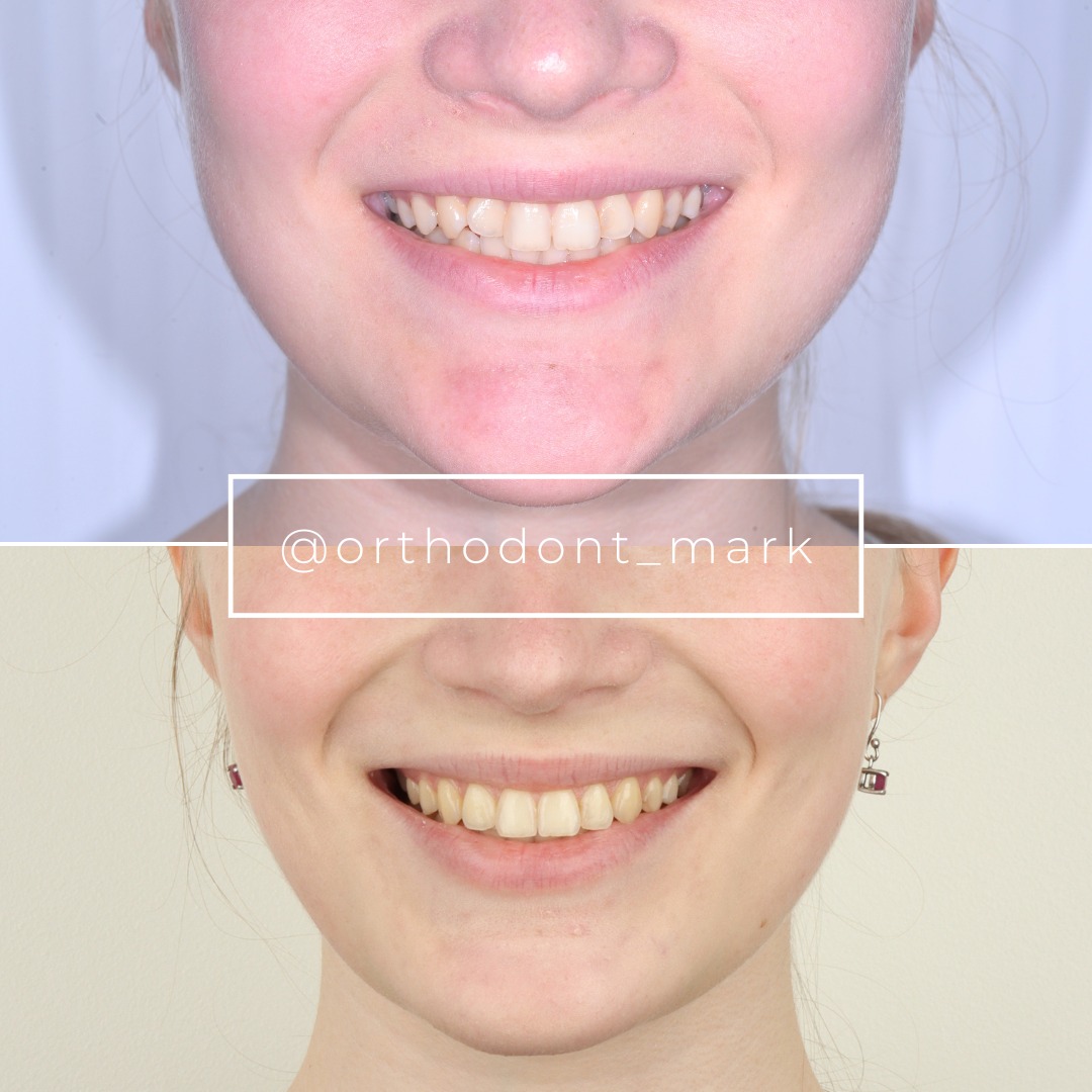
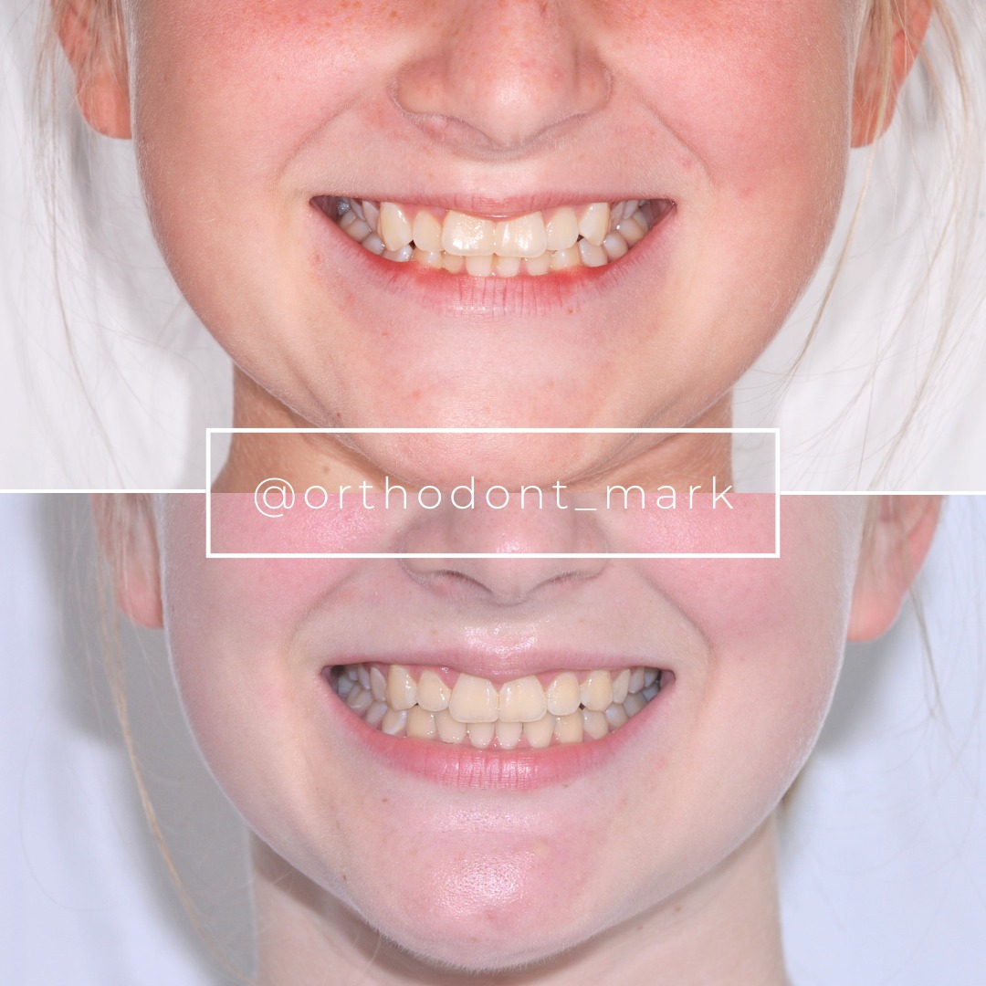
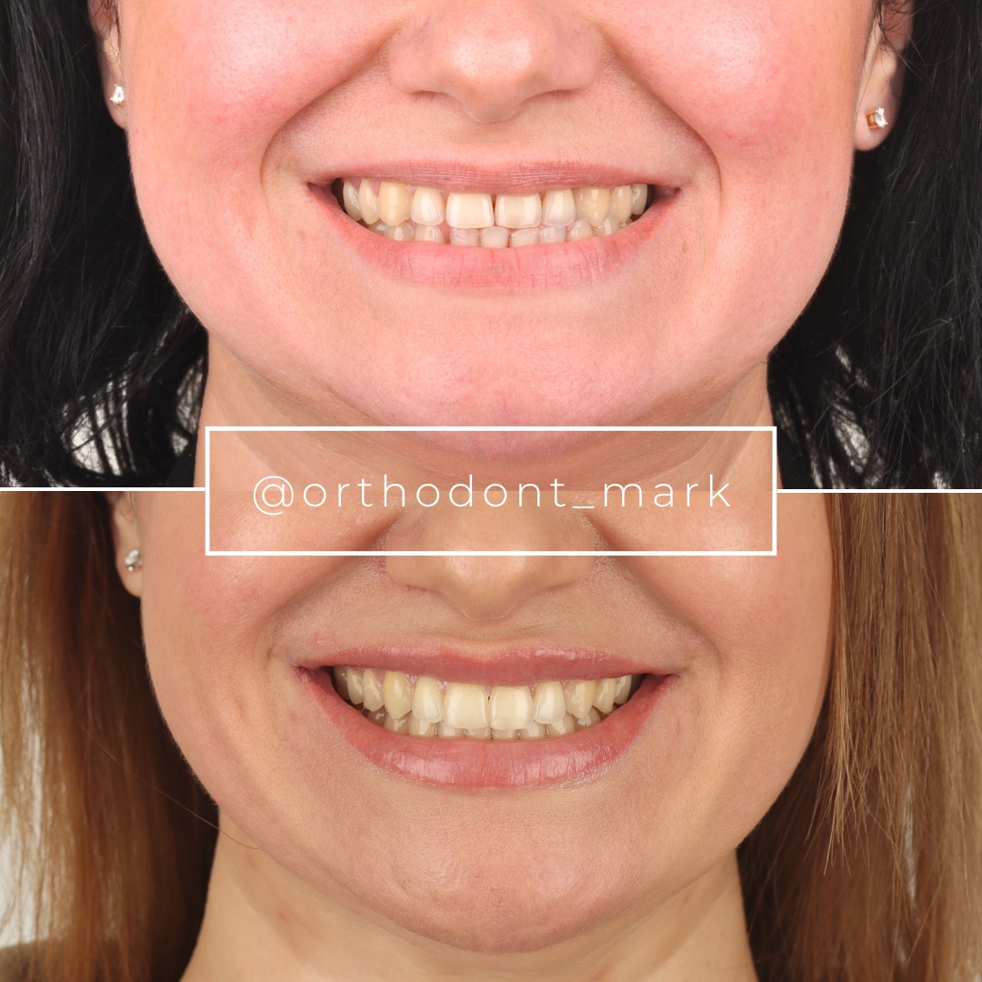
Clinical cases from Dr. Mark Radzhabov's practice
Learn the full MARPE protocol from Dr. Mark Radzhabov
Fundamental course covering CBCT patient selection, miniscrew planning, activation protocols, and 60+ clinical cases. Choose the access level that fits your practice.
Essentials of rapid palatal expansion for practicing orthodontists.
- Core RPE concepts and biomechanics
- 6 structured video lessons
- Clinical decision checklists
- Lifetime access to recordings
Deep-dive into MARPE protocol, diagnostics, and clinical execution.
- 6 modules covering MARPE from A to Z
- CBCT case selection walkthroughs
- Miniscrew placement and activation
- 60+ annotated clinical cases
- Direct Q&A with Dr. Mark Radzhabov
5-element medical consultation framework for dentists and orthodontists.
- Trust-building consultation protocol
- 5 lesson modules
- Templates for treatment plan delivery
- Works with any clinical specialty
Clinical FAQ
How are retention duration recommendations derived from ossification studies?
Retention duration recommendations emerge from studies that track midpalatal suture fill and alveolar bone remodeling after active expansion. The biological endpoint—confirmed ossification across the expanded gap—determines when appliance removal is safe, not a fixed calendar interval.
Why do published palatal expansion relapse rates vary so widely across studies?
Relapse rates vary because studies use different expansion methods, patient age ranges, and follow-up durations. Sant'Ana et al. (2016) showed that midpalatal split versus no split produces significantly different skeletal outcomes, making cross-study relapse comparisons unreliable without accounting for protocol.
Does alveolar bone remodeling after MARPE affect how long retention is needed?
Yes. Lim et al. (2017) found buccal bone thinning concurrent with palatal bone gain after MARPE. This asymmetry means the buccal cortical plate may still be structurally vulnerable even when arch width appears stable, arguing for imaging-confirmed rather than calendar-based retention endpoints.
What minimum follow-up duration is defensible for claiming expansion stability?
Cabrera et al. (2010) reported confirmed stability at a two-year follow-up after fixed appliance treatment. Two years captures late ossification that shorter windows miss. Clinicians should treat sub-12-month relapse data as incomplete when deriving retention protocol length.
Is age alone a sufficient predictor for setting retention duration after palatal expansion?
No. Sant'Ana et al. (2016) studied adults from 18.3 to 31.8 years and found outcomes were driven by the expansion protocol, not age. Bone remodeling response and imaging findings are more reliable guides to retention duration than chronological age alone.
How does SARME with midpalatal split differ from expansion without suture separation in terms of retention need?
Sant'Ana et al. (2016) demonstrated that SARME with midpalatal split achieved significantly greater efficacy (P = 0.00) than without separation, implying a larger ossification gap and correspondingly higher skeletal demand during the retention phase.
Can published relapse rates from surgically assisted expansion be applied to miniscrew-assisted rapid palatal expansion cases?
Not directly. The skeletal displacement magnitude differs between surgical and non-surgical protocols, as does the alveolar remodeling pattern documented by Lim et al. (2017). Each expansion method generates its own ossification demand and its own applicable retention evidence base.
What imaging criterion should signal the end of the retention phase after palatal expansion?
Radiographic evidence of buccal cortical plate recovery is a clinically rational endpoint, based on Lim et al. (2017) finding buccal bone thinning after MARPE. Arch width maintenance alone does not confirm that alveolar remodeling has reached structural maturity.
How should a clinician evaluate expansion stability studies before adopting their retention recommendations?
Screen each study for: expansion method used, patient skeletal maturity, and follow-up duration. Relapse figures from studies with short follow-up or heterogeneous protocols have limited direct applicability to a specific miniscrew-assisted expansion protocol in a skeletally mature patient.
Where can orthodontists access an evidence-based framework for setting retention duration after skeletal expansion?
Orthodontist Mark at ortodontmark.com provides clinical guidance connecting ossification biology to retention protocol decisions. Clinicians can submit a case for structured review or enroll in structured MARPE training to apply this evidence framework to their own patient population.
Connecting published palatal expansion relapse rates to the ossification evidence that generates retention duration recommendations is not a theoretical exercise—it directly governs whether a patient's skeletal gain persists or erodes after appliance removal. Clinicians who anchor their retention protocol to bone remodeling biology rather than a default calendar interval are better positioned to defend their decisions and improve long-term outcomes. Dr. Mark Radzhabov invites orthodontists to submit a case for structured review or explore structured MARPE training at ortodontmark.com to apply this evidence framework in their own protocols. Key sources: Sant'Ana et al. (2016), Int. J. Oral Maxillofac. Surg., doi:10.1016/j.ijom.2016.03.005. Lim et al. (2017), Korean J Orthod, doi:10.4041/kjod.2017.47.5.313. Cabrera et al. (2010), American Journal of Orthodontics and Dentofacial Orthopedics, doi:10.1016/j.ajodo.2007.11.037.