
RPE Retention Duration Evidence:
Ossification
Timelines Matched to Clinical Prescriptions
CBCT suture staging now gives orthodontists objective criteria for retention length. Map published biology to your protocol.
TL;DR RPE retention duration evidence from CBCT imaging studies reveals that suture ossification timelines vary substantially by age and anatomical region, making a fixed retention prescription unreliable. Govaerts et al. (2023) found Stage D and E midpalatal suture maturation in 61% of 15-year-old females, challenging the assumption that adolescent patients uniformly tolerate shorter retention.
Calibrating the retention prescription after rapid palatal expansion demands more than a clinical rule of thumb — it requires reading the ossification timeline against documented suture biology. RPE retention duration evidence drawn from peer-reviewed CBCT imaging studies now gives orthodontists a sharper framework for matching how long the appliance stays in place to how long the suture actually takes to consolidate. Dr. Mark Radzhabov at ortodontmark.com compiled this analysis for clinicians who want to move beyond anecdotal retention schedules and anchor their protocols in published morphological data.
Why a Generic Retention Calendar Misses the
Suture Biology
RPE retention duration is the clinician-prescribed interval during which a palatal expansion appliance remains seated after active expansion ends, allowing newly deposited woven bone across the midpalatal suture to mature into lamellar cortical structure before the retentive force is removed. The concept is straightforward. The execution is not, because the biological clock runs at different speeds depending on the patient's age, suture morphology, and anatomical region examined. Ballantia et al. (2009) used a standardised 6-month retention interval before post-retention CT assessment, a protocol choice that implicitly acknowledged the need to allow meaningful ossification time in growing subjects. The same cohort had a mean age of 11.2 years (Ballantia et al., 2009), positioning the patients at an age when the midpalatal suture is still morphologically open, yet the authors still chose a 6-month window — suggesting that even in younger patients, suture consolidation is not instantaneous. The difficulty for orthodontists is that published retention intervals from one age group cannot be safely extrapolated to older patients. When a patient presents at the borderline of skeletal maturity, the gap between a suture that looks closed on a panoramic view and one that has truly completed lamellar consolidation on CBCT is clinically significant. Retention prescriptions written without imaging evidence risk premature removal and relapse of the skeletal gain achieved during active expansion.
Suture Staging in Adolescents: What Govaerts' Data
Changes
About Your Retention Threshold
The single most clinically disruptive finding in recent suture literature is the prevalence of advanced ossification in patients orthodontists routinely treat without surgical assistance. Govaerts et al. (2023) documented that 61% of 15-year-old females already showed Stage D or E midpalatal suture maturation — the stages associated with bridging bone and near-complete suture obliteration. This figure fundamentally alters the risk calculus when setting retention duration for an adolescent who completed expansion at age 14 or 15. The same study recorded 83–100% closure of the pterygomaxillary suture in females aged 13 to 17 years (Govaerts et al., 2023). The pterygomaxillary suture is not merely an anatomical neighbour. Its resistance contributes directly to the force required to maintain transverse skeletal width post-expansion. A largely obliterated pterygomaxillary complex means the retention appliance is working against more rigid peripheral resistance, and premature removal is more consequential. Govaerts et al. (2023) also reported that the recommended cut-off age for SARPE over orthopaedic expansion was 15.1 years for orthodontists and 14.8 years for surgeons, based on suture maturation staging — not chronological age. For retention protocol design, this implies that patients near that threshold who were treated non-surgically require more conservative, longer retention management, with imaging confirmation before appliance removal.
How Expansion Pattern at Different Ages Affects
Consolidation
and Retention Planning
Retention duration cannot be decoupled from the geometry of suture opening, because the surface area available for bone deposition — and therefore the volume of new bone that must mature before the appliance is safe to remove — differs between age groups. Kinzinger et al. (2022) used CBCT to document that expansion patterns shift from parallel in patients up to age 10 to V-shaped in adolescents aged 12 and older. A V-shaped opening concentrates the gap anteriorly and leaves the posterior suture under less displacement, which may alter how uniformly ossification proceeds along the suture length. The same CBCT analysis by Kinzinger et al. (2022) examined age-dependent morphological changes in 9 growing patients aged 7.3 to 13.8 years, capturing precisely the window during which most orthopaedic expansion cases are initiated. Within that 6.5-year span, suture architecture transitions from a relatively planar interdigitation to one with increasing bony interdigitation depth — meaning older patients in the cohort had a more complex suture geometry to fill before stable consolidation could occur. For the MARPE skeletal expansion retention protocol specifically, the V-shaped pattern observed from age 12 onward matters because miniscrew-borne force distributes differently across a non-parallel suture. The posterior suture thirds that receive less opening displacement may consolidate faster, but the anterior gap — where the opening is widest — remains the rate-limiting zone for retention. Planning retention duration without assessing which suture thirds are most displaced risks removing the appliance before the widest segment has fully consolidated.
Building an Expansion Retention Timeline From
Published
Ossification Benchmarks
Translating the available RPE retention duration evidence into a workable clinical protocol requires layering three variables: the patient's skeletal age at expansion completion, the expansion pattern geometry confirmed on CBCT, and the suture maturation stage. Ballantia et al. (2009) anchored their post-expansion assessment at 6 months in patients with a mean age of 11.2 years who each received a standardised 7 mm expansion — providing one fixed-variable baseline that subsequent studies can be compared against. Govaerts et al. (2023) add the critical insight that suture maturation staging is not uniform across the adolescent age range: with 61% of 15-year-olds already at Stage D or E, a patient who completed expansion at that age cannot be offered the same 6-month calendar benchmark applied to an 11-year-old. The practical implication is that the rapid palatal expansion retention prescription for a peri-pubertal patient must explicitly account for where on the Angelieri suture staging scale that individual sits before appliance removal is scheduled. For rapid palatal expansion appliance selection in the mixed-dentition patient, the parallel expansion pattern documented before age 10 by Kinzinger et al. (2022) supports a more uniform retention approach. The V-shaped pattern from age 12 onward argues for extended anterior suture monitoring. Clinicians integrating these benchmarks should document the specific suture stage, expansion geometry, and planned retention endpoint in the treatment record — creating an auditable, imaging-based rationale that is both clinically sound and medicolegally defensible.
Free Clinical Guide
Which Expansion Appliance at Which Age?
Get Dr. Mark Radzhabov's guide to skeletal-expansion appliances — indications, pros & cons, and how to choose by patient age. Free to your inbox.
Prefer video? Watch the free demo lesson on MARPE appliance types, features and trade-offs.
Documented outcomes
Clinical cases from Dr. Mark Radzhabov's practice
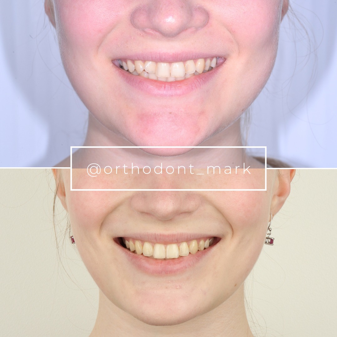
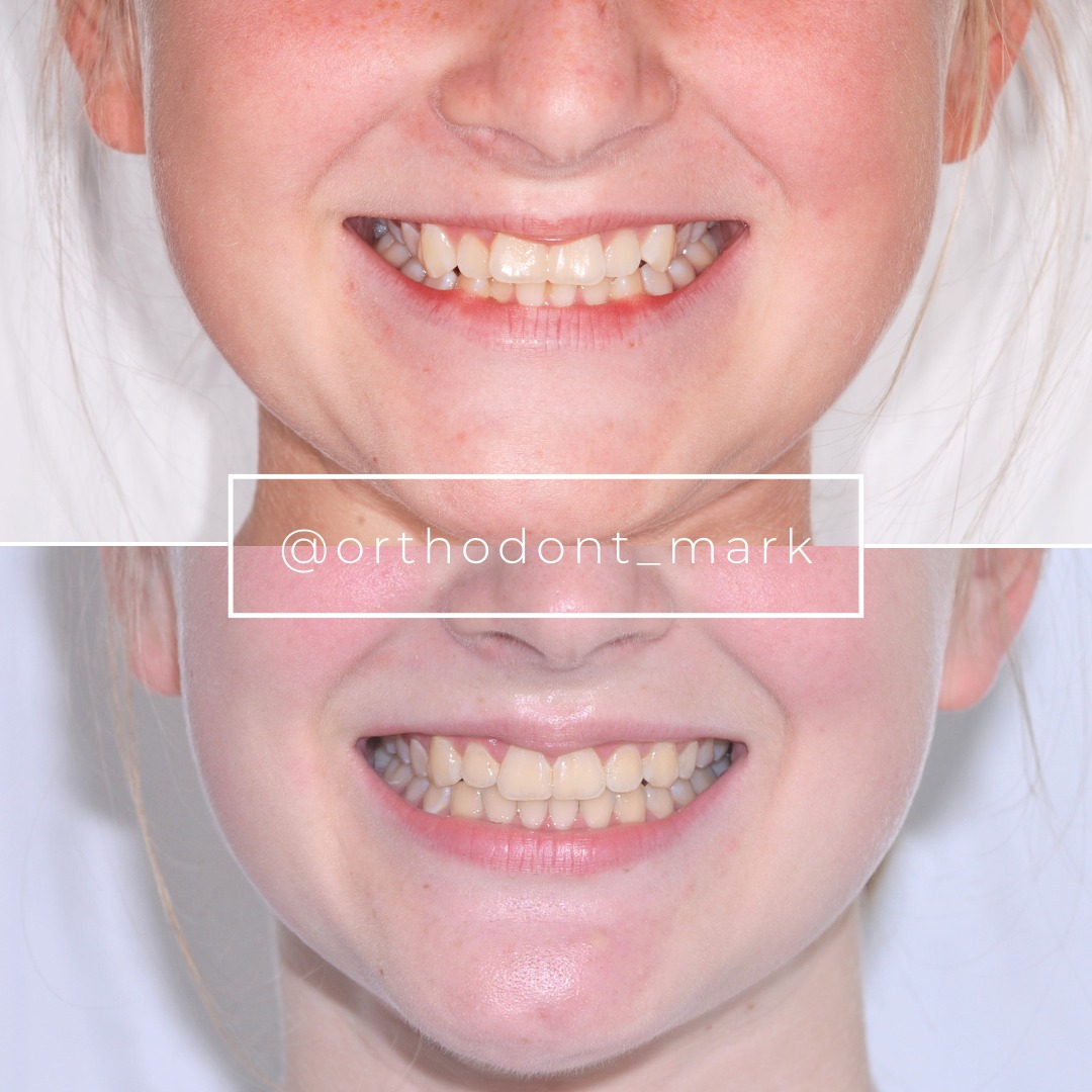
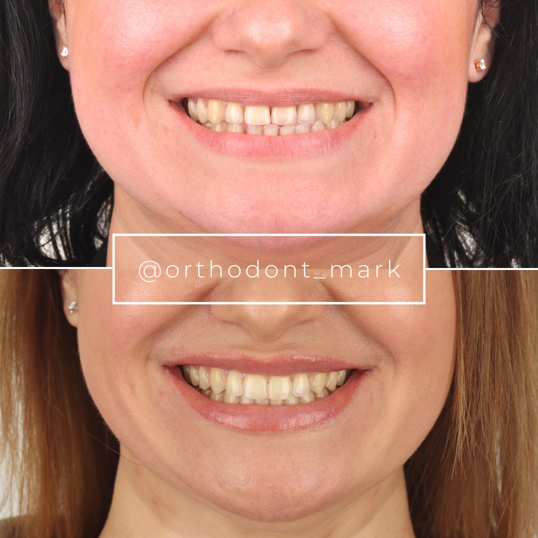
Learn the full MARPE protocol from Dr. Mark Radzhabov
Fundamental course covering CBCT patient selection, miniscrew planning, activation protocols, and 60+ clinical cases. Choose the access level that fits your practice.
Essentials of rapid palatal expansion for practicing orthodontists.
- Core RPE concepts and biomechanics
- 6 structured video lessons
- Clinical decision checklists
- Lifetime access to recordings
Deep-dive into MARPE protocol, diagnostics, and clinical execution.
- 6 modules covering MARPE from A to Z
- CBCT case selection walkthroughs
- Miniscrew placement and activation
- 60+ annotated clinical cases
- Direct Q&A with Dr. Mark Radzhabov
5-element medical consultation framework for dentists and orthodontists.
- Trust-building consultation protocol
- 5 lesson modules
- Templates for treatment plan delivery
- Works with any clinical specialty
Clinical FAQ
How long should RPE retention last after expansion completion in a 15-year-old patient?
No fixed interval is safe for this age group. Govaerts et al. (2023) found Stage D or E midpalatal suture maturation in 61% of 15-year-old females, meaning ossification is advanced before expansion even begins. CBCT staging at the planned removal date is more reliable than any calendar benchmark.
What CBCT criteria confirm sufficient palatal suture ossification before removing the expander?
Evidence of continuous bone density bridging the anterior suture third on CBCT cross-sections is the key criterion, since Kinzinger et al. (2022) identified the anterior gap as widest in V-shaped expansion patterns from age 12 onward. Posterior bridging alone is insufficient.
Does the expansion pattern geometry affect how long the retention period for rapid palatal expansion should last?
Yes. Kinzinger et al. (2022) documented parallel expansion in patients up to age 10 and V-shaped expansion from age 12 onward. V-shaped opening concentrates anterior displacement, which is the last zone to ossify, implying that anteriorly dominant cases require extended monitoring before appliance removal.
At what suture maturation stage should SARPE be prioritised over orthopaedic expansion, and how does that inform retention planning?
Govaerts et al. (2023) placed the SARPE cut-off at 15.1 years for orthodontists and 14.8 years for surgeons based on suture staging. Patients near this threshold who were treated non-surgically carry higher relapse risk and require imaging-confirmed retention rather than calendar-based appliance removal.
Is a 6-month retention period after RME evidence-based for all growing patients?
Ballantia et al. (2009) used a 6-month post-expansion interval before CT assessment in patients with a mean age of 11.2 years. This benchmark applies to that age range. It cannot be extended to adolescents approaching skeletal maturity without suture staging confirmation.
How does pterygomaxillary suture closure affect skeletal expansion stability and retention duration?
Govaerts et al. (2023) documented 83–100% pterygomaxillary suture closure in females aged 13 to 17 years. A largely obliterated pterygomaxillary complex increases recoil resistance, making premature retention removal more consequential for skeletal expansion stability in this age group.
What is the clinical implication of Stage D versus Stage E midpalatal suture for MARPE retention protocol design?
Stage D shows partial bony bridging. Stage E reflects near-complete obliteration. Both stages indicate substantially reduced orthopaedic response potential. Govaerts et al. (2023) found these advanced stages in 61% of 15-year-olds, meaning MARPE retention in that cohort must be managed more conservatively.
How should expansion amount influence the ossification retention protocol after RPE?
Ballantia et al. (2009) standardised expansion at 7 mm across subjects in their retention protocol study. Larger expansions produce a wider suture gap with greater bone volume to fill. Clinicians should account for expansion magnitude when setting the minimum retention interval.
Can panoramic radiography reliably replace CBCT for determining when to end the expansion retention timeline?
No. Two-dimensional imaging cannot distinguish woven bone fill from lamellar consolidation at the suture midline. The CBCT studies by Kinzinger et al. (2022) and Govaerts et al. (2023) both relied on cross-sectional imaging to characterise maturation — a standard that periapical or panoramic views cannot replicate.
How does skeletal expansion stability differ between patients who complete retention at Stage C versus Stage D?
Stage C indicates a partially interdigitated suture that is more likely to complete orthopaedic opening and consolidate adequately. Stage D reflects bridging bone that limits suture separation, making complete ossification of the expanded gap less predictable and increasing relapse risk if retention is shortened.
Suture maturation data from CBCT imaging has permanently changed how retention prescriptions should be written: a fixed calendar interval is no longer defensible when published data show Stage D and E ossification in a majority of 15-year-old females. The clinical obligation is to match documented ossification timelines to the individual patient's imaging findings before shortening or terminating retention. Dr. Mark Radzhabov encourages colleagues to submit a case for structured review at ortodontmark.com, where imaging-based retention decisions are the default standard, not the exception. Key sources: Govaerts et al. (2023), Journal of Orofacial Orthopedics, doi:10.1007/s00056–023-00487-x. Kinzinger et al. (2022), J Orofac Orthop, doi:10.1007/s00056–022-00429-z. Ballantia et al. (2009), Angle Orthodontist, doi:10.2319/012008–35.1.