
Buccal Corridor Change After MARPE:
Esthetic
Endpoints Beyond the Skeletal Split
A clinician's framework for measuring smile-display outcomes and soft-tissue adaptation after miniscrew-assisted rapid palatal expansion.
TL;DR Buccal corridor change after MARPE is a critical esthetic endpoint that extends well beyond confirming a midpalatal split. Skeletal expansion widens the maxillary arch and reduces dark lateral smile corridors, yet asymmetric expansion and soft-tissue adaptation complicate standardized assessment. Clinicians should integrate smile-display photography and transverse width analysis alongside CBCT verification to capture the full esthetic outcome.
When a patient's chief complaint is a narrow smile rather than a crossbite, the skeletal split confirmed on CBCT is only the beginning of the clinical story. Buccal corridor change after MARPE represents the visible, patient-perceived dividend of transverse maxillary expansion, yet most protocols stop at confirming suture separation and measuring intermolar width. Dr. Mark Radzhabov at ortodontmark.com argues that esthetic endpoints — smile-arc display, buccal corridor ratio, and soft-tissue drape — must be captured systematically to evaluate whether skeletal expansion has translated into a meaningful smile improvement.
Why Does the Buccal Corridor Deserve Its Own
Measurement Protocol?
Buccal corridor change after MARPE is the measurable reduction in the dark negative space visible between the buccal surfaces of posterior teeth and the corners of the lips during a posed social smile, resulting from transverse maxillary arch expansion. For decades, clinicians judged expansion success by millimeter gains at the first molar or premolar, yet the smile corridor — captured at natural lip posture with a standardized focal length and retractor position — tells a different story about what the patient actually perceives. The distinction matters because skeletal and dental effects do not always distribute symmetrically. Yoon et al. (2022) found that asymmetric expansion exceeding 1 millimeter occurred in 47.8% of MARPE patients, a prevalence high enough to produce detectable left-right differences in buccal corridor width at the commissures. When one side expands more than the other, the corridor narrows asymmetrically, creating a smile that reads as canted or lopsided even when the arch form appears adequate on a dental cast. A dedicated buccal corridor measurement protocol requires at minimum: a full-smile photograph taken at pupil height with the Frankfurt horizontal parallel to the floor, a relaxed lip posture confirmed before capture, and digital caliper analysis comparing the ratio of the inter-commissure width to the visible tooth display width. This ratio, rather than an absolute millimeter figure, standardizes comparison across patients with different facial widths. Linking this photographic record to the miniscrew-assisted skeletal expansion protocol at each recall interval gives the clearest picture of esthetic progress over time.
Which Arch-Width Gains Actually Translate Into
Visible Smile Improvement?
Not every millimeter of skeletal expansion is visible at the lips. Width gained at the palatal vault or nasal floor contributes to airway and dental torque corrections but does not directly reduce the buccal corridor. The gains that matter for smile esthetics are those expressed at the buccal cusp tips of the maxillary premolars and first molars, since these surfaces form the lateral boundary of the smile frame. Chun et al. (2022) demonstrated that MARPE achieved greater bilateral width at the first premolar and first molar compared to conventional tooth-borne RPE, and also produced greater increases in nasal width at the molar region. When expansion is distributed more broadly across the dental arch rather than concentrated at a single transverse point, the buccal corridor narrows more uniformly from premolar to molar — a more esthetically complete result than one where only the posterior arch widens. For clinicians using a tooth-borne rapid palatal expander comparison to frame their appliance selection, this distinction has direct implications. Bone-borne force vectors apply load through palatal cortex rather than through the periodontal ligament of anchor teeth, which means dental tipping is minimized and the skeletal arch width more reliably matches the surface measurement taken from buccal cusp to buccal cusp. That concordance between skeletal and dental width is precisely the measurement zone that governs buccal corridor reduction.
How Do MARPE Complications Confound
Smile Corridor
Assessment?
Gingival inflammation was the most frequent MARPE complication, occurring in 83.9% of patients according to Yoon et al. (2022). Inflamed and hypertrophied gingival tissue along the buccal aspects of the posterior teeth alters the apparent tooth-to-corridor boundary, making the buccal corridor appear narrower than the underlying arch width would predict. Post-expansion photographs taken before gingival inflammation resolves will systematically underestimate the esthetic benefit of expansion. Pain during and after expansion was reported in 45% of patients in the same analysis by Yoon et al. (2022), and discomfort influences lip posture during photography. A patient guarding against appliance sensitivity will often compress the orbicularis oris, reducing the inter-commissure width and artifactually narrowing the smile display. Standardizing photography to a pain-free recall appointment — rather than immediately after an activation visit — is a simple protocol adjustment with meaningful impact on measurement validity. Appliance breakage, occurring in 10% of MARPE cases per Yoon et al. (2022), introduces irregular expansion pauses that can produce the same asymmetric corridor outcome as uneven miniscrew loading. When a breakage event is documented in the clinical record, the post-repair photograph series should be restarted rather than compared directly to pre-breakage images. Recognizing these confounders protects the integrity of the transverse esthetic outcome data collected across the treatment arc.
Does Patient Biology Predict the Magnitude of
Buccal Corridor
Improvement?
The degree of esthetic improvement achievable through MARPE is upstream-limited by whether the midpalatal suture separates at all. Jeon et al. (2022) reported a suture separation success rate of 61.05% in males versus 94.17% in females, and noted that older male patients showed significantly higher rates of suture non-separation — while this age-related effect was not significant in females. A male patient with a fused or near-fused suture who achieves only dental tipping rather than skeletal separation will show minimal buccal corridor change, because the arch perimeter has not truly widened at the skeletal level. This sex-differentiated response has a direct implication for esthetic prognosis: female patients across a broader age range can be counseled with greater confidence that MARPE will deliver visible smile corridor improvement, while male patients — particularly those with more advanced skeletal maturity — require more conservative expectations and closer CBCT monitoring during the activation phase. Suture morphology on CBCT, not chronological age alone, should anchor the esthetic prognosis conversation. For complex cases where suture fusion risk is high, the combined presurgical MARPE and orthognathic approach documented by Facio et al. (2022) offers an alternative pathway: presurgical MARPE followed by bimaxillary orthognathic surgery was completed in 18 months, with the orthognathic component providing the definitive skeletal width and vertical repositioning that governs final smile-arc display. Clinicians managing this patient profile can explore the full decision tree through a case review and treatment planning consultation.
Building a Post-Expansion Smile Record
That Holds Up
to Scrutiny
A defensible buccal corridor measurement protocol begins before expansion activations start. A T1 full-smile photograph — taken with a 100 mm macro lens at pupil height, lips relaxed, teeth in maximum intercuspation — establishes the baseline corridor ratio. The same setup is repeated at T2 (end of active expansion) and T3 (end of retention, typically after new bone has consolidated in the midpalatal split site). Comparing the ratio of visible tooth display width to inter-commissure width across these three timepoints separates true arch-display improvement from transient soft-tissue adaptation. Overlay analysis using dental analysis software allows the commissure and buccal cusp landmarks to be digitized, providing a reproducible left-corridor and right-corridor measurement that is operator-independent. Given that Yoon et al. (2022) reported asymmetric expansion exceeding 1 millimeter in 47.8% of cases, bilateral corridor measurement — rather than a single composite score — is non-negotiable if the record is to be clinically meaningful. CBCT taken at T1 and T2 provides the skeletal reference: intermolar width at the alveolar crest and suture patency status. When the smile photograph shows persistent buccal corridors despite confirmed skeletal expansion, the differential includes buccal crown torque loss, lip-width mismatch, or residual gingival inflammation obscuring the tooth-display boundary. Mapping each photographic finding against the corresponding CBCT and dental cast measurement allows the clinician to identify which variable is limiting the esthetic result and direct the appropriate adjunctive intervention.
Free Clinical Guide
Which Expansion Appliance at Which Age?
Get Dr. Mark Radzhabov's guide to skeletal-expansion appliances — indications, pros & cons, and how to choose by patient age. Free to your inbox.
Prefer video? Watch the free demo lesson on MARPE appliance types, features and trade-offs.
Documented outcomes
Clinical cases from Dr. Mark Radzhabov's practice
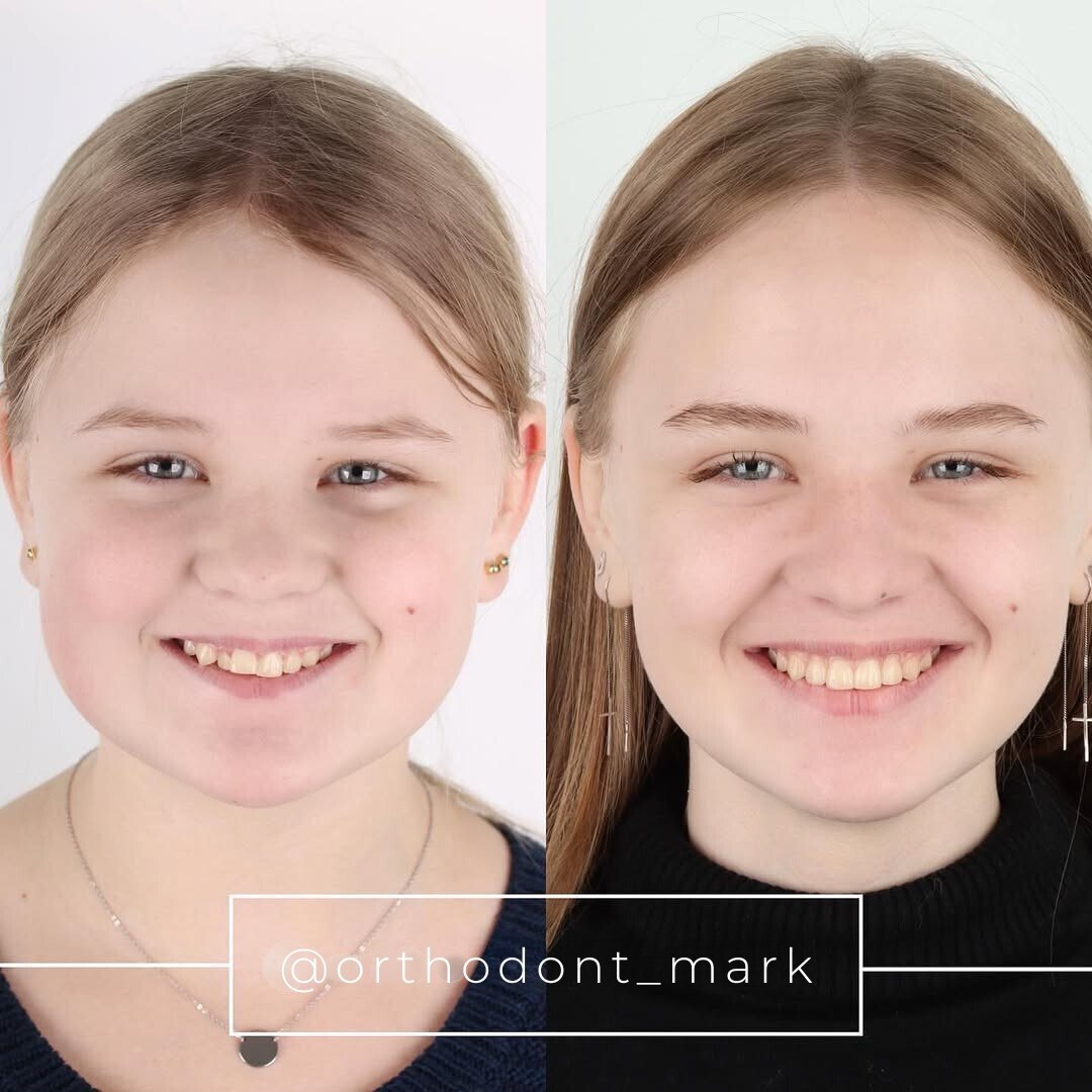

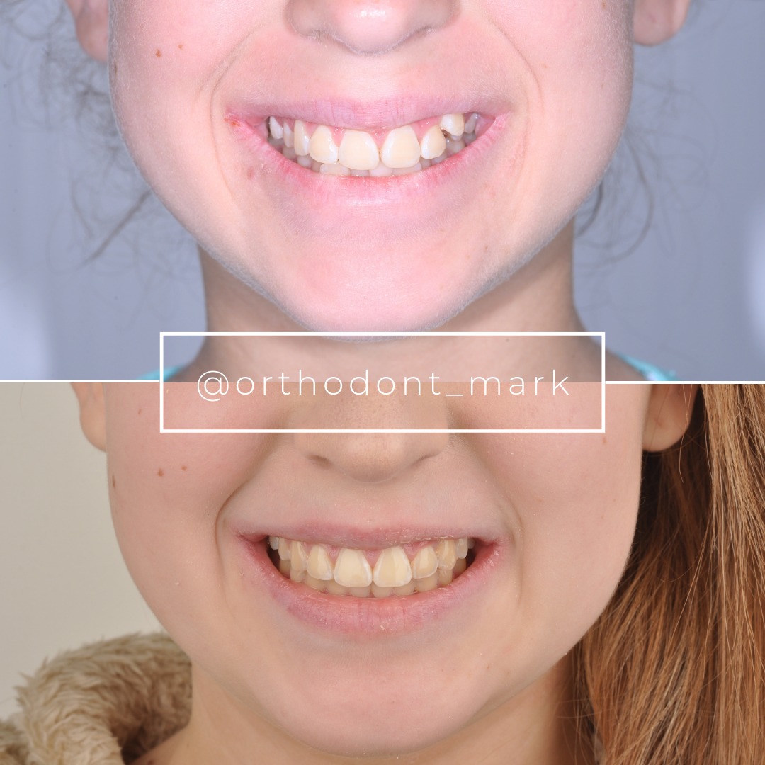
Learn the full MARPE protocol from Dr. Mark Radzhabov
Fundamental course covering CBCT patient selection, miniscrew planning, activation protocols, and 60+ clinical cases. Choose the access level that fits your practice.
Essentials of rapid palatal expansion for practicing orthodontists.
- Core RPE concepts and biomechanics
- 6 structured video lessons
- Clinical decision checklists
- Lifetime access to recordings
Deep-dive into MARPE protocol, diagnostics, and clinical execution.
- 6 modules covering MARPE from A to Z
- CBCT case selection walkthroughs
- Miniscrew placement and activation
- 60+ annotated clinical cases
- Direct Q&A with Dr. Mark Radzhabov
5-element medical consultation framework for dentists and orthodontists.
- Trust-building consultation protocol
- 5 lesson modules
- Templates for treatment plan delivery
- Works with any clinical specialty
Clinical FAQ
How is buccal corridor change after MARPE measured reliably in clinical practice?
Use standardized full-smile photography at pupil height with a 100 mm macro lens and relaxed lip posture. Measure the ratio of visible tooth display width to inter-commissure width bilaterally at T1, T2, and T3 to separate skeletal gains from soft-tissue adaptation.
Does MARPE improve smile esthetics more reliably than conventional tooth-borne expansion?
Chun et al. (2022) found MARPE achieved greater bilateral first premolar and molar maxillary width than RPE, along with greater nasal width gains — suggesting more complete arch-frame widening that is more likely to reduce buccal corridor display.
How often does asymmetric expansion complicate buccal corridor assessment after MARPE?
Yoon et al. (2022) reported that asymmetric expansion exceeding 1 millimeter occurred in 47.8% of MARPE patients. This prevalence makes bilateral, side-specific corridor measurement essential rather than optional in any post-expansion esthetic record.
Which patient sex shows more predictable transverse esthetic outcomes after MARPE?
Jeon et al. (2022) reported suture separation in 94.17% of female versus 61.05% of male patients. Female patients therefore have more predictable skeletal expansion and, by extension, more reliable buccal corridor improvement.
Why does gingival inflammation confound smile corridor measurement after MARPE?
Yoon et al. (2022) found gingival inflammation in 83.9% of MARPE patients. Hypertrophied buccal gingiva narrows the apparent tooth-to-commissure boundary, causing smile photographs to underestimate the actual esthetic benefit of expansion if taken during active inflammation.
When should post-expansion smile photographs be taken to avoid measurement error?
Schedule smile-display records at a pain-free, inflammation-resolved appointment. Yoon et al. (2022) reported pain in 45% of MARPE patients. Discomfort compresses orbicularis posture, artifactually narrowing the inter-commissure width and misrepresenting the corridor ratio.
What is the overall suture separation success rate that underpins meaningful esthetic outcomes with MARPE?
Jeon et al. (2022) reported an overall suture separation success rate of 79.53%, while Yoon et al. (2022) found 87.8% separation. Both figures confirm that a meaningful proportion of patients achieve the skeletal foundation required for genuine buccal corridor improvement.
How does presurgical MARPE followed by orthognathic surgery affect buccal corridor and smile outcomes?
Facio et al. (2022) documented that presurgical MARPE combined with bimaxillary orthognathic surgery was completed in 18 months. This sequenced approach is appropriate where suture fusion limits MARPE-only expansion, with orthognathic repositioning providing the definitive transverse and vertical smile-arc correction.
Does appliance breakage during MARPE affect the final buccal corridor outcome?
Yoon et al. (2022) reported appliance breakage in 10% of MARPE cases. Irregular activation pauses from breakage can produce the same asymmetric corridor discrepancy as uneven miniscrew loading. Post-repair photographs should restart the T2 comparison rather than continue an interrupted series.
Should CBCT or smile photography be the primary endpoint for evaluating dental arch width expansion esthetics?
Both are necessary but measure different things. CBCT confirms skeletal intermolar width and suture separation. Smile photography captures what the patient perceives — buccal corridor ratio, tooth display, and lip-frame balance. Neither alone constitutes a complete transverse esthetic evaluation.
Quantifying buccal corridor change after MARPE demands a dual-track protocol: radiographic confirmation of skeletal expansion paired with standardized smile photography at consistent focal length and lip posture. Clinicians who capture only the CBCT miss the esthetic dividend their patients actually perceive. Review your current post-expansion records against the measurement framework discussed here, and consider submitting a complex case for peer discussion. Dr. Mark Radzhabov welcomes case consultations through ortodontmark.com for practitioners refining their transverse esthetic evaluation workflow. Key sources: Chun et al., 2022, BMC Oral Health, doi:10.1186/s12903–022-02138-w. Jeon et al., 2022, Clinical Oral Investigations, doi:10.1007/s00784–021-04281–0. Yoon et al., 2022, retrospective analysis.

