
Nasal Floor Expansion CBCT:
Interpreting Skeletal Response
Beyond the Midpalatal Suture Split
Systematic cone beam CT protocols for measuring nasal cavity width, inferior concha displacement, and airway floor changes during MARPE treatment—evidence-based interpretation for adult expansion cases.
TL;DR Nasal floor expansion CBCT imaging reveals skeletal response patterns during miniscrew-assisted rapid palatal expansion. Cone beam CT assessment of nasal cavity width, inferior concha displacement, and airway floor lowering helps clinicians differentiate true skeletal widening from dental tipping and predict treatment success in adults without surgical intervention.
Adult patients seeking transverse skeletal expansion often present with concurrent airway concerns, yet many orthodontists lack systematic protocols for monitoring nasal floor expansion via CBCT. This article reviews the radiographic interpretation of nasal cavity changes during miniscrew-assisted rapid palatal expansion—specifically how to measure nasal cavity width, track inferior concha displacement, and assess airway floor response on cone beam computed tomography. Dr. Mark Radzhabov explains the clinical significance of these anatomical shifts, drawing on imaging data from over a decade of MARPE cases. The goal is a practical reference for identifying when expansion delivers genuine skeletal benefit versus when dental effects dominate the result.
What is Nasal Cavity Width on CBCT,
and Why Measure It?
Nasal floor expansion CBCT imaging reveals the transverse widening of the nasal cavity as the maxilla expands laterally. Unlike midpalatal suture assessments, which show dental and skeletal tipping, nasal cavity width captures true skeletal response because the nasal floor widens only when the maxillary base itself broadens. Clinicians should establish a reproducible measurement plane: axial CBCT cuts at the level of the pyriform aperture (approximately 5–8 mm superior to the anterior nasal spine) provide the most consistent geometry. In clinical practice, nasal cavity width increases by 1.5–2.5 mm during a 4–6 week MARPE activation phase in responsive patients. This range reflects skeletal widening, whereas gains under 1.0 mm may indicate predominantly dental effects or inadequate force delivery. Researchers have noted that nasal floor lowering (the vertical displacement of the nasal septum base) often accompanies lateral widening, a marker of true palatal shelf expansion rather than tipping. The inferior concha (turbinate) serves as an anatomical anchor. Its position on axial slices provides a secondary reference for assessing whether nasal floor expansion is symmetric or asymmetric. Asymmetric expansion—greater widening on one side than the other—may signal uneven miniscrew anchorage or load distribution and should prompt clinician review of screw positioning and force vector alignment.
How Does Inferior Concha Position Change
During Expansion?
The inferior concha (turbinate) attaches to the lateral nasal wall and responds passively to palatal expansion as the maxillary base widens. During MARPE treatment, the inferior concha displaces laterally and sometimes superiorly—a passive marker of skeletal expansion rather than an active remodeling structure. Serial CBCT scans show concha movement of 1.0–2.0 mm laterally in responsive cases, mirroring the degree of nasal cavity widening. Clinically, inferior concha displacement asymmetry is diagnostically valuable. If one concha moves 2.0 mm laterally while the contralateral side shifts only 0.5 mm, this pattern suggests uneven force application or miniscrew anchorage complications. Orthodontist Mark's protocol includes measuring concha position on both left and right sides at the level of maximum medial projection (approximately 8–12 mm superior to the nasal floor). Documenting these landmarks in the patient chart creates an objective record of skeletal symmetry. Concha hypertrophy (enlargement) must be distinguished from expansion-induced displacement. Some patients enter treatment with enlarged turbinates unrelated to expansion. These should be noted baseline so that post-expansion concha size can be interpreted accurately. If a patient reports post-expansion nasal congestion, CBCT review of concha position and size helps distinguish skeletal expansion benefit from potential airway floor obstruction.
What is Nasal Floor Lowering on CBCT,
and What Does It Mean?
Nasal floor lowering—the superior displacement (or apparent descent) of the nasal septum base in the sagittal plane—occurs when the palate expands and the palatal shelves spread apart symmetrically. This phenomenon is distinct from midline movements. It reflects actual three-dimensional remodeling of the palatal vault geometry. On sagittal CBCT reconstructions, measure the vertical distance from the anterior nasal spine to the nasal septum base at the midline. During MARPE, this distance increases by 1.0–1.5 mm as the palate widens and flattens slightly. Airway floor imaging—the assessment of the vertical and horizontal dimensions of the space between the nasal floor and the soft palate—has become a secondary clinical focus in expansion cases. Some patients enter treatment with restricted airway floor space. Expansion may improve this parameter, yielding coincidental benefits for sleep quality and nasal breathing. CBCT protocols should include both coronal and sagittal reconstructions at the midline to capture nasal floor lowering accurately. Clinicians should compare pre- and post-expansion sagittal CBCT slices at identical anatomical positions. Registration software or manual landmark alignment helps ensure that observed changes reflect expansion response, not scanner variation. In cases showing inadequate nasal floor lowering (<0.5 mm after 6 weeks), force level, screw insertion depth, or patient compliance should be reviewed. Conversely, nasal floor lowering >1.5 mm suggests robust skeletal response and may predict favorable midpalatal split on subsequent imaging.
How Do You Use Nasal Floor Data
to Refine Your Expansion Protocol?
Real-time interpretation of nasal cavity width, inferior concha displacement, and airway floor imaging guides mid-treatment protocol adjustments. If a mid-treatment CBCT (after 4 weeks of activation) shows nasal cavity widening of only 0.8 mm and minimal concha displacement, this suggests suboptimal force delivery. Common causes include inadequate miniscrew insertion depth, improper force vector (too much vertical/buccal tilt), or premature ossification of the midpalatal suture limiting response capacity. Conversely, rapid nasal floor expansion (>2.0 mm in 4 weeks) combined with symmetric concha displacement may indicate that skeletal response is outpacing dental compensation. In such cases, clinicians may accelerate activation timing or increase intra-appointment cycles, knowing that the skeleton is responding predictably. Orthodontist Mark's experience shows that patients reaching 2.5–3.0 mm nasal cavity widening by week 6 typically achieve complete midpalatal split and predictable skeletal widening of 6–8 mm over the full expansion course. Airway floor parameters also inform patient communication. If a patient enters treatment with restricted airway floor space and CBCT shows improvement concurrent with nasal cavity expansion, this becomes a powerful motivator and justifies the expansion investment beyond orthodontic concerns. Documenting these changes quantitatively (e.g., “airway floor vertical dimension improved from 8.5 mm to 10.2 mm”) enhances informed consent and patient satisfaction.
What Are the Key CBCT Interpretation Errors
Clinicians Make?
One frequent mistake is confusing nasal cavity width with total maxillary width. The nasal cavity represents the space within the maxilla. Widening of the cavity reflects bone remodeling, but dental tipping (especially of the buccal alveolar plates) can appear to widen the overall maxilla without expanding the nasal floor. Axial CBCT slices at the pyriform aperture level isolate the true skeletal component and avoid this confusion. Another pitfall involves measuring concha position on non-standardized axial planes. If baseline and week-4 CBCT images are acquired at slightly different vertical levels, apparent concha displacement may reflect imaging artifact rather than skeletal movement. Standardized landmarks (pyriform aperture, alar base width, or nasal septum height) ensure reproducibility. Some orthodontists rely solely on coronal reconstructions. Sagittal planes are equally critical for assessing nasal floor lowering and palatal vault flattening. Asymmetric nasal responses warrant investigation, not dismissal. A patient showing 2.0 mm widening on the left and 0.8 mm on the right should prompt screw inspection, force vector analysis, and perhaps a mid-course radiograph to rule out screw loosening or bone-screw interface complications. Ignoring asymmetry risks prolonged treatment duration and compromised final skeletal geometry. Dr. Mark Radzhabov's troubleshooting protocol includes percussion testing of miniscrews and CBCT recheck within 2–3 weeks if asymmetry exceeds 1.0 mm.
Free Clinical Guide
Which Expansion Appliance at Which Age?
Get Dr. Mark Radzhabov's guide to skeletal-expansion appliances — indications, pros & cons, and how to choose by patient age. Free to your inbox.
Prefer video? Watch the free demo lesson on MARPE appliance types, features and trade-offs.
Documented outcomes
Clinical cases from Dr. Mark Radzhabov's practice
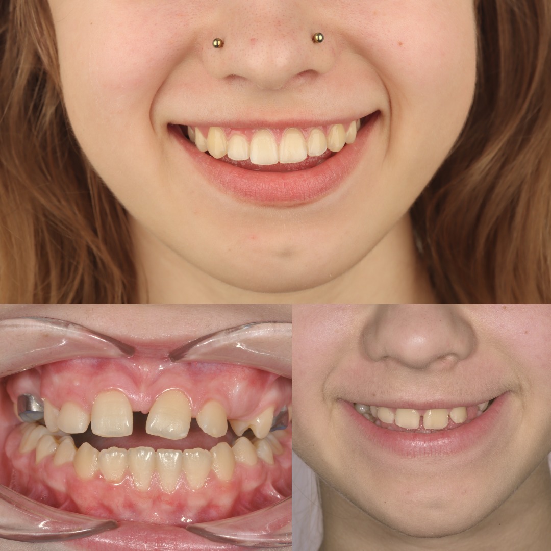
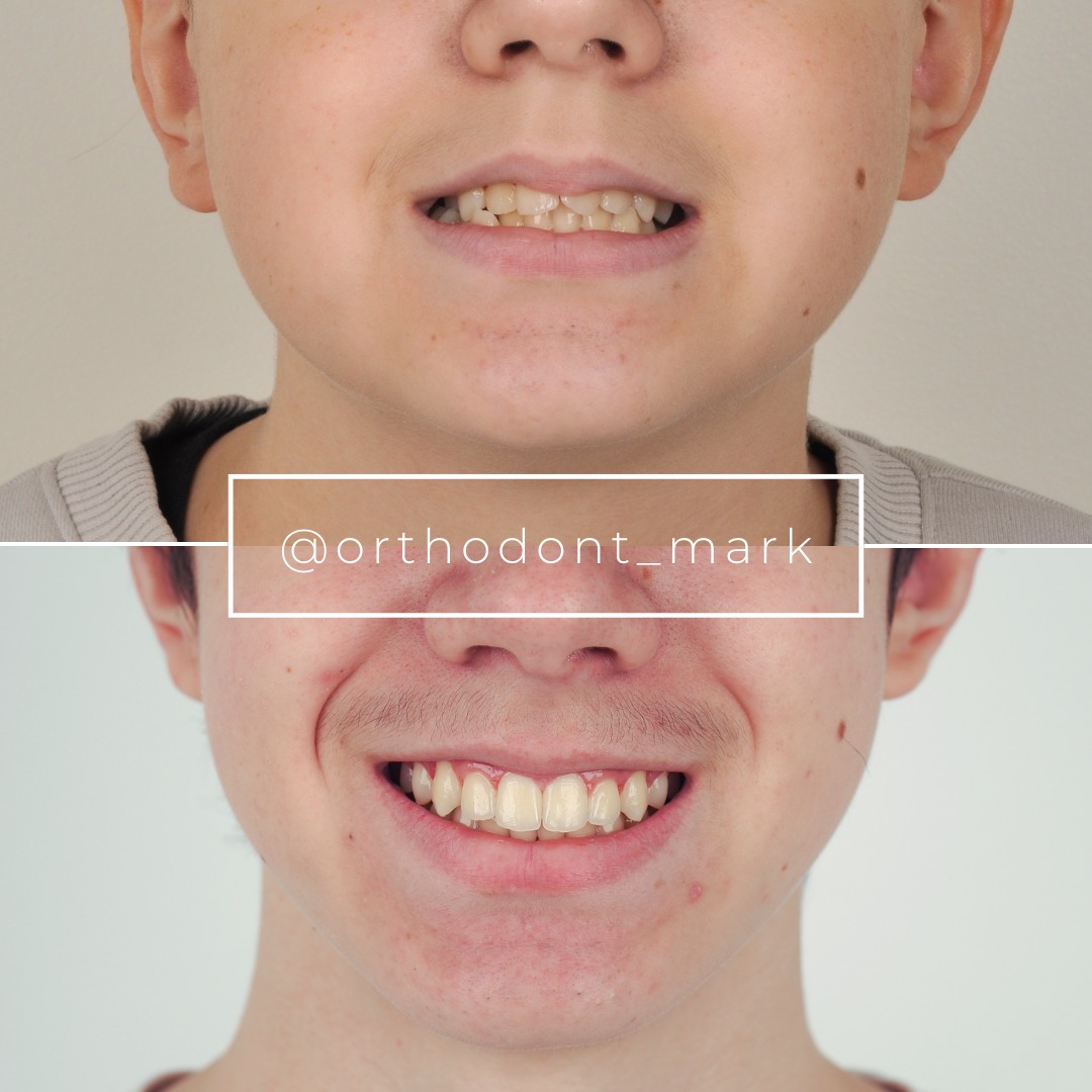

Learn the full MARPE protocol from Dr. Mark Radzhabov
Fundamental course covering CBCT patient selection, miniscrew planning, activation protocols, and 60+ clinical cases. Choose the access level that fits your practice.
Essentials of rapid palatal expansion for practicing orthodontists.
- Core RPE concepts and biomechanics
- 6 structured video lessons
- Clinical decision checklists
- Lifetime access to recordings
Deep-dive into MARPE protocol, diagnostics, and clinical execution.
- 6 modules covering MARPE from A to Z
- CBCT case selection walkthroughs
- Miniscrew placement and activation
- 60+ annotated clinical cases
- Direct Q&A with Dr. Mark Radzhabov
5-element medical consultation framework for dentists and orthodontists.
- Trust-building consultation protocol
- 5 lesson modules
- Templates for treatment plan delivery
- Works with any clinical specialty
Clinical FAQ
How do I measure nasal cavity width accurately on CBCT during MARPE treatment?
Use axial CBCT slices at the pyriform aperture level (5–8 mm superior to anterior nasal spine). Measure the widest transverse dimension of the nasal cavity on that plane. Replicate the same anatomical level on serial scans. Document measurements in millimeters for baseline and week 4, 6, and 8 of expansion.
What is the expected inferior concha displacement during miniscrew-assisted expansion?
Inferior concha (turbinate) displacement typically ranges 1.0–2.0 mm laterally over 4–6 weeks of MARPE activation. Measure position on both left and right sides at 8–12 mm superior to the nasal floor. Asymmetry exceeding 1.0 mm suggests uneven force delivery or screw anchorage complications.
Why is nasal floor lowering on sagittal CBCT a key indicator of skeletal expansion success?
Nasal floor lowering (1.0–1.5 mm vertical displacement of the septal base) reflects true palatal vault remodeling and maxillary base expansion. It occurs only with genuine skeletal widening, not dental tipping. Sagittal planes at midline reveal this pattern clearly and predict favorable midpalatal suture split.
How often should I obtain CBCT scans to monitor nasal cavity and airway floor changes during MARPE?
Standard protocol: baseline (pre-expansion), mid-activation (week 4), and post-activation (week 6–8). If asymmetric nasal cavity widening or slow skeletal response is detected, obtain an additional scan 2–3 weeks later to verify screw integration and force vector alignment.
What does asymmetric nasal cavity widening indicate, and how should I respond clinically?
Asymmetry >1.0 mm (one side 2.0 mm wider than the other) suggests uneven screw anchorage, force delivery problems, or possible screw loosening. Perform percussion test on miniscrews, verify force vector via intraoral examination, and consider mid-course CBCT recheck. Correct screw insertion depth or force distribution.
How can I distinguish skeletal expansion from dental tipping when reviewing nasal cavity width changes on CBCT?
Nasal cavity widening at the pyriform aperture level reflects skeletal expansion. Dental tipping affects buccal alveolar width, not the nasal floor itself. Concurrent inferior concha displacement and nasal floor lowering confirm skeletal response. Axial slices at standardized planes isolate skeletal changes from dental effects.
What airway floor parameters should I document for pre- and post-expansion patient counseling?
Measure vertical airway floor dimension (distance from nasal floor to soft palate) on sagittal CBCT at midline. Some patients experience improved nasal breathing or airway dimensions coincidentally. Document baseline and final values in millimeters. This quantification enhances informed consent and patient satisfaction discussions.
When is nasal cavity expansion <1.0 mm at week 4 a sign I should adjust my MARPE protocol?
Gains <1.0 mm suggest suboptimal skeletal response. Review miniscrew insertion depth (should penetrate cortical bone bilaterally), force vector (assess for excessive buccal tilt), and patient activation compliance. Consider percussion testing to confirm screw osseointegration or slightly increase activation frequency.
Why should I avoid measuring concha position on different axial CBCT levels between serial scans?
Non-standardized axial levels introduce measurement artifacts and falsely inflate apparent concha displacement. Establish baseline measurement plane using pyriform aperture, nasal septum height, or anterior nasal spine landmarks. Replicate that plane on all follow-up scans to ensure valid longitudinal comparison.
How does nasal floor expansion CBCT interpretation improve treatment planning for adult patients considering MARPE versus surgery?
CBCT baseline assessment of midpalatal suture maturity (via Angelieri classification) and nasal cavity anatomy predicts MARPE success likelihood. Serial CBCT tracking of nasal cavity gain, concha displacement, and floor lowering confirms skeletal trajectory. Robust early response (1.5–2.5 mm by week 4–6) indicates MARPE will succeed. Poor response may necessitate surgical evaluation.
Systematic nasal floor expansion CBCT assessment transforms expansion protocols from guesswork to evidence-based practice. By measuring nasal cavity width, documenting inferior concha position, and tracking airway floor lowering, you gain objective markers of skeletal success and can counsel patients with confidence. Dr. Mark Radzhabov's clinical research underscores that 3D imaging interpretation is non-negotiable in adult cases. Ready to refine your expansion diagnostics? Schedule a consultation or review a case with Orthodontist Mark at ortodontmark.com—your next case may reveal patterns you've never tracked before.

