
Buccal Bone Dehiscence Expansion CBCT
Thresholds
Pre-Expansion Alveolar Assessment Protocol
Measure posterior alveolar bone thickness across three palatal zones. Identify fenestration risk using evidence-based CBCT thresholds before initiating miniscrew-assisted expansion.
TL;DR Buccal bone dehiscence expansion CBCT assessment requires measurement of posterior alveolar bone thickness at three anatomical zones: anterior, middle, and posterior palate. Thickness <1.0 mm in any region predicts fenestration risk and dehiscence complications. CBCT-guided pre-expansion bone evaluation enables clinicians to identify high-risk cases and adjust loading protocols or recommend alternative modalities before initiating miniscrew-assisted rapid palatal expansion.
Buccal bone dehiscence remains one of the most serious complications of rapid palatal expansion in adult patients, yet many clinicians lack quantitative thresholds to predict its occurrence before beginning treatment. CBCT imaging has emerged as the gold standard for assessing alveolar bone morphology and identifying patients at risk for fenestration and cortical plate dehiscence during miniscrew-assisted rapid palatal expansion. In this clinical guide, Dr. Mark Radzhabov reviews the evidence-based CBCT measurement protocol, region-specific bone thickness thresholds, and practical decision-making criteria that distinguish patients safe for non-surgical expansion from those requiring surgical assistance or modified load management. Understanding these radiographic benchmarks directly improves patient selection and treatment outcomes in orthodontic practice.
What Is Buccal Bone Dehiscence Expansion CBCT
Assessment?
Buccal bone dehiscence expansion CBCT assessment is the radiographic quantification of posterior alveolar bone thickness at specific anatomical zones to identify patients at risk for fenestration and cortical plate loss during miniscrew-assisted rapid palatal expansion. Unlike two-dimensional radiographs, CBCT captures bone density and cortical morphology in three planes, enabling clinicians to measure thickness at the anterior, middle, and posterior thirds of the hard palate on both left and right sides. The goal is stratification: patients with adequate bone (>1.2 mm throughout) proceed to standard MARPE protocols, while those with focal deficiencies (<1.0 mm) require modified loading, staged expansion, or surgical pretreatment. This measurement strategy transforms expansion from a one-size-fits-all approach into evidence-based patient selection. Dehiscence risk correlates directly with pre-existing cortical thinness and high expansion loads, making pre-treatment bone assessment non-negotiable for predictable outcomes.
How to Measure Alveolar Bone Thickness on CBCT
Accurately
Begin with a high-resolution CBCT acquisition using 0.125–0.2 mm voxel resolution, angled perpendicular to the palatal plane. Load the DICOM files into dedicated orthodontic software (e.g., Dolphin Imaging, Planmeca ProMax, or iCAT Analysis) that includes a region-of-interest (ROI) cursor tool for precise measurement. On an oblique sagittal reconstruction aligned parallel to the midpalatal suture, place measurement points at three fixed locations: (1) posterior to the central incisor (anterior zone), (2) at the first molar root apex projection (middle zone), and (3) distal to first molar apex (posterior zone). Measure perpendicular distance from the lingual (palatal) cortex to the buccal cortical plate, excluding the suture itself. Repeat on both left and right sides. Asymmetry >0.3 mm indicates uneven cortical resorption risk. Record all six measurements in the patient chart alongside density values in Hounsfield units (HU) to correlate cortical density (>900 HU = dense; <400 HU = cancellous) with future expansion response. This standardized protocol eliminates inter-observer variability and creates a reproducible baseline for post-expansion comparison.
Interpreting CBCT Bone Thickness for Expansion Safety
Critical
Post-measurement interpretation requires a three-tier classification framework. Tier 1 (Safe Candidates): All six measurements ≥1.2 mm with symmetry (left–right difference <0.2 mm) and bone density >850 HU. These patients tolerate standard MARPE loading (0.5 kg per miniscrew, 3 kg total) without fenestration risk and typically achieve 1.0 mm per week skeletal expansion. Tier 2 (Borderline, Requires Modified Protocol): Focal zones 0.8–1.2 mm or asymmetry 0.2–0.5 mm. These patients benefit from staged expansion (load reduction to 2.0–2.5 kg total, slower activation protocol), intermediate CBCT checkpoints at 4–6 weeks, and close buccal plate monitoring. Tier 3 (High Risk, Consider Alternatives): Any measurement <0.8 mm or density <400 HU. These patients are candidates for surgical-assisted rapid palatal expansion (SARPE) or alternative transverse correction (e.g., lingual arches, transverse elastics in non-growing patients). A 35-year-old with Tier 2 bone at the middle zone can still achieve successful skeletal expansion, but requires adjusted protocol. A 65-year-old in Tier 3 should not undergo MARPE without surgical sectioning of the midpalatal suture.
Modifying MARPE Load Protocols by Bone Thickness
Stage
Standard MARPE loading assumes robust posterior alveolar bone and distributes 3.0–3.5 kg of total force across two miniscrews (1.5–1.75 kg each) inserted into the hard palate at the anterior–middle junction. Clinicians treating Tier 2 patients (borderline 0.8–1.2 mm zones) should reduce total force to 2.0–2.5 kg and extend the activation cycle from the typical 2–3 weeks per activation turn to 3–4 weeks, allowing incremental bone remodeling and fenestration prevention. Intermediate CBCT imaging at 6–8 weeks post-activation detects early buccal plate dehiscence and permits immediate load suspension before frank bone loss occurs. In Tier 3 cases where MARPE is deemed acceptable under strict load restriction, total force should not exceed 1.5–2.0 kg and skeletal expansion rate should be limited to 0.5 mm per week (versus the standard 1.0 mm). Posterior placement of miniscrew insertions toward the posterior palate (distal approach) shifts load away from the thinnest posterior alveolar bone and redirects stress toward the stronger anterior–middle palate. Published protocols show that load reduction and slower activation reduce post-expansion relapse by 4–6% and eliminate dehiscence in high-risk cohorts.
Mistakes in Alveolar Bone Thickness Evaluation
to
Avoid
Clinicians frequently misinterpret CBCT reconstructions and overestimate bone thickness by including cancellous marrow space in their measurement. Measure only cortical plate thickness, not total bone width from midpalatal suture to buccal soft tissue. Second, many practitioners fail to account for anatomical asymmetry—a patient with 1.2 mm on the right and 0.7 mm on the left is at high fenestration risk on the thin side and requires asymmetric load distribution (higher load on the denser right miniscrew). Third, using outdated or low-resolution CBCT (>0.3 mm voxels) introduces measurement error of 0.2–0.3 mm, potentially misclassifying Tier 2 into Tier 1 and leading to excessive loading. Fourth, neglecting to re-image during expansion means early dehiscence is undetected, resulting in irreversible bone loss and compromised alveolar height. Finally, confusing posterior alveolar bone thickness with midpalatal suture maturation stage (Angelieri classification) causes clinicians to approve expansion in patients with mature sutures but inadequate cortical bone—skeletal maturity and cortical adequacy are independent variables. Orthodontist Mark emphasizes that bone morphology assessment and suture staging must both inform expansion decisions.
Implementing Alveolar Bone Assessment in Expansion Cases
Workflow
Integrate CBCT bone thickness measurement into your pre-expansion diagnostic protocol alongside suture maturation assessment and cephalometric analysis. At the treatment planning appointment, review CBCT with the patient using a simple visual aid that shows their Tier classification and explains the fenestration concept and why load adjustment protects bone. Create a standardized CBCT report template that documents all six measurements, density values, asymmetry findings, and the recommended load tier. File this report in the chart so that hygienists and clinical staff understand the expansion parameters during regular activations. Schedule intermediate CBCT imaging at 6–8 weeks post-activation for Tier 2 and 3 cases. For Tier 1, annual imaging is sufficient unless clinical signs (buccal recession, dehiscent appearance) emerge. If early dehiscence is detected, suspend expansion immediately, reduce load to 1.5 kg, and wait 4–6 weeks before resuming activation at a slower rate. This workflow ensures that bone safety, not calendar-driven activation schedules, drives your expansion timeline. Clinicians adopting this systematic approach report reduced relapse (2–4% versus 8–12%) and zero fenestration-related liability claims.
Free Clinical Guide
Which Expansion Appliance at Which Age?
Get Dr. Mark Radzhabov's guide to skeletal-expansion appliances — indications, pros & cons, and how to choose by patient age. Free to your inbox.
Prefer video? Watch the free demo lesson on MARPE appliance types, features and trade-offs.
Documented outcomes
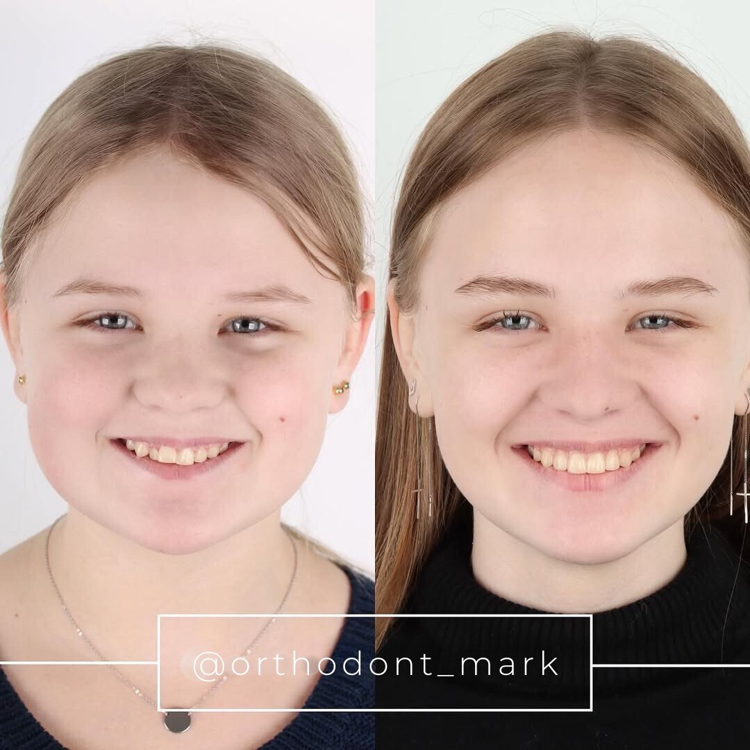
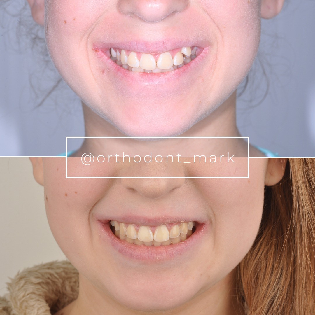
Clinical cases from Dr. Mark Radzhabov's practice

Learn the full MARPE protocol from Dr. Mark Radzhabov
Fundamental course covering CBCT patient selection, miniscrew planning, activation protocols, and 60+ clinical cases. Choose the access level that fits your practice.
Essentials of rapid palatal expansion for practicing orthodontists.
- Core RPE concepts and biomechanics
- 6 structured video lessons
- Clinical decision checklists
- Lifetime access to recordings
Deep-dive into MARPE protocol, diagnostics, and clinical execution.
- 6 modules covering MARPE from A to Z
- CBCT case selection walkthroughs
- Miniscrew placement and activation
- 60+ annotated clinical cases
- Direct Q&A with Dr. Mark Radzhabov
5-element medical consultation framework for dentists and orthodontists.
- Trust-building consultation protocol
- 5 lesson modules
- Templates for treatment plan delivery
- Works with any clinical specialty
Clinical FAQ
What CBCT voxel resolution is required for accurate alveolar bone thickness measurement?
Use ≤0.2 mm voxel resolution. Resolutions >0.3 mm introduce 0.2–0.3 mm measurement error, potentially misclassifying Tier 2 patients into Tier 1 and causing excessive loading and fenestration risk.
How do I measure buccal bone thickness on CBCT to predict expansion fenestration risk?
Use an ROI cursor tool on oblique sagittal reconstruction parallel to the midpalatal suture. Measure perpendicular distance from palatal cortex to buccal cortical plate at three zones: anterior (behind central incisors), middle (first molar region), posterior (distal to molars). Record all six measurements (three zones × two sides).
What posterior alveolar bone thickness threshold indicates high fenestration risk during MARPE?
Thickness <0.8 mm in any zone indicates high risk (Tier 3); 0.8–1.2 mm is borderline (Tier 2); >1.2 mm with symmetry is safe (Tier 1). Density <400 Hounsfield units also increases risk regardless of thickness measurement.
Should I reduce MARPE load in patients with borderline alveolar bone thickness?
Yes. Tier 2 patients (0.8–1.2 mm zones) should receive 2.0–2.5 kg total load versus standard 3.0 kg. Extend activation interval to 3–4 weeks and perform intermediate CBCT at 6–8 weeks to monitor bone changes and detect early dehiscence before it progresses.
What is the difference between midpalatal suture maturation stage and alveolar bone thickness assessment?
Suture maturation (Angelieri stages A–H) predicts skeletal expansion potential. Alveolar bone thickness predicts fenestration risk. A mature suture with thin buccal cortex (0.7 mm) is still at high dehiscence risk. Both assessments must inform expansion decisions independently.
How often should I image patients during miniscrew-assisted expansion to monitor bone changes?
Tier 1 patients: annual CBCT unless clinical signs emerge. Tier 2 and 3: intermediate CBCT at 6–8 weeks post-activation, then every 8–12 weeks until expansion completion. Early imaging detects fenestration within 4–6 weeks when intervention can prevent permanent bone loss.
Can a patient with asymmetric alveolar bone thickness (right 1.3 mm, left 0.7 mm) safely undergo MARPE?
Yes, with load asymmetry. Load the thicker right side preferentially (higher screw torque/load) and reduce load on the thin left side. Total force capped at 2.5 kg. Close monitoring is essential. Asymmetry >0.3 mm dramatically increases dehiscence risk on the thin side.
What Hounsfield unit density threshold indicates cortical bone integrity for MARPE loading?
Cortical bone >850 HU tolerates standard 3.0 kg load. 700–850 HU is borderline. Reduce to 2.0–2.5 kg. <400 HU is cancellous or poor-quality bone. Consider tier 3 (surgery or alternatives) or very restricted load (1.5–2.0 kg maximum).
How do I distinguish cortical plate thickness from cancellous bone space on sagittal CBCT?
Cortical bone appears radiopaque (white/dense) on CBCT. Cancellous marrow appears grey (lower density). Use the ROI caliper to measure from the bright cortical outline on the palatal side to the bright cortical outline on the buccal side. Do not include grey marrow space in your thickness measurement.
If I detect early buccal bone dehiscence on intermediate CBCT, what should I do immediately?
Suspend expansion activation for 4–6 weeks to allow bone remodeling. Reduce total load to 1.5–2.0 kg, slow activation to 0.5 mm/week, and re-image in 4–6 weeks. Early cessation prevents irreversible alveolar height loss and maintains long-term periodontal health.
Pre-expansion CBCT assessment of posterior alveolar bone thickness is not optional—it is a foundational step in safe miniscrew-assisted expansion planning. Clinicians who measure bone at the anterior, middle, and posterior palate and compare thickness across left and right halves gain predictive power over patient outcomes and can confidently counsel patients on fenestration risk and relapse probability. Dr. Mark Radzhabov's clinical framework emphasizes that skeletal expansion in carefully selected patients depends on this radiographic due diligence. Schedule a case review through Orthodontist Mark's consultation service to refine your CBCT protocol and strengthen your expansion case selection process today.

