
Rhinomanometry MARPE Airway:
Quantifying
Nasal Resistance Before and After Expansion
An evidence-based clinical framework for integrating rhinomanometry into MARPE records — so skeletal airway gains become documented, defensible outcomes.
TL;DR Rhinomanometry MARPE airway assessment quantifies nasal resistance changes that CBCT alone cannot capture. Skeletal expansion widens the nasal floor, reducing airway resistance in a measurable, reproducible way. Clinicians who integrate rhinomanometry pre- and post-expansion gain objective outcome data to guide case selection, counsel referring physicians, and document functional improvement alongside skeletal change.
A patient presents with confirmed transverse maxillary deficiency, mouth-breathing habit, and chronically elevated nasal resistance on anterior rhinomanometry — yet no sleep study, no ENT referral, and no functional baseline on the orthodontic chart. This gap between structural diagnosis and functional documentation is exactly what rhinomanometry MARPE airway protocols are designed to close. Dr. Mark Radzhabov at ortodontmark.com reviews the current evidence on quantifying nasal airway resistance change before and after miniscrew-assisted expansion, explaining why clinicians who add rhinomanometry to their pre-treatment records gain a defensible, peer-reviewed metric for both case justification and outcome reporting.
Why Is Nasal Airway Resistance the Right Metric After
Skeletal Expansion?
Rhinomanometry is an objective pressure-flow measurement technique that quantifies nasal airway resistance by recording differential pressure and airflow volume simultaneously at each nostril, providing a reproducible functional baseline before and after skeletal expansion. Unlike CBCT volumetric analysis, rhinomanometry captures dynamic resistance under physiological breathing conditions — the variable that correlates most directly with the patient's symptomatic complaint. The anatomical rationale is straightforward: the nasal floor shares a direct bony boundary with the palate, so lateral displacement of the maxillary halves during expansion widens the pyriform aperture and the nasal cavity floor simultaneously. Ramires et al. (2008) documented a mean 6.5 mm increase in nasal cavity width across 30 patients with nasal insufficiency who underwent rapid maxillary expansion, translating a skeletal change directly into a measurable anatomical gain in the airway. In a separate dataset, Ramires et al. (2008) reported nasal width increases ranging from 0.4 to 5.7 mm across 40 maxillary expansion cases, with a mean of 17 mm of total expansion delivered. This variance underlines why rhinomanometry — rather than a single anatomical measurement — is the appropriate outcome tool: it integrates the net effect of all structural changes on actual airflow resistance, not just one linear dimension.
Does MARPE Produce a More Favorable Airway Change
Than RPE?
The appliance architecture determines where force is delivered and how cleanly the midpalatal suture opens — both of which influence the magnitude and symmetry of the nasal floor displacement captured by rhinomanometry. Bone-borne miniscrew-assisted expansion bypasses dental tipping, directing orthopedic load through the palatal bone toward the suture itself, which should in principle produce a more parallel, less rotational separation of the maxillary halves. Chun et al. (2022) confirmed this biomechanical advantage in a randomized clinical trial: MARPE demonstrated greater maxillary width at premolar and molar regions compared to RPE at a statistically significant level (p < 0.05), and produced greater nasal width increases at the molar region alongside a greater palatine foramen dimension (p < 0.05). A wider nasal floor at the molar level corresponds to the posterior nasal aperture, the zone rhinomanometry is most sensitive to during nasal inspiration. Successful midpalatal suture separation — confirmed radiographically by the characteristic triangular diastema at the central incisors and a midpalatal radiolucency on CBCT — occurred in 95% of MARPE patients versus 90% of RPE patients in Chun et al. (2022), each group receiving 35 turns of expansion. A higher suture separation rate means more cases deliver the full skeletal stimulus to the nasal floor, reducing the proportion of patients who show purely dental tipping with minimal airway benefit on post-expansion rhinomanometry.
How Should Rhinomanometry Be Integrated Into the
MARPE Workflow?
Reliable pre-post rhinomanometry requires the same standardization applied to CBCT acquisition: consistent patient positioning, controlled ambient temperature, a documented decongestant washout period, and calibrated equipment verified against manufacturer norms at each session. Without standardization, intra-patient variability swamps the signal produced by expansion. For miniscrew-assisted expansion cases, the consolidation phase is the critical measurement window. Chun et al. (2022) used a 3-month consolidation period with CBCT recorded at baseline, immediately post-expansion, and after consolidation. Rhinomanometry should follow the same schedule: the immediate post-expansion reading reflects mucosal edema and appliance bulk, while the 3-month reading captures the stabilized skeletal contribution to resistance reduction. Comparing these two post-expansion points lets the clinician separate transient soft-tissue change from durable airway gain. When documenting outcomes for referring ENT physicians or insurance justification, report resistance values at both 75 Pa and 150 Pa pressure differentials — the two reference points recognized in international rhinomanometry standards — alongside the CBCT nasal floor width measurement. This dual-format report links the objective airway resistance number to the anatomical change that caused it, creating a clinically complete record for interdisciplinary communication.
Which Patients Show the Largest Nasal Resistance Reduction
After MARPE?
Patients presenting with documented nasal obstruction — high rhinomanometry resistance at baseline, confirmed transverse deficiency on CBCT, and a narrow nasal floor — represent the population most likely to show clinically meaningful resistance reduction after skeletal expansion. The structural substrate for improvement is present, and the appliance delivers force directly to the bone responsible for nasal floor width. Airway-related sleep-disordered breathing adds another layer of clinical context. Ferati et al. (2024) reported that OSAS prevalence in children ranges from 0.69% to 5.7% depending on diagnostic criteria employed, reflecting the wide variation in how airway obstruction severity is classified across published studies. This epidemiological spread underscores the importance of objective functional measurement — rhinomanometry provides a criterion-independent baseline that does not fluctuate with diagnostic threshold changes the way prevalence estimates do. Ferati et al. (2024) also noted that adenotonsillar hypertrophy peaks in occurrence between ages 2 and 6, which identifies younger patients presenting for early orthodontic evaluation as a group with potentially compounded upper airway obstruction. In these cases, rhinomanometry at initial presentation establishes whether the nasal component of resistance is already elevated before adenoid involvement is addressed — a finding that shapes both the sequencing of treatment and the timing of palatal expansion within the overall airway management plan.
How Do Rhinomanometry Reports Support ENT Referral
and Outcome Defense?
An orthodontist who delivers a paired rhinomanometry report — baseline resistance, post-expansion resistance, percent change, and the corresponding CBCT nasal floor width measurement — creates a document that an ENT physician can integrate directly into the patient's airway management record. This is qualitatively different from a clinical note describing 'improved nasal breathing,' and it positions the orthodontist as a functional contributor to the airway care team. The variability in nasal width response documented by Ramires et al. (2008) — ranging from 0.4 to 5.7 mm across 40 expansion cases — illustrates why a single post-expansion CBCT width measurement is an insufficient outcome marker. Two patients with identical skeletal width gains can have divergent resistance outcomes depending on mucosal status, turbinate size, and septal anatomy. Rhinomanometry captures that divergence. A CBCT measurement alone does not. For clinicians building a MARPE airway outcomes record across their patient population, the functional measurement also enables genuine outcome analysis. Because Chun et al. (2022) demonstrated that MARPE produced greater skeletal width gains at premolar and molar regions compared to RPE at p < 0.05, appliance selection itself becomes a variable in predicting rhinomanometry outcomes — making the pre-treatment appliance choice and the post-treatment resistance measurement part of the same clinical argument for miniscrew-assisted expansion cases.
Free Clinical Guide
Which Expansion Appliance at Which Age?
Get Dr. Mark Radzhabov's guide to skeletal-expansion appliances — indications, pros & cons, and how to choose by patient age. Free to your inbox.
Prefer video? Watch the free demo lesson on MARPE appliance types, features and trade-offs.
Documented outcomes
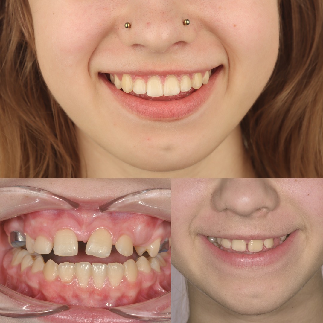
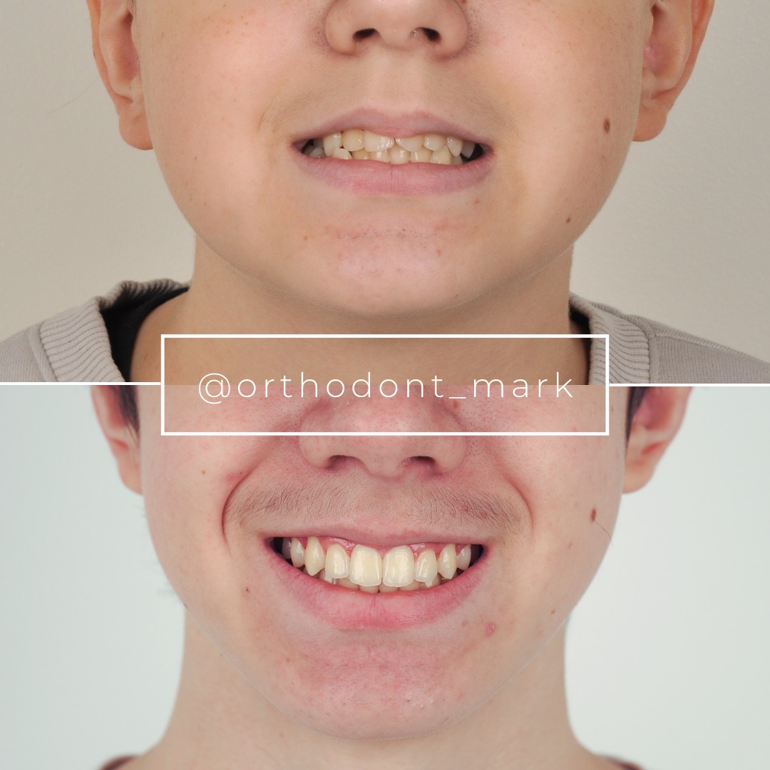
Clinical cases from Dr. Mark Radzhabov's practice

Learn the full MARPE protocol from Dr. Mark Radzhabov
Fundamental course covering CBCT patient selection, miniscrew planning, activation protocols, and 60+ clinical cases. Choose the access level that fits your practice.
Essentials of rapid palatal expansion for practicing orthodontists.
- Core RPE concepts and biomechanics
- 6 structured video lessons
- Clinical decision checklists
- Lifetime access to recordings
Deep-dive into MARPE protocol, diagnostics, and clinical execution.
- 6 modules covering MARPE from A to Z
- CBCT case selection walkthroughs
- Miniscrew placement and activation
- 60+ annotated clinical cases
- Direct Q&A with Dr. Mark Radzhabov
5-element medical consultation framework for dentists and orthodontists.
- Trust-building consultation protocol
- 5 lesson modules
- Templates for treatment plan delivery
- Works with any clinical specialty
Clinical FAQ
What does rhinomanometry measure that CBCT cannot in a MARPE airway assessment?
Rhinomanometry records dynamic nasal airway resistance under active breathing — capturing the net effect of mucosal status, turbinate size, and septal anatomy on airflow. CBCT measures static bony dimensions. Only rhinomanometry detects whether the skeletal width gain translates into reduced functional resistance.
At what timepoints should rhinomanometry be performed relative to MARPE treatment?
Record rhinomanometry at baseline before appliance insertion, immediately after full expansion, and after the consolidation phase. This three-point schedule — mirroring the CBCT protocol used by Chun et al. (2022) — separates acute mucosal changes from stable skeletal airway gains.
Does MARPE produce greater nasal airway resistance reduction than conventional RPE?
Chun et al. (2022) demonstrated MARPE produced greater nasal width increases at the molar region and greater maxillary width at premolar and molar regions compared to RPE at p < 0.05, suggesting MARPE delivers a larger structural substrate for nasal resistance reduction, though direct rhinomanometry comparison requires further study.
How does nasal cavity width change after rapid palatal expansion, and how variable is it?
Ramires et al. (2008) reported nasal width increases ranging from 0.4 to 5.7 mm across 40 expansion cases, illustrating substantial inter-patient variability. This range reinforces why rhinomanometry — rather than a single anatomical width — is the appropriate functional outcome metric.
What is the clinical significance of midpalatal suture separation rate for airway outcomes after MARPE?
Chun et al. (2022) reported 95% suture separation in MARPE patients versus 90% in RPE patients. A confirmed suture opening indicates true skeletal displacement of the nasal floor. Failed separation produces dental tipping with minimal nasal floor widening and negligible rhinomanometry benefit.
How should rhinomanometry resistance values be reported to ENT physicians after skeletal expansion?
Report nostril-specific resistance at both 75 Pa and 150 Pa pressure differentials, express the change as a percentage of the patient's own baseline rather than a population norm, and attach the CBCT nasal floor width measurement from the same visit to link functional and anatomical data explicitly.
Which patients are least likely to show nasal airway resistance improvement after MARPE on rhinomanometry?
Patients with concurrent septal deviation, hypertrophic inferior turbinates, or persistent mucosal inflammation retain significant non-skeletal resistance sources after expansion. Pre-treatment lateralized rhinomanometry identifies these cases, allowing realistic counseling before MARPE is initiated.
How does adenotonsillar hypertrophy affect the rhinomanometry baseline in younger expansion candidates?
Ferati et al. (2024) noted adenotonsillar hypertrophy peaks between ages 2 and 6. In these patients, adenoid bulk contributes to nasopharyngeal resistance that rhinomanometry at the nostril does not isolate — making sequential assessment after adenoid resolution important before attributing resistance change solely to expansion.
What expansion protocol was used in the MARPE versus RPE randomized trial reporting airway changes?
Chun et al. (2022) administered 35 turns of expansion in both MARPE and RPE groups, with CBCT recorded at baseline, immediately after expansion, and after a 3-month consolidation period, providing a reproducible protocol reference for clinicians designing their own rhinomanometry measurement schedule.
How does pre-treatment nasal insufficiency status affect the expected magnitude of functional airway improvement after expansion?
Ramires et al. (2008) reported a mean 6.5 mm nasal cavity width increase specifically in 30 patients presenting with nasal insufficiency — a higher-risk subgroup. Patients with documented pre-treatment obstruction and elevated baseline rhinomanometry resistance appear to yield larger, more clinically detectable post-expansion functional improvements.
Integrating rhinomanometry into a MARPE workflow transforms airway outcomes from a narrative into a measurable clinical variable — one that strengthens interdisciplinary communication with ENT colleagues and supports outcome-based documentation. Suture separation rates and nasal cavity width changes reported in the literature underscore that skeletal expansion produces real, quantifiable airway benefit. Clinicians ready to implement this protocol can explore structured MARPE training at ortodontmark.com, or submit a complex airway-expansion case for a direct consultation with Dr. Mark Radzhabov to map out an evidence-aligned pre- and post-expansion rhinomanometry protocol. Key sources: Ferati et al., 2024, Diagnostics, doi:https://doi.org/10.3390/diagnostics14030289. Chun et al., 2022, BMC Oral Health, doi:https://doi.org/10.1186/s12903–022-02138-w. Ramires et al., 2008, Rev Bras Otorrinolaringol.

