
Nasal Cavity Width CBCT:
Landmarks
Measurement Protocols and Reproducibility After MARPE
Published measurement planes, airway volume benchmarks, and reproducibility standards for post-expansion cone-beam CT — a practical imaging reference for clinicians.
TL;DR Nasal cavity width CBCT assessment after palatal expansion depends on consistent anatomical landmarks and validated measurement planes. CBCT is the universal imaging standard across published expansion studies. Nasopharyngeal volume gains after MARPE range from 6.4% to 20.7%, per Benetti et al. (2024), while suture separation rates differ meaningfully between appliance types.
Post-expansion CBCT interpretation is where appliance selection decisions are ultimately validated or questioned, yet nasal cavity width measurement landmarks remain poorly standardized across published protocols. Clinicians ordering follow-up scans after MARPE or RPE often face inconsistent reference planes, making inter-study comparison unreliable. At ortodontmark.com, Dr. Mark Radzhabov addresses this gap directly by reviewing published measurement frameworks, reproducibility criteria, and the anatomical boundaries that distinguish skeletal nasal widening from dental tipping artifacts on post-expansion cone-beam CT.
Why Does Every Published Expansion Study
Use CBCT
for Nasal Cavity Assessment?
Nasal cavity width CBCT measurement is the standardized radiographic quantification of transverse nasal floor and lateral nasal wall dimensions on cone-beam computed tomography taken before and after palatal expansion. Conventional periapical or panoramic imaging cannot isolate the nasal structures from dental and skeletal anatomy in three planes simultaneously, making volumetric airway tracking impossible without tomographic data. Benetti et al. (2024) confirmed that all studies included in their scoping review used CBCT imaging as the exclusive measurement methodology for upper airway changes following palatal expansion. This uniform adoption is not coincidental: CBCT provides the sub-millimetre voxel resolution needed to distinguish lateral nasal wall displacement from bony remodelling at the nasal floor, and it allows retrospective re-slicing to match published reference planes exactly — a prerequisite for reproducible landmark placement. For the clinician ordering a post-expansion scan, this universality carries a practical obligation. Because every published benchmark for nasal airway change was generated on CBCT, your own measurements are only comparable to the literature — and to your own baseline scan — when the same imaging modality, field of view, and voxel size are used consistently. Deviating to a medical CT or to digital volume tomography with different reconstruction kernels introduces systematic measurement error that cannot be corrected in post-processing.
How Much Does Nasal Airway Volume Actually
Change
After MARPE, and Why Do Studies Disagree?
Published volumetric data after MARPE shows considerable spread, and understanding that spread is clinically more useful than quoting a single figure. Benetti et al. (2024) reported nasal cavity volume increases ranging from 9.9% to 31% across studies, with nasopharyngeal volume increases ranging from 6.4% to 20.7% in the same body of literature. The width of those ranges reflects genuine biological variation across patient age, skeletal maturity, and appliance design — but it also reflects inconsistent segmentation boundaries and non-uniform landmark definitions between research groups. The nasal floor landmark is the most contested reference point. Some protocols place the inferior boundary at the hard palate surface visible on the axial CBCT slice passing through the greater palatine foramen, while others define it at the nasal mucosa-air interface on the coronal slice at the level of the first maxillary molar. Chun et al. (2022) specifically identified the molar region and the greater palatine foramen as anatomical reference zones where MARPE produced greater nasal width increase than RPE, underscoring that these two landmarks are not interchangeable — each captures a distinct portion of the skeletal response. For inter-clinician reproducibility, the most defensible approach anchors the superior nasal boundary at the inferior turbinate attachment and the lateral boundary at the medial surface of the maxillary sinus wall, both of which are identifiable on standardized coronal CBCT slices and carry higher inter-rater agreement than soft-tissue airway margins. Documenting exact slice position in Hounsfield-unit-based bone window settings further reduces ambiguity when scans are reviewed by a second clinician or submitted for peer-reviewed case documentation.
Does Appliance Choice Affect the Nasal
Suture Signal
You See on Post-Expansion CBCT?
The nasal width change captured on post-expansion CBCT is downstream of midpalatal suture separation, so the appliance's ability to open the suture defines the ceiling for detectable nasal widening. Chun et al. (2022) reported midpalatal suture separation in 90% of RPE patients (18 of 20) and 95% of MARPE patients (19 of 20) in their prospective randomized clinical trial — a difference that appears modest in percentage terms but has direct implications for how confidently you can attribute nasal floor displacement to a true skeletal response rather than dental tipping. When suture separation is confirmed on the axial CBCT slice — visible as a low-density line coursing from the anterior nasal spine toward the posterior nasal spine — the nasal floor width measurement in the coronal plane at the molar level reflects genuine skeletal change. Absent that radiographic sign, a measurable increase in inter-nasal-wall distance may represent appliance-driven buccal crown torque projecting upward through alveolar bone rather than a true transverse skeletal gain. This distinction matters when counselling patients on airway benefit and when comparing your outcomes to published benchmarks anchored in confirmed suture separation. Chun et al. (2022) also found that MARPE produced greater nasal width increase at the molar region and at the greater palatine foramen immediately after expansion and during the consolidation period compared to RPE. For the MARPE protocol and skeletal expansion planning workflow, this regional specificity means that a single mid-palatal coronal slice is insufficient — both the molar-level and the greater palatine foramen-level slices should be included in your standard post-expansion measurement set to capture the full spatial distribution of the nasal response.
How Does Suture Maturation Change What
CBCT Shows
in the Nasal Cavity After Expansion?
The spatial pattern of midpalatal suture opening is not uniform across age groups, and that non-uniformity projects directly into post-expansion nasal width CBCT readings. Kinzinger et al. (2022) demonstrated that the opening width of the median palatine suture decreases in the cranial and dorsal directions, with age-dependent variation across their dental cast and CBCT analysis of 60 children. As the suture becomes more interdigitated in its posterior and cranial portions with advancing skeletal maturity, the widening effect at the nasal floor in those regions becomes progressively less symmetrical and harder to reproduce between measurement operators. This has a direct consequence for landmark reproducibility. In younger patients with a patent, fusiform suture visible on the axial slice as a uniformly dark radiolucent band, nasal floor measurements in the coronal plane align predictably with the midpalatal opening. In patients with advanced suture maturation — where the suture appears irregular, partially fused, or shows asymmetric interdigitation on CBCT — the lateral nasal wall displacement may be eccentric, and a single coronal landmark may underestimate or overestimate the true transverse change depending on the slice position chosen. For reproducibility purposes, this argues for a multi-slice measurement strategy: record nasal width at a minimum of two standardized coronal positions (molar level and greater palatine foramen level) and note the degree of suture patency on the axial view as a qualitative modifier. When the posterior suture shows early fusion on CBCT, interpret the nasal floor change conservatively and compare it against the RPE and tooth-borne expansion imaging baseline rather than against benchmarks derived from fully patent sutures.
Which Measurement Variables Most Undermine
Reproducibility
in Clinical Post-Expansion CBCT Protocols?
Inter-operator reproducibility in nasal cavity width CBCT measurement breaks down at three predictable steps: reference plane orientation, boundary definition at the nasal mucosa-air interface, and slice thickness selection. Each can be controlled with a written, appliance-agnostic measurement standard applied before the first post-expansion scan is acquired. Reference plane angulation is the highest-impact variable. A coronal slice that deviates even a few degrees from perpendicular to the palatal plane will intersect a different cross-section of the lateral nasal wall, artificially widening or narrowing the measured distance. Locking the measurement plane to a bony landmark — the posterior nasal spine on the mid-sagittal scout, for example — rather than to a soft-tissue airway wall removes this operator-dependent step from the workflow. The nasal floor measurement from the inferior turbinate attachment to the nasal septum on the same coronal slice then becomes a secondary, confirmatory dimension rather than a primary airway metric. Segmentation boundary variability accounts for most of the spread between published volume figures. Benetti et al. (2024) found nasopharyngeal volume increases from 6.4% to 20.7% across studies, a range that partially reflects differing superior and inferior segmentation cut-offs — whether the nasopharynx is bounded superiorly at the sella or at the posterior choanae, for instance, changes the denominator of the percentage calculation substantially. Standardizing to a published segmentation protocol and documenting the exact boundary coordinates in your CBCT software's region-of-interest settings ensures that your pre- and post-expansion volumes are computed over identical anatomical extents, making your data internally consistent and externally comparable.
Free Clinical Guide
Which Expansion Appliance at Which Age?
Get Dr. Mark Radzhabov's guide to skeletal-expansion appliances — indications, pros & cons, and how to choose by patient age. Free to your inbox.
Prefer video? Watch the free demo lesson on MARPE appliance types, features and trade-offs.
Documented outcomes
Clinical cases from Dr. Mark Radzhabov's practice
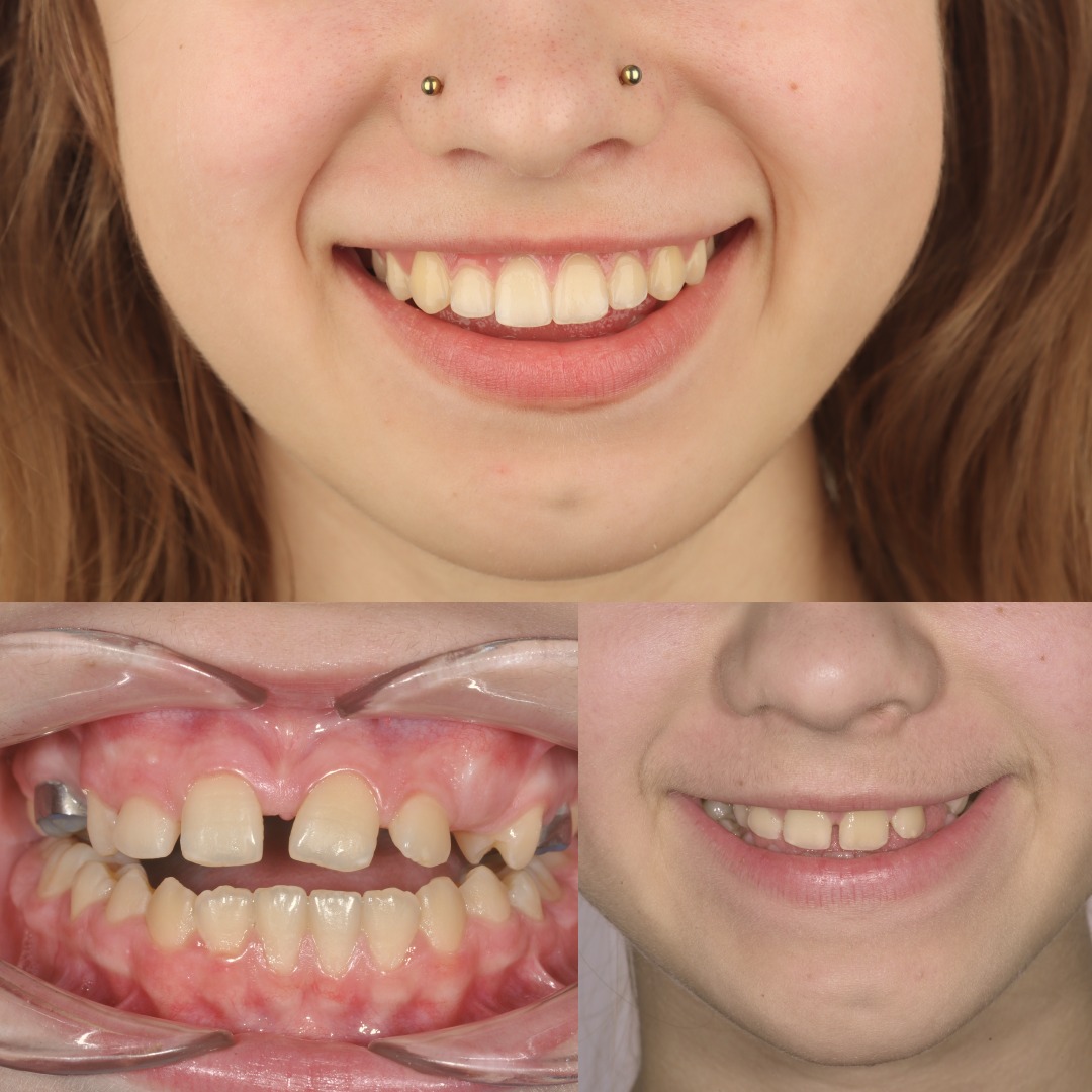
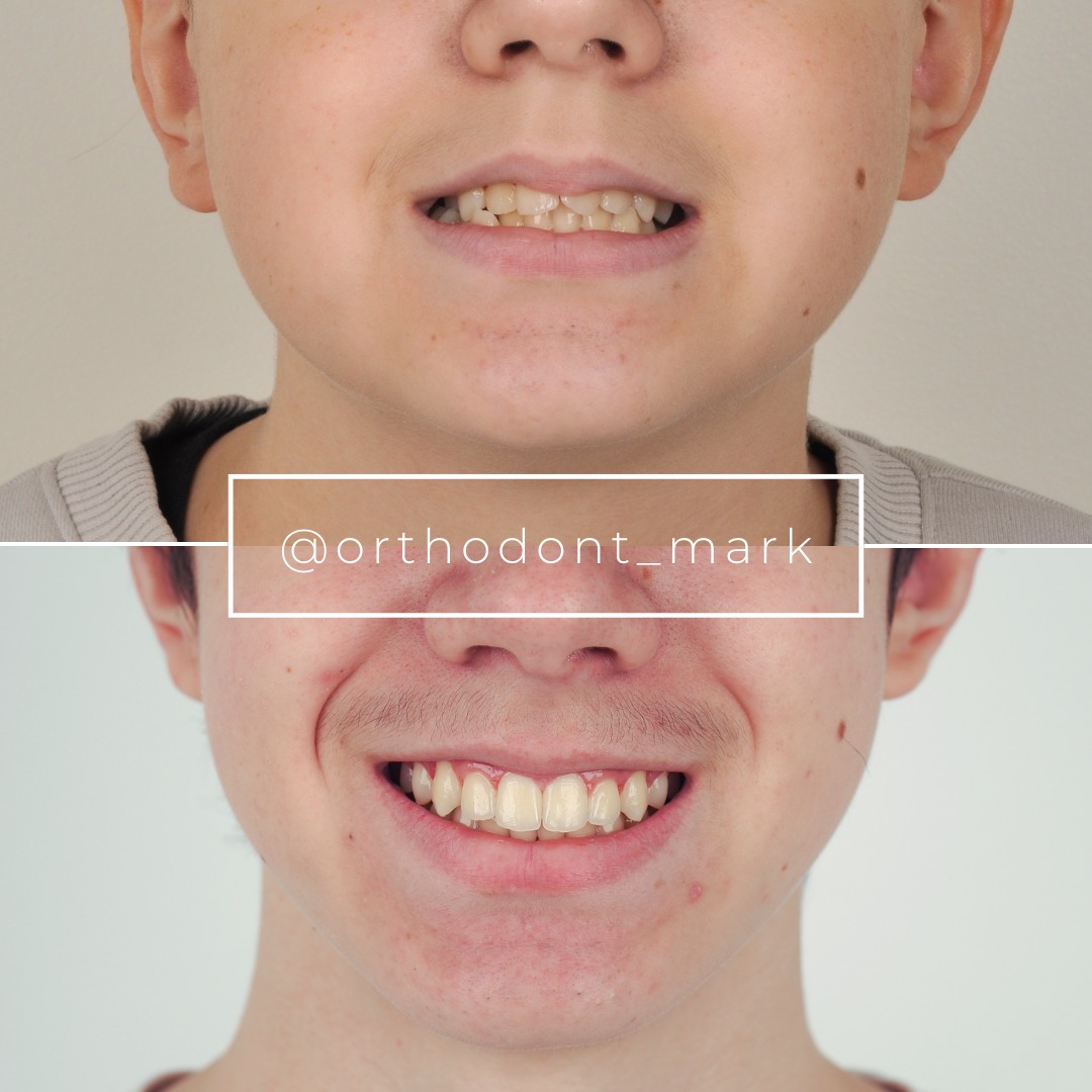
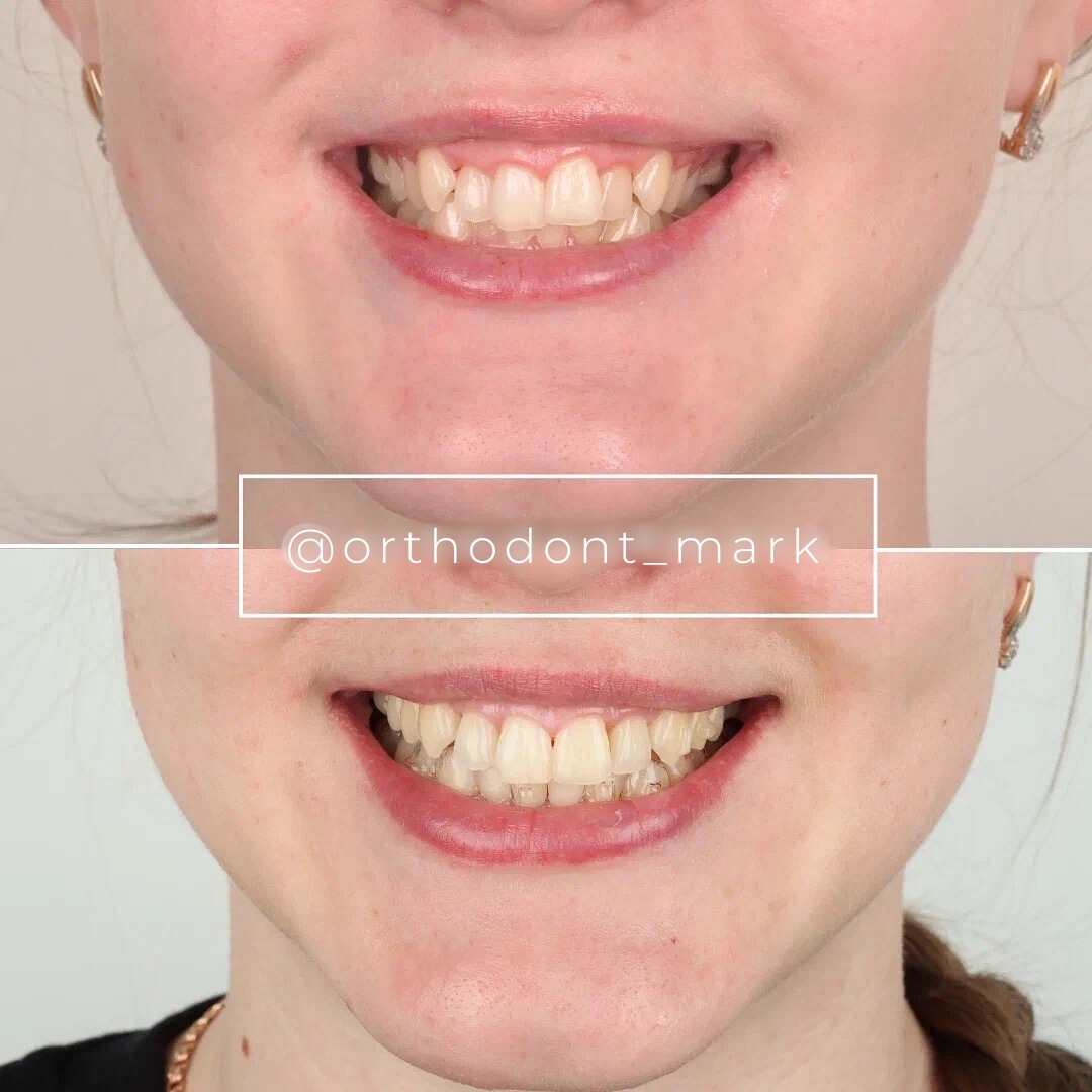
Learn the full MARPE protocol from Dr. Mark Radzhabov
Fundamental course covering CBCT patient selection, miniscrew planning, activation protocols, and 60+ clinical cases. Choose the access level that fits your practice.
Essentials of rapid palatal expansion for practicing orthodontists.
- Core RPE concepts and biomechanics
- 6 structured video lessons
- Clinical decision checklists
- Lifetime access to recordings
Deep-dive into MARPE protocol, diagnostics, and clinical execution.
- 6 modules covering MARPE from A to Z
- CBCT case selection walkthroughs
- Miniscrew placement and activation
- 60+ annotated clinical cases
- Direct Q&A with Dr. Mark Radzhabov
5-element medical consultation framework for dentists and orthodontists.
- Trust-building consultation protocol
- 5 lesson modules
- Templates for treatment plan delivery
- Works with any clinical specialty
Clinical FAQ
Which anatomical landmarks are most reproducible for nasal cavity width measurement on post-expansion CBCT?
The lateral nasal wall at the inferior turbinate attachment and the nasal floor at the greater palatine foramen level offer the highest inter-rater agreement because both are identifiable on bony window CBCT settings. Anchoring the coronal slice to the posterior nasal spine eliminates reference plane angulation error.
How do I measure nasal cavity width on CBCT after MARPE in a way comparable to published studies?
Use a coronal slice perpendicular to the palatal plane at the molar level and a second slice at the greater palatine foramen. Chun et al. (2022) identified both as sites of differential MARPE response — measuring only one underestimates the spatial distribution of skeletal widening.
Why does nasal cavity volume increase vary so widely across MARPE studies in the literature?
Benetti et al. (2024) reported increases from 9.9% to 31%, with the range driven by differing segmentation boundaries, patient age, and skeletal maturity variation. Standardizing your ROI cut-offs to a published protocol reduces this inter-study gap in your own data.
What is the clinical significance of nasopharyngeal volume change after MARPE on CBCT?
Benetti et al. (2024) reported nasopharyngeal volume increases from 6.4% to 20.7% after MARPE. This range reflects genuine anatomical variation across patients and differing superior segmentation boundaries between studies — both factors must be accounted for before counselling patients on airway benefit.
Does suture separation rate affect how I interpret nasal floor displacement on CBCT?
Yes. Chun et al. (2022) reported suture separation in 95% of MARPE patients versus 90% of RPE patients. Nasal floor widening is attributable to true skeletal change only when separation is confirmed on axial CBCT. Absent that sign, dental tipping may inflate the apparent nasal width gain.
How does patient age affect the CBCT pattern of midpalatal suture opening and nasal measurement reproducibility?
Kinzinger et al. (2022) showed suture opening width decreases in the cranial and dorsal directions with age. This means nasal floor displacement becomes anteriorly biased in older patients, requiring clinicians to document posterior suture morphology as a qualifier when interpreting nasal width measurements.
Should I use a single coronal slice or a volumetric segmentation for reproducible nasal cavity assessment after palatal expansion?
Both serve different purposes. A standardized coronal slice at two defined landmarks provides rapid, reproducible linear data for clinical tracking. Volumetric segmentation, validated by Benetti et al. (2024) across all reviewed studies, is needed for airway change quantification and inter-study comparison.
How does MARPE compare to RPE in the nasal width increase visible on cone-beam CT?
Chun et al. (2022) found MARPE produced greater nasal width increase at the molar region and the greater palatine foramen both immediately after expansion and during consolidation, compared to RPE in a prospective randomized clinical trial — a difference that is anatomically localized, not global.
What segmentation boundary decisions most affect nasal cavity volume calculations on post-expansion CBCT?
The superior boundary of the nasopharyngeal segment is the highest-impact decision — placing it at the sella versus the posterior choanae changes the volume denominator substantially. Documenting exact ROI coordinates in your CBCT software removes this ambiguity and keeps pre- and post-expansion volumes internally consistent.
How do I differentiate true skeletal nasal widening from dental tipping artifacts on post-expansion CBCT?
Confirm midpalatal suture separation on the axial slice as a low-density line from anterior to posterior nasal spine. Chun et al. (2022) used this separation criterion to validate skeletal response. Without confirmed suture opening, measured increases in inter-nasal-wall distance may reflect buccal crown torque rather than skeletal change.
Reliable nasal cavity width CBCT measurement after MARPE requires pre-defined coronal reference planes, bilateral landmark symmetry checks, and volumetric verification — not a single linear calliper reading. Clinicians who standardize their imaging protocol before appliance placement will capture the most defensible before-and-after data for case documentation and consent. For a structured approach to integrating post-expansion CBCT interpretation into your workflow, Dr. Mark Radzhabov offers peer-reviewed case analysis and structured MARPE training at ortodontmark.com. Key sources: Benetti et al. (2024), Dentistry Journal, doi:10.3390/dj12030060. Chun et al. (2022), BMC Oral Health, doi:10.1186/s12903–022-02138-w.

