
MARPE-Induced Zygomatic Suture Changes:
Imaging
What the Evidence Really Shows
A clinician-focused review of CBCT and animal-model data on midfacial suture response to bone-borne and tooth-borne palatal expansion.
TL;DR MARPE-induced zygomatic suture changes are documented through CBCT imaging, yet the full circummaxillary suture response remains incompletely mapped in humans. Animal models confirm distraction-induced expansion far exceeds physiologic growth, while prospective trials show bone-borne anchorage reduces dental tipping versus tooth-borne designs. Suture maturation, not device type alone, governs predictability.
When a clinician evaluates a skeletally mature patient for orthopedic expansion, the midpalatal suture draws immediate attention — but the zygomatic, frontomaxillary, and broader circummaxillary suture network transmits force simultaneously, and imaging evidence on those secondary sites remains sparse. Dr. Mark Radzhabov at ortodontmark.com reviews what peer-reviewed imaging literature actually demonstrates about midfacial suture response to MARPE and MSE, contrasting animal-model distraction data with prospective CBCT trials, so clinicians can calibrate expectations and refine case selection protocols before committing to a bone-borne expansion approach.
Why the Circummaxillary Suture System Matters
Beyond
the Midpalatal Suture
Circummaxillary suture disarticulation is the process by which orthopedic expansion forces propagate beyond the midpalatal suture into adjacent articulations — including the zygomaticomaxillary, frontomaxillary, and pterygomaxillary sutures — producing measurable skeletal displacement at multiple midfacial sites. Understanding this network is clinically essential because resistance at peripheral sutures, not just palatal bone density, modulates how predictably a given appliance will open the midpalatal complex. The force path differs meaningfully between tooth-borne rapid palatal expansion appliances and bone-borne MARPE designs. Tooth-borne hyrax-style expanders transmit a significant portion of their orthopedic load through the periodontal ligament of the premolars and molars before reaching bone, which introduces a dental tipping component. Bone-borne appliances apply force directly to the palatal cortex and circumvent that dental intermediary, altering both the magnitude and the vector of load reaching the zygomatic and frontomaxillary sutures. CBCT has become the diagnostic standard for evaluating these responses because conventional radiography cannot resolve sutural width changes at the millimetre or sub-millimetre scale. Axial and coronal reconstructions allow clinicians to assess midpalatal suture morphology, palatine foramen dimensions, and nasal cavity width simultaneously — creating a multi-landmark picture of how expansion force is distributed across the midfacial skeleton. Without that volumetric data, decisions about appliance choice and activation rate rest on inference rather than direct measurement.
What Rabbit Distraction Models Reveal About
Sutural
Expansion Capacity
Animal studies provide the mechanistic foundation for understanding suture response that prospective human trials cannot ethically replicate at the histological level. Klytong (2010) conducted a rabbit model investigation at Prince of Songkla University that remains a key reference for understanding distraction-induced sutural expansion physiology and its limits relative to physiologic growth norms. In that study, expansion devices were activated twice daily for 7 consecutive days, beginning 3 days after surgery, according to Klytong (2010). The resulting sutural expansion across different retention periods ranged from 3.72 mm to 4.45 mm, compared to an expected distance of 5.6 mm, as Klytong (2010) reported. This gap between achieved and expected separation underscores that device-driven distraction, even under controlled conditions, does not perfectly replicate ideal physiologic suture width — a finding with direct implications for consolidation planning in human patients. Critically, Klytong (2010) confirmed that distraction-induced sutural expansion greatly exceeded the normal physiologic growth of 0.48 mm observed in control rabbits, establishing that mechanical activation delivers a qualitatively different stimulus to sutural cells than natural growth signals. For the clinician interpreting CBCT data after MARPE activation, this means that radiographic suture widening reflects a mechanically driven process with its own healing and remodelling trajectory — one that requires adequate consolidation time before appliance removal to prevent relapse.
How CBCT Trials Map Nasal Width and
Palatine
Foramen Response
Moving from animal distraction models to prospective human trials, Chun et al. (2022) published a randomized clinical trial in BMC Oral Health that used CBCT imaging at baseline, immediately after 35 turns of identical expansion, and after a 3-month consolidation period to compare RPE and MARPE outcomes across multiple skeletal and dental landmarks. MARPE produced significantly greater increases in molar-region nasal width and greater palatine foramen dimensions than RPE, according to Chun et al. (2022), with between-group differences reaching statistical significance at p < 0.05. These findings are relevant to the frontomaxillary suture discussion because nasal width changes at the molar level reflect expansion propagating superiorly through the maxillary walls and into the nasal floor — anatomical territory that abuts the circummaxillary suture network. Greater nasal cavity widening with MARPE suggests more efficient orthopedic load transfer up the maxillary pillars toward the zygomatic and frontal articulations. On the dental side, Chun et al. (2022) demonstrated that MARPE significantly reduced buccal displacement of premolar and molar anchor teeth compared to RPE (p < 0.05), confirming that removing the tooth-borne component of force delivery changes the skeletal-to-dental effect ratio in a clinically meaningful direction. For the orthodontist evaluating a case where zygomatic suture disarticulation is a treatment goal, this means that skeletal expansion protocol and device selection directly influences how much of the applied force reaches the upper suture complex versus being absorbed as dental tipping.
Does Appliance Design Predict Suture
Separation
Consistency Across Groups?
Midpalatal suture separation rates in the imaging literature set a baseline against which clinicians can contextualize upper suture findings. Chun et al. (2022) reported midpalatal suture separation in 90% of RPE patients and 95% of MARPE patients across their randomized sample, demonstrating that both appliance types reliably open the midpalatal complex in the study population, though MARPE's rate was marginally higher. The remaining cases without confirmed radiographic separation highlight that even with standardized activation, biological variability — including suture maturation stage — can prevent full opening. Sarraj et al. (2021) examined MSE and BAME designs in a CBCT comparative study. Both groups achieved complete midpalatal suture separation, and the study identified that skeletal expansion protocol and device selection influences suture response patterns. However, the article does not specify a differential response at the zygomatic or frontomaxillary sutures between the two bone-borne designs, which underscores the current evidence gap: while palatal suture outcomes are relatively well-documented by CBCT, secondary suture responses at the zygomatic and frontomaxillary articulations require further dedicated imaging studies to characterise fully. For clinical interpretation, the discrepancy between the near-universal separation seen in the Sarraj et al. (2021) bone-borne groups and the Chun et al. (2022) mixed-design population reinforces that study design, appliance type, and patient selection criteria all influence what a CBCT scan will show post-expansion. Clinicians should not extrapolate from palatal suture separation rates to infer equivalent predictability at peripheral midfacial sutures — those articulations mature at different rates and respond to expansion vectors with different mechanical thresholds.
Applying the Imaging Data to Activation and
Consolidation
Decisions
Translating imaging findings into protocol decisions requires integrating data from both animal distraction models and prospective clinical trials. The Klytong (2010) rabbit model showed that the gap between achieved sutural expansion and the expected distance persists across retention periods, which in human terms argues for conservative consolidation timelines and staged CBCT review rather than appliance removal based on activation count alone. The Chun et al. (2022) trial design — imaging at baseline, after 35 turns, and after a 3-month consolidation period — offers a practical temporal template. Evaluating nasal width change at the molar region and palatine foramen dimensions at the post-expansion CBCT gives the clinician an indirect read on how efficiently orthopedic force reached the upper midfacial suture complex. If molar-region nasal width gain is below expectation despite confirmed midpalatal separation, upper suture resistance may be limiting the skeletal response — a clinical scenario where force magnitude or anchorage design warrants re-evaluation. Clinicians who want to develop a systematic approach to imaging interpretation and appliance selection for circummaxillary suture disarticulation cases will find detailed protocol frameworks in the structured MARPE training available through ortodontmark.com. Integrating the evidence from Klytong (2010), Chun et al. (2022), and Sarraj et al. (2021) into a coherent pre-treatment and post-expansion imaging checklist is more reliable than relying on activation count or patient age as the sole expansion endpoint.
Free Clinical Guide
Which Expansion Appliance at Which Age?
Get Dr. Mark Radzhabov's guide to skeletal-expansion appliances — indications, pros & cons, and how to choose by patient age. Free to your inbox.
Prefer video? Watch the free demo lesson on MARPE appliance types, features and trade-offs.
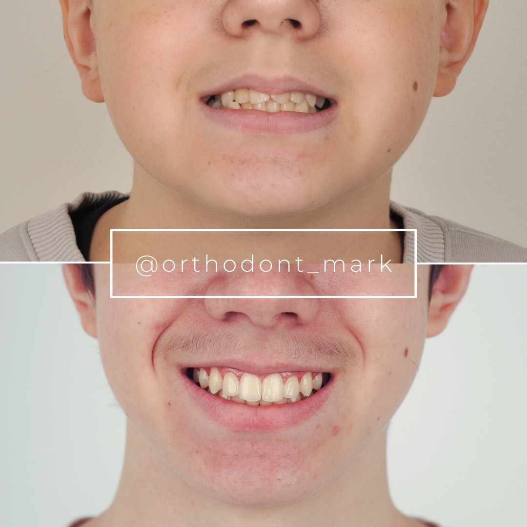
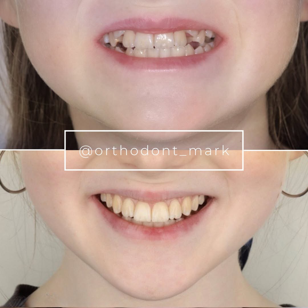
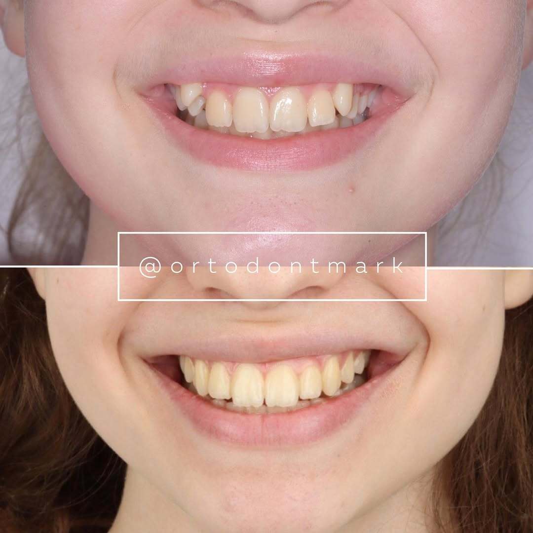
Documented outcomes
Clinical cases from Dr. Mark Radzhabov's practice
Learn the full MARPE protocol from Dr. Mark Radzhabov
Fundamental course covering CBCT patient selection, miniscrew planning, activation protocols, and 60+ clinical cases. Choose the access level that fits your practice.
Essentials of rapid palatal expansion for practicing orthodontists.
- Core RPE concepts and biomechanics
- 6 structured video lessons
- Clinical decision checklists
- Lifetime access to recordings
Deep-dive into MARPE protocol, diagnostics, and clinical execution.
- 6 modules covering MARPE from A to Z
- CBCT case selection walkthroughs
- Miniscrew placement and activation
- 60+ annotated clinical cases
- Direct Q&A with Dr. Mark Radzhabov
5-element medical consultation framework for dentists and orthodontists.
- Trust-building consultation protocol
- 5 lesson modules
- Templates for treatment plan delivery
- Works with any clinical specialty
Clinical FAQ
What does CBCT imaging show about MARPE-induced zygomatic suture changes compared to RPE?
Current CBCT evidence documents greater nasal width and palatine foramen changes with MARPE than RPE at the molar level, per Chun et al. (2022), but dedicated imaging quantification of zygomatic suture width changes during expansion remains an evidence gap in the published literature.
How does bone-borne anchorage affect frontomaxillary suture response during orthopedic expansion?
Bone-borne devices bypass periodontal force absorption, directing more orthopedic load toward the maxillary pillars and the frontomaxillary articulation. Chun et al. (2022) documented greater nasal width gain with MARPE, suggesting more efficient force transmission superiorly, though direct frontomaxillary suture width data are not yet well-established.
What activation protocol did the prospective CBCT trials use for comparing MSE and RPE suture outcomes?
Chun et al. (2022) standardised 35 turns of identical expansion before interim CBCT assessment, followed by a 3-month consolidation period and a final scan — providing a reproducible imaging timeline that separates expansion-phase from consolidation-phase suture responses.
How much does distraction-induced sutural expansion exceed physiologic growth in animal models?
Klytong (2010) found that distraction-induced expansion greatly exceeded the 0.48 mm normal physiologic sutural growth seen in control rabbits, with device-activated expansion reaching between 3.72 mm and 4.45 mm, though still below the 5.6 mm expected distance.
What activation frequency was used in the animal distraction model relevant to MARPE suture physiology?
Klytong (2010) activated devices twice daily for 7 consecutive days, starting 3 days after surgery. This protocol provides a reference point for understanding the rate-dependency of sutural cell stimulation during orthopedic expansion in a controlled biological model.
Does appliance type — MSE versus BAME — produce different midpalatal suture separation outcomes on CBCT?
Sarraj et al. (2021) documented complete midpalatal suture separation in both MSE and BAME groups in their CBCT comparative study, indicating that different bone-borne anchorage configurations can achieve reliable palatal opening, though differential effects at secondary sutures were not the primary reported outcome.
Why does the gap between achieved sutural expansion and expected suture distance matter for consolidation planning?
Klytong (2010) showed that achieved expansion across retention periods ranged from 3.72 mm to 4.45 mm against an expected distance of 5.6 mm, suggesting incomplete mineralisation under shorter consolidation. Clinicians should use CBCT evidence of suture fill rather than activation count alone to time appliance removal.
How does MARPE reduce dental side effects compared to tooth-borne RPE in CBCT studies?
Chun et al. (2022) demonstrated that MARPE significantly reduced buccal displacement of premolar and molar anchor teeth compared to RPE during expansion and consolidation (p<0.05). Reducing dental tipping means a greater proportion of the applied force is available to drive skeletal suture disarticulation.
What secondary CBCT landmarks should clinicians evaluate alongside midpalatal suture separation during MARPE?
Chun et al. (2022) measured nasal width at the molar region and palatine foramen dimensions as secondary landmarks beyond the midpalatal suture. These sites reflect upper midfacial suture response and can serve as indirect indicators of circummaxillary disarticulation efficiency when direct zygomatic suture imaging is unavailable.
Is there published imaging evidence specifically quantifying frontomaxillary suture width change during MARPE or MSE?
Dedicated quantification of frontomaxillary suture width change during MARPE or MSE is not well-represented in the current available evidence. Existing studies such as Chun et al. (2022) and Sarraj et al. (2021) focus on palatal and nasal landmarks, leaving upper suture characterisation as an open research question.
The imaging literature on MARPE-induced zygomatic and frontomaxillary suture changes is still developing, but the available evidence already informs smarter case selection: CBCT staging of suture maturity, careful comparison of bone-borne versus tooth-borne dental side effects, and realistic consolidation timelines all belong in the pre-treatment workup. Clinicians looking to sharpen their interpretation skills and expand their skeletal expansion protocols are encouraged to explore the structured MARPE training resources and case review materials available at ortodontmark.com, curated by Dr. Mark Radzhabov to bridge the gap between published evidence and daily clinical decisions. Key sources: Chun et al. (2022), BMC Oral Health, doi:10.1186/s12903–022-02138-w. Sarraj et al. (2021), APOS Trends in Orthodontics, doi:10.25259/APOS_160_2020. Klytong (2010), Prince of Songkla University.