
Appraising MARPE Skeletal Dental Evidence:
Conflicting
A Framework for Reconciling Conflicting Literature
Learn to distinguish skeletal expansion from dental compensation in published MARPE outcomes — a clinical decision tool for orthodontists.
TL;DR Appraising MARPE skeletal dental evidence requires a structured framework because outcome studies measure the same appliance yet reach divergent conclusions. Measurement landmarks, suture maturation staging, and the skeletal-to-dental ratio all shift reported results markedly. Clinicians who apply consistent radiographic criteria can distinguish genuine skeletal widening from dental compensation before selecting a protocol.
When appraising MARPE skeletal dental evidence, the gap between studies reporting robust suture opening and those highlighting predominant dental tipping often traces back to inconsistent measurement methodology rather than genuine biological disagreement. Dr. Mark Radzhabov at ortodontmark.com built this critical appraisal framework to equip orthodontists with the tools to interrogate landmark selection, suture staging criteria, and outcome variable definitions before accepting any headline figure at face value. A clinician who cannot distinguish a skeletal measurement from a dental one cannot confidently translate published results into protocol decisions for the next patient seated in the chair.
Why Do Two MARPE Studies Reach
Opposite
Conclusions on the Same Appliance?
A critical appraisal framework for orthodontic expansion studies is a systematic set of radiographic, anatomical, and methodological criteria used to evaluate whether reported outcomes reflect true skeletal widening, dental tipping, or a combination of both. The divergence between positive and cautionary expansion studies rarely originates in the biology — it originates in what each team chose to measure, and where on the maxilla they chose to measure it. Landmark selection is the first variable to interrogate. Intermolar crown width and intermolar skeletal width respond differently to the same expansion force, and a study reporting only crown-level data will consistently overestimate the dental component of change. Chun et al. (2022) addressed this directly by measuring skeletal widening at the molar region and at the greater palatine foramen separately, demonstrating that MARPE produced greater skeletal nasal widening at both anatomical sites compared to RPE — a distinction that crown-level measurements alone would have obscured. The second variable is outcome timing. Consolidation-phase measurements capture a different biological state than post-debond records, and post-retention records reveal yet another. Kurt et al. (2010) followed both orthopedic and surgically-assisted maxillary expansion for 3 years post-retention, confirming that stability assessments conducted before that window closes may misrepresent long-term skeletal gain. Clinicians appraising a study should confirm that the follow-up endpoint aligns with the clinical question they are actually asking.
How Suture Staging Criteria Change
Everything
About the Patient Pool You Are Reading
When two expansion studies enroll patients by chronological age alone, they may be comparing biologically dissimilar populations. Angelieri et al. (2013) demonstrated that early suture maturation stages A and B occurred up to age 13, while the intermediate stage C was observed primarily from age 11 to 17 years — meaning a 15-year-old in stage B and a 15-year-old in stage C are not interchangeable study subjects, yet both would appear identical in an age-matched sample. This maturation overlap is clinically consequential for appraisal. Angelieri et al. (2013) also found that complete midpalatal suture fusion before age 11 occurred only in girls, while partial fusion in the palatine region was documented in males aged 14–17 — underscoring that suture status is not reliably inferred from sex or age without direct imaging. A study that stratifies by age but not by CBCT suture stage is reporting a mixed population, and its mean outcome figures may not apply to the specific maturation subgroup presenting in your clinic. For a reader appraising skeletal versus dental expansion ratios, the practical implication is to check whether the source study controlled for suture stage. If it did not, the reported skeletal gain is an average across biologically heterogeneous cases. Clinicians making miniscrew-assisted expansion protocol decisions should weight evidence from suture-staged cohorts more heavily than age-only stratified samples when translating findings to individual case selection.
Reading the Alveolar Bone Response
Correctly
in MARPE Outcome Reports
Buccal tipping reporting in rapid palatal expansion literature has historically relied on angular measurements of anchor teeth, but alveolar bone architecture captures a mechanically distinct and clinically relevant dimension of the same event. Lim et al. (2017) documented that MARPE caused buccal alveolar bone thickness to decrease while palatal alveolar bone thickness increased — a directional remodeling pattern that angular tipping measurements alone do not capture and that has direct implications for periodontal risk assessment. The same retrospective cohort recorded apical movement of the alveolar crest at the first premolar region following MARPE (Lim et al., 2017). This finding matters for appraisal because studies that restrict outcome variables to molar angulation or intermolar width will not detect crest-level changes at the premolar, producing an artificially favorable skeletal-to-dental ratio for the appliance. When two studies report different tipping rates, confirming whether both measured alveolar bone morphology — not just tooth position — is a necessary appraisal step. Chun et al. (2022) found that MARPE demonstrated significantly less buccal tipping of anchor teeth compared to RPE at a significance threshold of p < 0.05, reinforcing that force distribution through cortical bone anchorage reduces dental side effects. Reading that finding correctly, however, requires acknowledging that 'less tipping' is a relative statement: the comparator matters as much as the appliance, and a study without an RPE control group cannot produce the same comparison.
What Suture Separation Percentages
Do and Do Not
Tell the Reader
Suture separation rate is frequently the headline figure in MARPE literature, but appraising it requires separating the occurrence of a radiographic gap from the magnitude and distribution of skeletal change that gap represents. Chun et al. (2022) reported midpalatal suture separation in 90% of RPE patients and 95% of MARPE patients — a difference of five percentage points that is statistically significant at p < 0.05. A surface-level reading treats the two figures as nearly equivalent; a deeper appraisal asks where along the suture the opening occurred and whether it was symmetric. Skeletal widening at the greater palatine foramen is anatomically posterior to molar-level measurements and captures a region that tooth-borne appliances address less efficiently. Chun et al. (2022) found greater skeletal nasal widening in both the molar region and at the greater palatine foramen for MARPE compared to RPE, even though both groups achieved high separation rates. This means a suture separation percentage, taken alone, is an incomplete outcome metric: two appliances can share a similar separation rate while producing meaningfully different spatial distributions of skeletal change. For appraisal purposes, clinicians should look for studies that report widening at multiple anatomical levels — anterior palate, molar region, and posterior landmark such as the greater palatine foramen — rather than collapsing everything into a single intermolar figure. A study that reports only one measurement plane has not described the three-dimensional skeletal response, and meta-analyses pooling single-plane figures across studies are mixing geometrically non-equivalent data.
Translating Appraised Evidence into
Case Selection
Without Importing the Study's Bias
The final step in appraising MARPE skeletal dental evidence is determining whether the study population matches the patient in front of you closely enough to justify direct protocol transfer. Suture maturation stage, anchor tooth selection, and the skeletal-to-dental outcome ratio must all align before a published protocol is adopted wholesale. A finding from a population of patients with stage B sutures does not translate automatically to a patient presenting with stage D fusion, regardless of chronological age similarity. Stability data add another filter. Kurt et al. (2010) confirmed that both orthopedic and surgically-assisted maxillary expansion remained stable with postretention relapse after 3 years, but this long follow-up is rarely replicated in shorter studies. When a paper reports favorable outcomes at a shorter interval, appraising whether the follow-up window is sufficient to capture postretention behavior is not optional — it is the difference between a durable clinical decision and one that will require revision. Applying this framework consistently does not mean rejecting studies with methodological limitations. It means weighting them appropriately within a hierarchy of evidence. A prospective randomized trial with suture staging, multi-landmark skeletal measurement, and a minimum three-year follow-up sits at the top of that hierarchy. Studies without one or more of those features remain useful for generating hypotheses and identifying trends, but their headline figures should not be the sole basis for changing an established protocol. Clinicians who want to submit a case for consultation and review the appraisal framework interactively can do so at ortodontmark.com.
Free Clinical Guide
Which Expansion Appliance at Which Age?
Get Dr. Mark Radzhabov's guide to skeletal-expansion appliances — indications, pros & cons, and how to choose by patient age. Free to your inbox.
Prefer video? Watch the free demo lesson on MARPE appliance types, features and trade-offs.
Documented outcomes
Clinical cases from Dr. Mark Radzhabov's practice
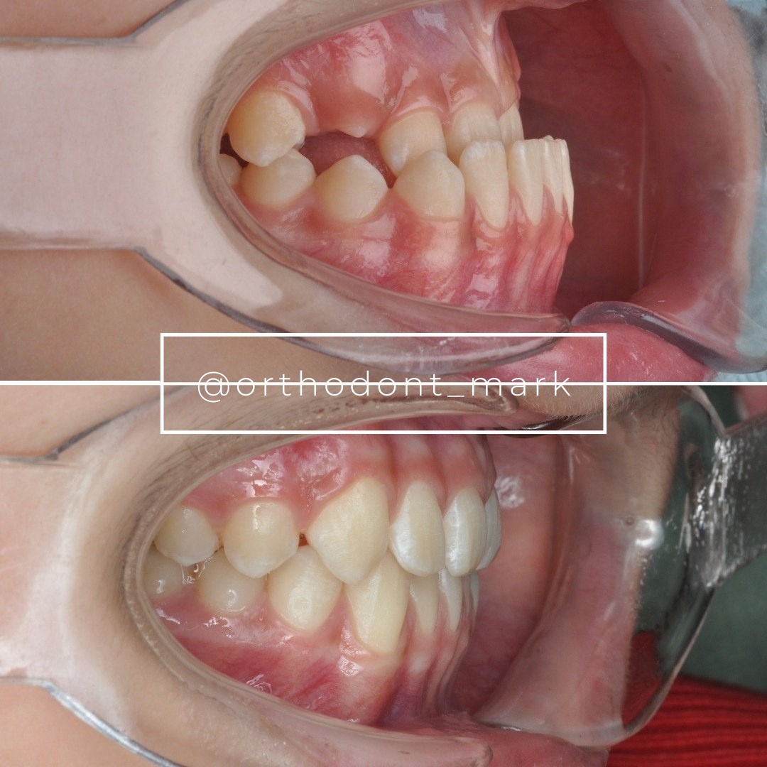
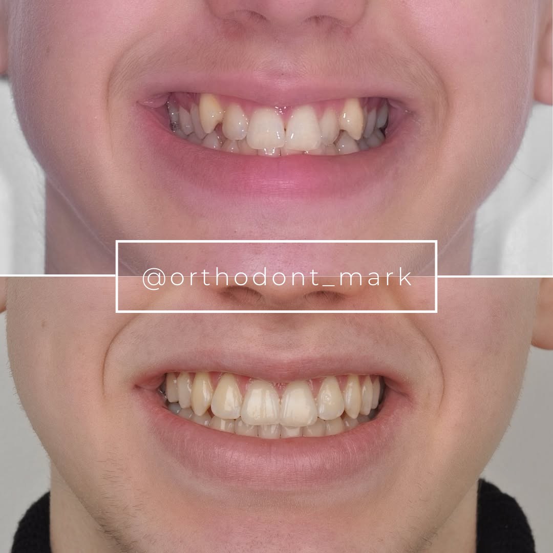
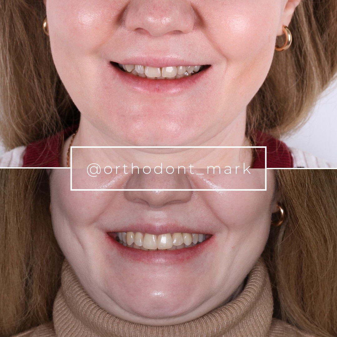
Learn the full MARPE protocol from Dr. Mark Radzhabov
Fundamental course covering CBCT patient selection, miniscrew planning, activation protocols, and 60+ clinical cases. Choose the access level that fits your practice.
Essentials of rapid palatal expansion for practicing orthodontists.
- Core RPE concepts and biomechanics
- 6 structured video lessons
- Clinical decision checklists
- Lifetime access to recordings
Deep-dive into MARPE protocol, diagnostics, and clinical execution.
- 6 modules covering MARPE from A to Z
- CBCT case selection walkthroughs
- Miniscrew placement and activation
- 60+ annotated clinical cases
- Direct Q&A with Dr. Mark Radzhabov
5-element medical consultation framework for dentists and orthodontists.
- Trust-building consultation protocol
- 5 lesson modules
- Templates for treatment plan delivery
- Works with any clinical specialty
Clinical FAQ
How do I identify buccal tipping reporting bias when reading a MARPE outcome study?
Check whether the study measured alveolar bone thickness changes alongside crown-level angular tipping. Lim et al. (2017) showed buccal alveolar bone decreased while palatal bone increased — a remodeling pattern invisible to angular measurements alone, which systematically underreport the osseous component of dental side effects.
Why do two MARPE studies using the same appliance reach different conclusions on skeletal expansion?
Landmark selection and suture staging criteria differ between studies. A trial measuring only intermolar crown width captures a different signal than one measuring at the greater palatine foramen. Chun et al. (2022) found MARPE produced greater skeletal widening at the foramen even when both groups showed similar separation rates.
How does suture maturation staging affect the validity of a rapid palatal expansion study population?
Angelieri et al. (2013) showed that stages A through C span ages 11 to 17, meaning age-matched samples can contain biologically heterogeneous patients. A study stratifying by age alone pools different maturation stages, making mean skeletal gain figures unreliable for applying to a specific patient's suture status.
What follow-up duration is adequate when appraising expansion stability data?
Kurt et al. (2010) tracked both orthopedic and surgically-assisted expansion for 3 years post-retention, capturing postretention relapse that shorter endpoints miss. Studies reporting stability at intervals shorter than that window may misclassify a case as stable when late relapse has not yet manifested.
How should I weigh a study that reports suture separation rate without regional skeletal widening data?
Treat it as partial evidence. Chun et al. (2022) showed 90% and 95% separation for RPE and MARPE respectively, yet MARPE produced meaningfully greater widening at the greater palatine foramen. Separation rate alone does not describe spatial distribution of skeletal change and cannot substitute for landmark-specific measurement.
What anatomical landmarks should a well-designed MARPE skeletal expansion study include?
At minimum: molar-region skeletal width, a posterior landmark such as the greater palatine foramen, and alveolar bone thickness at buccal and palatal aspects of anchor teeth. Chun et al. (2022) and Lim et al. (2017) together illustrate why single-plane or single-landmark designs produce an incomplete skeletal-to-dental ratio.
How do I critically appraise MARPE skeletal dental evidence when the study lacks a control group?
Interpret tipping and widening figures as absolute values only, not comparative ones. Chun et al. (2022) demonstrated that 'less buccal tipping' for MARPE is a relative finding against an RPE control. Without that comparator, the same tipping measurement cannot be labeled favorable or unfavorable on its own.
Does chronological age reliably substitute for CBCT suture staging in reading expansion literature?
No. Angelieri et al. (2013) found complete fusion before age 11 only in girls, and partial palatine fusion was documented in males aged 14 to 17. Age predicts maturation probabilistically, not individually. Studies reporting only age-based inclusion criteria may contain a wide range of suture stages within a narrow age band.
What alveolar bone changes should orthodontists look for when evaluating MARPE versus RPE outcome data?
Buccal alveolar bone thickness, palatal alveolar bone thickness, and alveolar crest position at the premolars. Lim et al. (2017) recorded apical crest movement at the first premolar and inverse thickness changes buccally versus palatally — variables omitted from most RPE comparison studies, which biases the apparent safety profile.
How does evidence quality hierarchy apply when selecting between miniscrew-assisted expansion protocols?
Prospective randomized trials with suture staging, multi-landmark skeletal measurement, and adequate follow-up sit highest. Studies missing one or more criteria are useful for trend identification but should not drive protocol changes unilaterally. Apply the appraisal framework to weight each source before translating findings into clinical decisions for individual patients.
Appraising MARPE skeletal dental evidence is not an academic exercise — it directly governs whether a patient receives bone-borne expansion, tooth-borne expansion, or surgical referral. Suture maturation staging, landmark-specific widening data, and alveolar bone response must all be weighed together before a protocol is chosen. Dr. Mark Radzhabov encourages clinicians to bring a specific case or review structured MARPE training at ortodontmark.com, where appraisal skills are integrated into every stage of the clinical curriculum. Sharper evidence literacy produces sharper case selection. Key sources: Chun et al. (2022), BMC Oral Health, doi:https://doi.org/10.1186/s12903–022-02138-w. Angelieri et al. (2013), doi:doi:10.1016/j.ajodo.2013.04.022. Lim et al. (2017), Korean Journal of Orthodontics, doi:https://doi.org/10.4041/kjod.2017.47.5.313.