
RPE Mistakes Clinicians Make
in Diagnosis, Activation, and Retention
Evidence-based thresholds for RPE case selection, activation calibration, and retention scheduling—so the skeletal gain holds.
TL;DR RPE mistakes at the diagnosis, activation, and retention stages are among the most preventable causes of failed maxillary expansion in clinical practice. Misreading skeletal readiness, miscalibrating daily activation, and ending retention prematurely each compound downstream. This article outlines the evidence-based thresholds every clinician should apply before and after RPE placement.
RPE mistakes are more common than the literature acknowledges, and they rarely announce themselves until relapse, anchorage loss, or an unresolved airway finding forces a second look. At ortodontmark.com, Dr. Mark Radzhabov has distilled the recurring diagnostic, activation, and retention failures seen across clinical practice and peer-reviewed evidence into a single actionable framework. Clinicians who read this article will leave with concrete decision thresholds—imaging criteria, activation rates, and retention windows—grounded in published findings rather than convention. Mastering this sequence protects both the patient's structural outcome and the clinician's treatment trajectory.
Which Patients Are Misclassified as Ready
for Rapid Palatal Expansion?
Rapid palatal expansion (RPE) is an orthopedic procedure that applies transverse force across the midpalatal suture to increase maxillary arch width, with outcomes shaped critically by accurate diagnosis, calibrated activation, and adequate retention. The single most preventable diagnostic error is selecting a candidate without first characterizing midpalatal suture maturity through high-resolution imaging. Relying on chronological age alone—without confirming suture morphology—exposes the clinician to resistance patterns the appliance is not designed to overcome, converting what should be a straightforward orthopedic case into a biomechanical struggle. Research focused on the pediatric age window reinforces why cohort selection matters. Mummolo et al. (2014) note that RPE and slow palatal expansion studies are typically conducted in children aged 6 to 8 years, underlining that the biological window of greatest suture compliance is narrow and well-defined. Enrolling patients outside that developmental band without adjusting appliance selection or activation strategy is a diagnostic failure, not a mechanical one. A second frequently missed diagnostic variable is the upper airway. Clinicians often frame RPE as a purely dental-skeletal intervention and overlook the adenotonsillar findings that may accompany transverse maxillary deficiency. Yoon et al. (2022) documented that tissue reduction following RPE averages 16.8% for adenoids and 38.5% for tonsils, confirming that the airway responds measurably to expansion. Ignoring this finding at the intake stage means missing a co-treatment opportunity and an important outcome metric. Thorough pre-treatment documentation of airway dimensions alongside skeletal and dental indices is therefore not optional—it is part of a defensible diagnostic record.
How Activation Rate Errors Undermine
the Skeletal Response to RPE
Activation errors fall into two directions—too fast and too slow—and both compromise the skeletal result. Excessive daily turns generate force peaks the suture cannot dissipate gradually, increasing the risk of dental tipping, buccal cortical bending, and pain that drives non-compliance. Insufficient activation, by contrast, allows fibrous tissue to reorganize across the suture before adequate width is achieved, effectively wasting the expansion window. Yoon et al. (2022) specify an activation protocol of 0.25 mm per day, maintained for a period of 4 to 6 weeks. This range provides a practical clinical anchor: the daily increment is small enough to permit sutural micro-fracture and remodeling without overwhelming the adjacent bony architecture, while the duration is long enough to accumulate the transverse gain the case requires. Departing from this cadence—whether by doubling turns on missed days or stopping early once a midline diastema appears—introduces variability that the retention phase cannot correct. For the McNamara RPE device specifically, Giannasi et al. (2014) describe a compressed activation window of 7 to 15 days of active expansion, after which the appliance transitions to passive retention. In that protocol, the speed of active expansion is higher, making precision in turn sequence even more critical. Giannasi et al. (2014) also report a mean transverse maxillary expansion of 7.2 mm with this approach, establishing what a correctly executed McNamara protocol can realistically achieve. Clinicians who apply a generic activation schedule across dissimilar appliance designs—treating a McNamara expander with the same multi-week cadence as a conventional Hyrax—are making a protocol mismatch error that distorts the expected skeletal gain.
When Is It Safe to Discontinue RPE Retention
Without Risking Transverse Relapse?
Retention errors are the most quietly destructive RPE mistakes because they emerge weeks to months after the appliance is removed—long after the clinical interaction that caused them. Two categories dominate: retention that ends too soon, before mineralization across the sutural gap is adequate, and retention in which follow-up imaging is scheduled too late to catch early relapse while it is still correctable. For the McNamara RPE device, Giannasi et al. (2014) specify a passive retention phase of 6 to 8 months following active expansion. This interval is not arbitrary—it reflects the time required for woven bone to mature into lamellar bone across the expanded suture. Clinicians who remove the appliance at three or four months, citing patient discomfort or hygiene concerns, are trading short-term compliance for long-term structural instability. On the imaging side, Yoon et al. (2022) recommend scheduling follow-up assessment at approximately 13.8 months post-treatment to allow adequate retention evaluation. An imaging review conducted at the standard six-month recall misses the window during which late sutural remodeling—and the airway changes documented in the same study—fully consolidate. For clinicians managing RPE outcomes in patients with co-existing sleep-disordered breathing, the airway data from that same follow-up point are clinically significant: Yoon et al. (2022) recorded a 38.5% mean tonsillar tissue reduction, a finding that warrants documentation at the correct post-treatment interval rather than at a convenient but premature one. Aligning retention duration with appliance-specific evidence and scheduling imaging at the published timepoint are the two protocol adjustments most likely to reduce palatal expansion retention loss in everyday practice.
Why Clinicians Should Track Airway Changes
Alongside Skeletal Expansion Metrics
One of the more consequential RPE diagnostic errors is treating the procedure as a purely transverse skeletal event while ignoring the nasopharyngeal and oropharyngeal dimensions that change alongside arch width. This is not a theoretical concern—it affects how the case is documented, how co-treatment with ENT is timed, and how the outcome is communicated to the referring clinician. Yoon et al. (2022) provide quantified benchmarks: adenoid tissue reduced by a mean of 16.8% and tonsillar tissue by a mean of 38.5% following RPE in their retrospective cohort. These figures give the clinician a reference range against which individual patient responses can be compared. A patient who shows minimal tonsillar reduction at the 13.8-month follow-up despite adequate skeletal expansion may warrant ENT reassessment, whereas one who exceeds these averages may experience accelerated resolution of sleep-disordered breathing symptoms. Integrating airway assessment into the RPE workflow requires only two additions to standard practice: a pre-treatment lateral cephalometric or CBCT-based airway measurement at defined pharyngeal landmarks, and a matched measurement at the post-treatment imaging appointment. Without the baseline, the follow-up figure is uninterpretable. Clinicians who want to build this into a replicable clinical protocol will find that the diagnostic logic for skeletal expansion decision-making with MARPE follows the same staged imaging framework—baseline, active phase, retention, and long-term follow-up—that makes RPE outcomes trackable and defensible.
How Does Appliance Choice Affect RPE Activation Errors
in Mixed and Permanent Dentition Cases?
Selecting the correct expander design is inseparable from selecting the correct activation protocol, yet many RPE mistakes originate precisely at this junction—where the clinician chooses an appliance from habit rather than from a protocol-matched rationale. A Hyrax, a bonded acrylic expander, and a McNamara-style device each distribute force differently across the mid-palatal suture and the dentoalveolar complex, and each demands a distinct activation cadence. Mummolo et al. (2014) situate the evidence base for RPE squarely in children aged 6 to 8 years, the developmental range in which suture compliance is highest and dental side effects of expansion are easiest to manage. This framing matters for appliance selection: in this age range, tooth-borne appliances with a conventional screw can achieve significant width change without the biomechanical reinforcement that bone-borne designs provide in older patients. Using a bone-borne or miniscrew-assisted design in a 6-year-old purely out of clinician preference rather than indication-driven logic is as much a mismatch error as applying a conventional Hyrax to a skeletally mature adult. The McNamara protocol described by Giannasi et al. (2014) compresses the active phase to 7 to 15 days, achieving a mean skeletal gain of 7.2 mm—results that depend entirely on the clinician executing the turn sequence correctly within that brief window. Applying the same device with a conventional slow-expansion cadence defeats the biomechanical premise of the appliance. Conversely, applying a rapid protocol to a slow-expander design risks excessive dental tipping. Matching appliance to protocol to patient developmental stage is the clinical decision that prevents the activation errors described in the preceding section from occurring in the first place.
Free Clinical Guide
Which Expansion Appliance at Which Age?
Get Dr. Mark Radzhabov's guide to skeletal-expansion appliances — indications, pros & cons, and how to choose by patient age. Free to your inbox.
Prefer video? Watch the free demo lesson on MARPE appliance types, features and trade-offs.
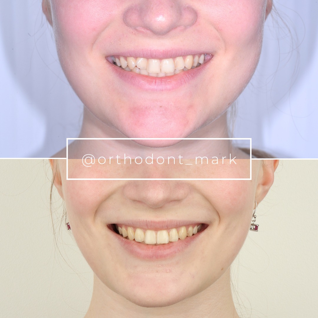
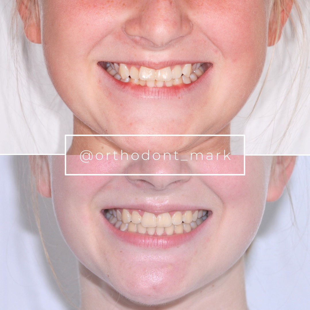
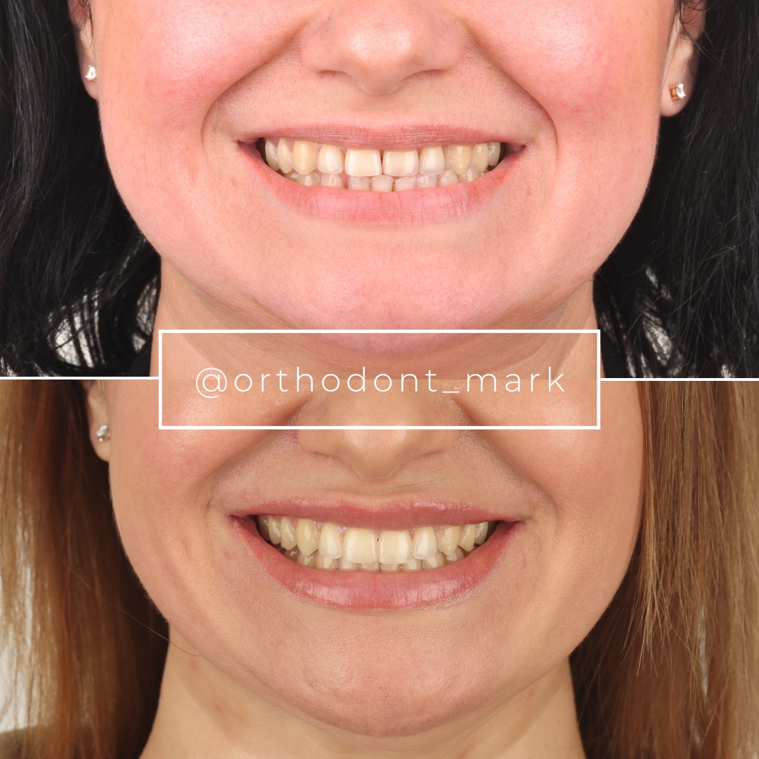
Documented outcomes
Clinical cases from Dr. Mark Radzhabov's practice
Learn the full MARPE protocol from Dr. Mark Radzhabov
Fundamental course covering CBCT patient selection, miniscrew planning, activation protocols, and 60+ clinical cases. Choose the access level that fits your practice.
Essentials of rapid palatal expansion for practicing orthodontists.
- Core RPE concepts and biomechanics
- 6 structured video lessons
- Clinical decision checklists
- Lifetime access to recordings
Deep-dive into MARPE protocol, diagnostics, and clinical execution.
- 6 modules covering MARPE from A to Z
- CBCT case selection walkthroughs
- Miniscrew placement and activation
- 60+ annotated clinical cases
- Direct Q&A with Dr. Mark Radzhabov
5-element medical consultation framework for dentists and orthodontists.
- Trust-building consultation protocol
- 5 lesson modules
- Templates for treatment plan delivery
- Works with any clinical specialty
Clinical FAQ
What is the correct RPE activation protocol to avoid daily rate errors?
Yoon et al. (2022) specify 0.25 mm per day maintained over 4 to 6 weeks. Deviating from this cadence—doubling turns on missed days or stopping early after diastema appearance—introduces force variability the retention phase cannot compensate for.
How long should RPE retention last to prevent transverse relapse?
For the McNamara RPE device, Giannasi et al. (2014) prescribe 6 to 8 months of passive retention after active expansion. Removing the appliance earlier risks relapse before lamellar bone has adequately mineralized across the expanded sutural gap.
At what post-treatment interval should follow-up imaging be scheduled after RPE?
Yoon et al. (2022) recommend follow-up imaging at approximately 13.8 months post-treatment. Earlier reviews miss late sutural remodeling and do not capture the full airway tissue response that consolidates over the retention period.
What airway changes can clinicians document following rapid palatal expansion?
Yoon et al. (2022) recorded a mean adenoid tissue reduction of 16.8% and a mean tonsillar reduction of 38.5% after RPE. Clinicians should document pharyngeal dimensions at a consistent anatomical landmark before treatment to make post-treatment comparison valid.
How does the McNamara RPE activation window differ from a conventional Hyrax protocol?
Giannasi et al. (2014) describe the McNamara device as completing active expansion in 7 to 15 days, achieving a mean of 7.2 mm of transverse gain, before transitioning to retention. A conventional Hyrax protocol typically uses a slower, multi-week cadence at a lower daily increment.
What patient age group provides the strongest evidence base for RPE outcomes?
Mummolo et al. (2014) note that RPE and slow palatal expansion research is typically conducted in children aged 6 to 8 years. Applying protocols extrapolated from this cohort to patients outside that developmental window requires adjusted appliance selection and activation logic.
How do common RPE diagnostic errors affect skeletal expansion outcomes?
Selecting candidates without imaging-confirmed suture maturity and omitting a pre-treatment airway baseline are the two most prevalent diagnostic errors. Both limit the clinician's ability to measure the true skeletal and airway response and to identify early relapse.
Does appliance type affect the risk of RPE activation errors in mixed dentition?
Yes. Each expander design has a specific activation cadence. Applying a rapid protocol to a device calibrated for slow expansion generates unpredictable force magnitudes. Matching appliance type to the patient's developmental stage and the intended activation rate is essential for avoiding protocol mismatch errors.
What are the most common palatal expansion retention failure causes in clinical practice?
Early appliance removal before adequate sutural mineralization and follow-up imaging scheduled too soon after treatment are the dominant causes. Both errors are preventable by anchoring retention duration and imaging timing to published protocol benchmarks rather than calendar convenience.
How does the RPE protocol differ between tooth-borne and bone-borne expanders in terms of diagnostic requirements?
Tooth-borne designs in younger patients can achieve clinically significant expansion with conventional activation, whereas bone-borne designs in older patients require suture maturity assessment to justify the added procedural complexity. Prescribing a bone-borne device without confirming indication through imaging is a diagnostic mismatch error.
Avoiding RPE mistakes demands more than technical skill—it demands a staged clinical logic that runs from the first CBCT slice through the final retention check. Diagnostic shortcuts, aggressive activation, and abbreviated retention schedules each erode the skeletal gain that expansion is designed to produce. Dr. Mark Radzhabov encourages clinicians to review their current RPE workflow against the evidence-based thresholds discussed here, then seek structured RPE training to close any protocol gaps. Consistent application of these principles is what separates a stable skeletal result from a retreatment case. Key sources: Yoon et al., 2022, Sleep Medicine, doi:10.1016/j.sleep.2022.02.011. Giannasi et al., 2014, CRANIOH: The Journal of Craniomandibular & Sleep Practice, doi:10.1179/2151090314Y.0000000029.