MARPE Treatment Sequencing:
Expansion
Before or After Comprehensive Therapy?
An evidence-based framework for staging skeletal expansion relative to fixed appliances or aligners—built on published imaging and mechanical data.
TL;DR MARPE treatment sequencing should resolve transverse skeletal deficiency before comprehensive appliance therapy begins. Expansion pattern, force magnitude, and dental side effects all shift depending on patient age and appliance design. Evidence from peer-reviewed imaging and mechanical studies informs when and how to stage expansion relative to fixed or aligner treatment.
A patient with documented transverse maxillary deficiency arrives having already started aligner therapy elsewhere, and the arch width problem is only getting harder to disguise. Decisions about when to expand—before, during, or after comprehensive treatment—carry real biomechanical and stability consequences. On ortodontmark.com, Dr. Mark Radzhabov draws on peer-reviewed imaging studies and in-vitro mechanical data to build a decision framework for MARPE treatment sequencing that clinicians can apply at the case-planning stage, before a single wire or aligner is placed.
Why Does the Order of Expansion and
Comprehensive Treatment
Change the Skeletal Outcome?
MARPE treatment sequencing is the clinical process of determining whether miniscrew-assisted palatal expansion should precede, overlap with, or follow comprehensive fixed appliance or aligner therapy, based on suture maturity, expansion geometry, and appliance force characteristics. This decision is not cosmetic—it directly determines whether the transverse correction is skeletal or merely dental, and whether it will remain stable once space closure begins. The expansion pattern itself changes with age in a way that affects sequencing logic. Stefanm et al. (2022) demonstrated through CBCT analysis that the RME expansion pattern is parallel in children under 10 years but becomes V-shaped in adolescents aged 12 and older. A V-shaped opening widens more at the anterior than the posterior palate, which means arch-form coordination with brackets placed before expansion is unreliable—the posterior segments will not respond symmetrically to leveling and aligning mechanics. This geometry shift was observed across a dental cast sample in which Stefanm et al. (2022) analysed 60 children divided into three equal groups: under 10 years (n=20), 10–12 years (n=20), and 12 years and older (n=20). The finding reinforces the clinical principle that sequencing expansion first—particularly in adolescents—prevents the arch form from being shaped by bracket prescription around a transverse discrepancy that is simultaneously changing shape. Brackets placed into a V-opening midpalatal suture will need significant torque and angulation correction that could have been avoided.
Does Appliance Design Affect Which Phase
Comes First
in the Treatment Sequence?
Appliance force magnitude is directly relevant to sequencing because high-force devices produce greater skeletal displacement but also impose higher loads on anchor teeth—a problem that multiplies when fixed appliances are already in place. Camporesi et al. (2013) measured that the Hyrax and A2620 expanders generated forces exceeding 20 kg, while the Palatal Split screw generated approximately 16 kg. Rapid maxillary expansion devices as a category produce forces ranging from 7.54 to 15.8 kg, according to Camporesi et al. (2013). The A2620 and Hyrax screws demonstrated significantly greater rigidity and force production across all activation levels compared to the Palatal Split screw, as Camporesi et al. (2013) confirmed in in-vitro mechanical testing. When brackets and archwires are present during activation of a high-force device, the dental anchorage units resist expansion forces through the wire rather than transmitting them directly to the suture. This dilutes skeletal effect and increases the risk of tipping anchor premolars and molars—precisely the side effect that miniscrew-assisted expansion was designed to reduce. For this reason, sequencing the expansion phase before archwire placement preserves the clean force path from miniscrews through the palatal bone to the suture. Clinicians choosing between a tooth-borne rapid palatal expander and a bone-borne miniscrew-assisted expansion system should factor in whether arch leveling has already begun, because the interaction between archwire stiffness and expander force magnitude is not negligible at forces exceeding 20 kg, as documented by Camporesi et al. (2013).
How Does MARPE Reduce Tipping When Sequenced
Before Fixed Appliances
Are Placed?
One of the strongest arguments for completing skeletal expansion before bracket bonding is the dental side-effect profile of different expander types. Chun et al. (2022) found that MARPE produced significantly less buccal tipping of anchor teeth compared to RPE during the expansion and consolidation period. When fixed appliances are present, even a small degree of uncorrected tipping becomes a torque error that must be resolved with auxiliary mechanics. Chun et al. (2022) conducted a randomized controlled trial in which both RPE and MARPE groups received identical activation of 35 screw turns, with CBCT imaging at baseline, immediately after expansion, and after 3 months of consolidation. This controlled protocol confirms that the tipping difference is appliance-specific, not activation-specific—MARPE's skeletal anchorage redistributes force away from the crowns of the premolars and molars regardless of the turn count. The same study showed that MARPE produced significantly greater increases in nasal width and greater palatine foramen opening compared to RPE (Chun et al., 2022). These skeletal dimensional changes—nasal width and palatine foramen displacement—are more difficult to achieve after fixed appliances begin altering dentoalveolar positions. Clinicians planning aligner therapy after expansion should note that the nasal width increase documented by Chun et al. (2022) represents a true skeletal shift that aligner attachments cannot replicate through dental tipping alone, reinforcing the case for sequencing miniscrew-assisted expansion first.
Does MARPE Deliver Reliable Suture Opening
Regardless
of When It Is Sequenced?
Understanding suture separation probability helps clinicians decide whether to expand before or after committing to an appliance sequence—because a failed expansion mid-treatment creates far more disruption than a properly staged pre-treatment expansion phase. Chun et al. (2022) reported midpalatal suture separation in 90% of RPE patients and 95% of MARPE patients following 35 identical screw turns, with both groups assessed by CBCT at baseline, immediately after expansion, and after 3 months of consolidation. The separation rate advantage for MARPE—95% versus 90% per Chun et al. (2022)—is clinically meaningful when weighed against the downstream consequences of incomplete opening. A partially separated suture at the start of comprehensive treatment leaves the clinician managing residual transverse discrepancy through dental tipping, which undermines the entire rationale for skeletal expansion. Sequencing MARPE before archwire placement ensures that any need for re-activation or supplementary procedures is identified and resolved before the comprehensive phase introduces competing mechanics. For aligner sequencing specifically, the 3-month consolidation window documented by Chun et al. (2022) provides a natural break point: digital impressions or intraoral scans taken after consolidation capture the stabilised skeletal width, producing a more accurate digital arch model than scans taken during active expansion. Clinicians planning aligner therapy after MARPE should integrate this consolidation interval into their overall treatment timeline rather than scanning immediately post-expansion.
Free Clinical Guide
Which Expansion Appliance at Which Age?
Get Dr. Mark Radzhabov's guide to skeletal-expansion appliances — indications, pros & cons, and how to choose by patient age. Free to your inbox.
Prefer video? Watch the free demo lesson on MARPE appliance types, features and trade-offs.
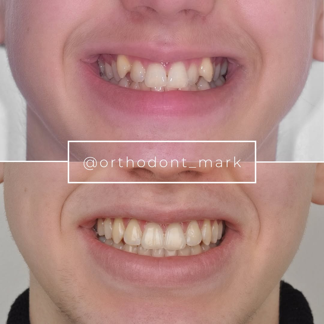
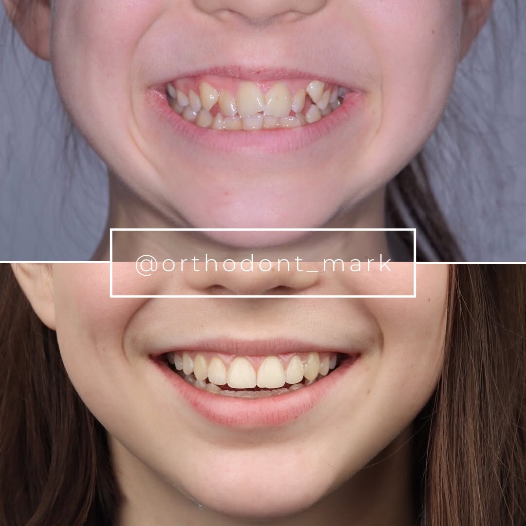
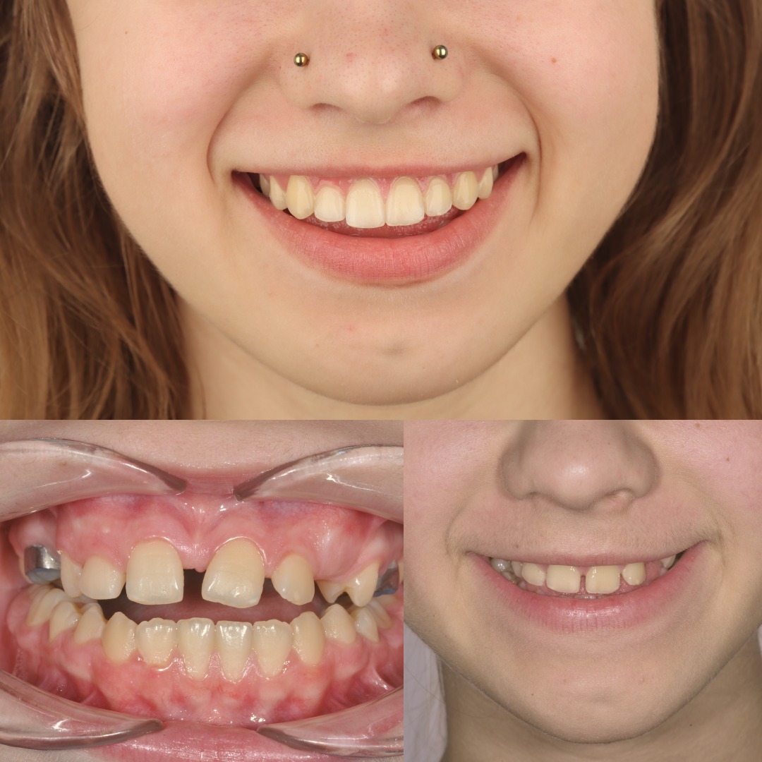
Documented outcomes
Clinical cases from Dr. Mark Radzhabov's practice
Learn the full MARPE protocol from Dr. Mark Radzhabov
Fundamental course covering CBCT patient selection, miniscrew planning, activation protocols, and 60+ clinical cases. Choose the access level that fits your practice.
Essentials of rapid palatal expansion for practicing orthodontists.
- Core RPE concepts and biomechanics
- 6 structured video lessons
- Clinical decision checklists
- Lifetime access to recordings
Deep-dive into MARPE protocol, diagnostics, and clinical execution.
- 6 modules covering MARPE from A to Z
- CBCT case selection walkthroughs
- Miniscrew placement and activation
- 60+ annotated clinical cases
- Direct Q&A with Dr. Mark Radzhabov
5-element medical consultation framework for dentists and orthodontists.
- Trust-building consultation protocol
- 5 lesson modules
- Templates for treatment plan delivery
- Works with any clinical specialty
Clinical FAQ
Should MARPE treatment sequencing always place expansion before bracket bonding?
In most cases, yes. Completing expansion before bonding preserves the direct miniscrew-to-suture force path. Archwires present during activation alter load distribution across anchor teeth and reduce skeletal efficiency, particularly at high force levels documented by Camporesi et al. (2013).
How does the V-shaped expansion pattern affect aligner treatment planning after MARPE?
Stefanm et al. (2022) found a V-shaped expansion pattern in patients aged 12 and older, meaning anterior width increases more than posterior. Digital scans for aligner setup must be taken after consolidation—not during active expansion—to capture the final transverse dimension accurately.
What is the suture separation rate with MARPE compared to RPE when activation is identical?
Chun et al. (2022) reported midpalatal suture separation in 95% of MARPE patients versus 90% of RPE patients, with both groups receiving 35 identical screw turns and CBCT assessment at baseline, post-expansion, and after 3 months of consolidation.
Does MARPE produce less anchor tooth tipping than RPE during the consolidation period?
Yes. Chun et al. (2022) confirmed significantly less buccal tipping of anchor teeth in the MARPE group compared to RPE across the entire expansion and consolidation period, an advantage that directly reduces the torque correction load once fixed appliances are placed.
What consolidation interval should clinicians observe before scanning for aligner fabrication?
Chun et al. (2022) used a 3-month consolidation period with CBCT imaging at that interval. This window allows new bone to form within the suture gap, stabilising arch width. Scanning before this period risks capturing a transient dimension that will narrow after retainer removal.
How does expander force magnitude interact with existing archwires during expansion?
Camporesi et al. (2013) documented forces exceeding 20 kg for Hyrax and A2620 devices. At these levels, an archwire in place transmits part of the expansion force through the dental units rather than the suture, reducing skeletal effect and increasing crown tipping on anchor molars and premolars.
Is parallel versus V-shaped expansion relevant when sequencing expansion with fixed appliances in adolescents?
Critically relevant. Stefanm et al. (2022) showed the pattern becomes V-shaped at age 12 and older. Brackets bonded before expansion in this age group will be positioned on a transverse relationship that will change asymmetrically, creating arch-form discrepancies requiring additional correction.
Does MARPE produce greater nasal width change than RPE, and does sequencing affect this?
Chun et al. (2022) found MARPE produced significantly greater nasal width increase and greater palatine foramen opening than RPE. These skeletal changes are best captured before fixed appliances alter dentoalveolar positions, reinforcing pre-treatment sequencing of the expansion phase.
At what age does the RME expansion pattern shift from parallel to V-shaped according to imaging evidence?
Stefanm et al. (2022) identified the shift at age 12. Children under 10 demonstrated parallel expansion. Those aged 12 and older showed a V-shaped pattern. The transition zone between ages 10 and 12 warrants individual CBCT assessment before finalising an expansion and appliance sequencing plan.
How should clinicians sequence rapid palatal expansion when the patient has already begun aligner therapy?
The aligner series should be paused before MARPE activation to allow accurate arch-form tracking. The expansion phase, including consolidation, should be completed and a new intraoral scan taken before aligner fabrication resumes. Proceeding without pausing risks delivering aligners designed around a pre-expansion transverse dimension.
Correct sequencing of skeletal expansion relative to comprehensive orthodontic treatment is among the highest-leverage decisions in a complex case plan. The evidence reviewed here—covering suture separation rates, V-shaped versus parallel expansion geometry, and appliance force ranges—gives clinicians a concrete checklist before committing to an appliance sequence. Submit a case to Dr. Mark Radzhabov for a structured case review at /consultation, or explore his structured MARPE training at ortodontmark.com to build the imaging and protocol skills needed to sequence these cases with confidence. Key sources: Chun et al., 2022, BMC Oral Health, doi:10.1186/s12903–022-02138-w. Stefanm et al., 2022, doi:10.1007/s00056–022-00429-z. Camporesi et al., 2013, BioMedical Engineering OnLine.