Screw Angulation MARPE
Divergence
Decisions in Hyperdivergent Patients
A clinical framework for aligning miniscrew trajectories with occlusal plane orientation and facial divergence in MARPE treatment planning.
TL;DR Screw angulation for MARPE placement must account for facial divergence and the spatial orientation of the palatal vault relative to the miniscrew trajectory. Hyperdivergent patients present altered palatal inclination that shifts optimal insertion vectors. Evidence confirms asymmetric expansion occurs in nearly half of all MARPE cases, making precise angulation planning non-negotiable for predictable skeletal outcomes.
When a hyperdivergent patient presents with a steep mandibular plane and an anteriorly tipped occlusal plane, the insertion trajectory for MARPE miniscrews cannot follow a generic protocol. Screw angulation for MARPE in divergent skeletal patterns demands case-specific planning because the palatal vault inclination, cortical bone thickness, and available vertical height all shift with facial type. Dr. Mark Radzhabov at ortodontmark.com examines how occlusal plane orientation and facial divergence interact with miniscrew angulation decisions, drawing on the most current peer-reviewed imaging and clinical trial evidence to give orthodontists a concrete, decision-ready framework.
Why Does Screw Angulation for MARPE
Start With Facial Pattern?
Screw angulation for MARPE is the three-dimensional insertion trajectory applied to palatal miniscrews, determined by the clinician's assessment of palatal vault inclination, cortical bone density, and the patient's vertical skeletal pattern before appliance activation. In a normodivergent patient, the palatal plane and the occlusal plane maintain a relatively consistent angular relationship, allowing a near-perpendicular miniscrew insertion into the paramedian palate. In a hyperdivergent patient, the steeper mandibular plane rotates the entire maxillary complex so that the palatal vault becomes more steeply inclined anteriorly — a shift that changes both the available cortical depth at each insertion site and the mechanical advantage of the applied expansion force. The practical consequence is that a trajectory that achieves adequate bicortical engagement in a normodivergent patient may underperform or strip cortical bone in a high-angle case if left unadjusted. CBCT-based pre-treatment planning allows the clinician to trace the palatal slope at the intended insertion sites — typically the paramedian region between the first and second premolars — and calculate the angular correction needed to maintain perpendicularity to the local cortical surface rather than to a generic axial reference. This site-specific approach reduces the mechanical error that accumulates when a standardized angulation is applied across divergent skeletal phenotypes. For a detailed overview of the miniscrew-assisted skeletal expansion protocol and appliance configuration options, the MARPE section at ortodontmark.com provides a structured reference before moving into angulation-specific decision-making. Building that foundational understanding first makes the divergence-related adjustments discussed below significantly easier to apply chairside.
How Angulation Error Drives
Asymmetric Expansion
in High-Angle Patients
Asymmetric expansion is the most clinically visible consequence of mismatched screw trajectories. When the left and right miniscrews engage cortical bone at unequal depths or at diverging angles relative to the midpalatal suture, the resultant force couple is asymmetric and one palatal half translates more than the other. Yoon et al. (2022) found that asymmetric expansion greater than 1 mm occurred in 47.8% of MARPE patients in a retrospective analysis of 256 patients with a mean age of 18.8 ± 8.6 years comprising 126 females and 130 males. The frequency underlines that angulation mismatch is not a rare edge case — it is the modal outcome when trajectory planning is insufficiently individualized. In hyperdivergent patients, the anterior tipping of the palatal vault creates a structural asymmetry risk even when the screws appear clinically parallel, because the cortical bone on the steeper anterior slope is thinner and more prone to differential engagement. A CBCT-guided angulation protocol in which the insertion angle is measured against the local cortical surface at each of the four intended sites — rather than against a single horizontal reference — distributes anchorage force more evenly and keeps the midpalatal suture opening vector closer to the sagittal plane. Pain during and after MARPE expansion was reported by 45% of patients in the same cohort reported by Yoon et al. (2022), and gingival inflammation around the appliance was the most common complication, occurring in 83.9% of patients. Both outcomes are worsened by suboptimal screw angulation: a poorly angled screw generates eccentric loading that inflames the surrounding mucosa and concentrates stress at the cortical interface, amplifying patient discomfort throughout the activation period.
Does Suture Maturation Change
Angulation Strategy
in Adolescent vs Adult Cases?
Angulation decisions cannot be separated from suture maturation status because the mechanical response of the midpalatal suture to the applied force vector changes dramatically as ossification progresses. Govaerts et al. (2023) found that midpalatal suture closure at stages D and E was present in 61% of 15-year-old females, and that pterygomaxillary suture closure reached 83–100% and transpalatal suture closure reached 78–85% from age 15 in females aged 13–17 years. A patient whose suture is already in advanced ossification will resist opening regardless of how precisely the miniscrew trajectory is optimized, shifting the clinical question from angulation refinement toward surgical referral. For the subset of adolescent patients where suture patency is confirmed by CBCT — identifiable as a continuous radiolucent midline gap without bridging trabecular bone — the angulation strategy can be more aggressive in targeting the posterior palatal vault, where cortical bone is denser and better positioned to resist the moment arms generated by a steep mandibular plane. In high-angle patients with confirmed open sutures, posteriorly positioned miniscrews at a slightly more apically directed insertion angle allow the expansion force to engage a thicker cortical layer while keeping the activation vector aligned with the suture plane. Govaerts et al. (2023) also documented that orthodontists recommended SARPE at a mean age of 15.1 years while maxillofacial surgeons recommended it at 14.8 years for females with transverse maxillary deficiency — a divergence in threshold that reflects how differently clinicians weight suture imaging findings versus age-based criteria when deciding whether miniscrew anchorage alone is sufficient. The practical implication: angulation optimization is only worth pursuing once imaging has confirmed that the suture remains capable of mechanical separation.
How Does Facial Divergence Affect
Dental Side Effects
Across Expansion Appliance Designs?
The choice between bone-borne MARPE and conventional tooth-borne rapid palatal expansion carries distinct consequences for high-angle patients because facial divergence amplifies the dental tipping that occurs when expansion forces are transmitted through anchor teeth. Chun et al. (2022) demonstrated that MARPE produced significantly less buccal displacement of anchor teeth than conventional RPE across both the expansion and consolidation periods, with both groups receiving identical expansion protocols of 35 activation turns and CBCT imaging at baseline, immediately after expansion, and at a 3-month consolidation point. In a hyperdivergent patient where posterior vertical dimension is already a management priority, additional buccal tipping of the upper molars and premolars worsens the arch-to-arch vertical discrepancy and can deepen the curve of Spee on the lower arch. The practical implication for angulation planning is that the miniscrew trajectory in a high-angle case should be optimized to maximize direct skeletal force transmission rather than relying on the tooth-band assembly to redirect force toward the suture. When the screws are angled too palatally or too buccally relative to the midpalatal suture plane, a greater share of the activation force is absorbed by the band-and-screw interface rather than transferred to the basal bone, effectively converting a bone-borne appliance into a hybrid with unintended dental side effects. For a comparison of rapid palatal expander biomechanics and the force paths that distinguish tooth-borne from bone-borne designs, the rapid palatal expander biomechanics section at ortodontmark.com provides a systematic breakdown that contextualizes why angulation precision matters more in bone-borne systems. Suture separation occurred in 95% of MARPE patients versus 90% of RPE patients as reported by Chun et al. (2022), confirming that the bone-borne approach achieves a higher rate of true skeletal opening — but only when the screws are positioned and angled to deliver force directly to the suture.
A CBCT-Driven Angulation Workflow
for Divergent
Skeletal Patterns
A reproducible angulation workflow for hyperdivergent MARPE patients begins with a standardized CBCT reconstruction oriented to the palatal plane rather than the Frankfort horizontal, because the Frankfort reference exaggerates the apparent palatal slope in high-angle cases and inflates the calculated insertion angle. Once the reconstruction is reoriented, the clinician identifies the four intended miniscrew sites — typically paramedian, straddling the midpalatal suture at the premolar region — and draws a cortical surface tangent at each point using cross-sectional slices in both the coronal and sagittal planes. The target insertion angle for each screw is then set perpendicular to its local tangent rather than to any global palatal plane, which typically results in anterior screws being directed more apically and posterior screws being directed more coronally in a hyperdivergent patient. Hounsfield unit measurement at each site informs screw length selection: sites with lower bone density require longer insertion depth to achieve equivalent primary stability, and this depth decision interacts with angulation because a longer screw at a steeper angle can perforate the nasal floor in patients with reduced vertical palatal height — a risk that is heightened in high-angle cases due to the narrow vault geometry that often accompanies vertical maxillary excess. Marking the intended depth on the miniscrew before insertion and confirming with an intraoperative periapical radiograph or CBCT scout prevents floor perforation without requiring the clinician to abandon an otherwise biomechanically sound trajectory. Activation protocol monitoring should include bilateral caliper measurements at each appointment to detect asymmetric expansion early. Given that asymmetric expansion greater than 1 mm occurred in 47.8% of patients as Yoon et al. (2022) reported, early detection at the first two to three activation visits allows differential activation — turning the appliance additional quarter-turns on the lagging side — before asymmetry becomes structurally fixed in consolidating bone.
Free Clinical Guide
Which Expansion Appliance at Which Age?
Get Dr. Mark Radzhabov's guide to skeletal-expansion appliances — indications, pros & cons, and how to choose by patient age. Free to your inbox.
Prefer video? Watch the free demo lesson on MARPE appliance types, features and trade-offs.
Documented outcomes
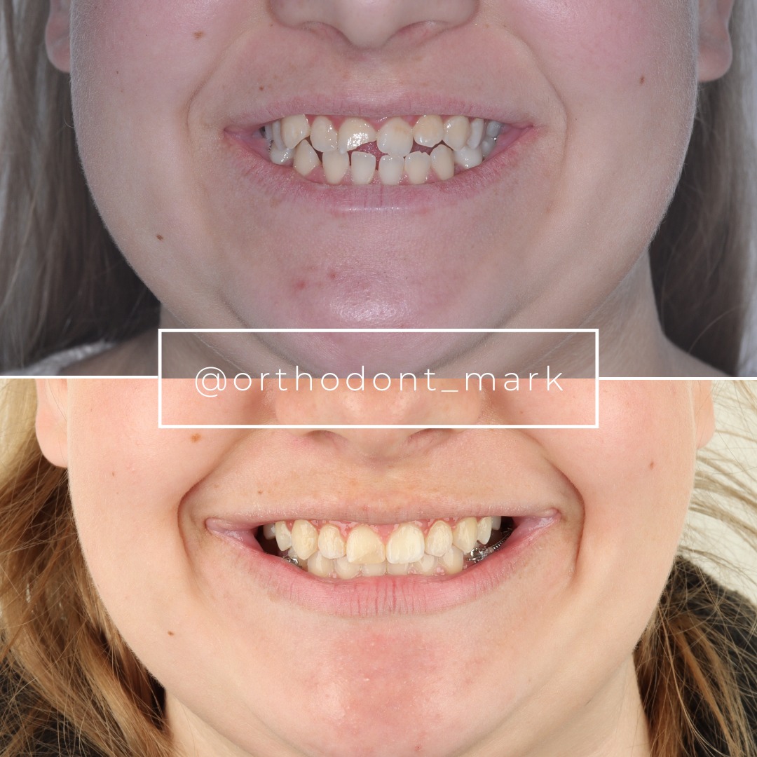
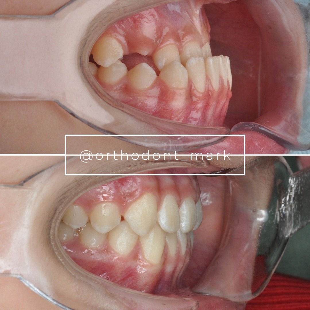
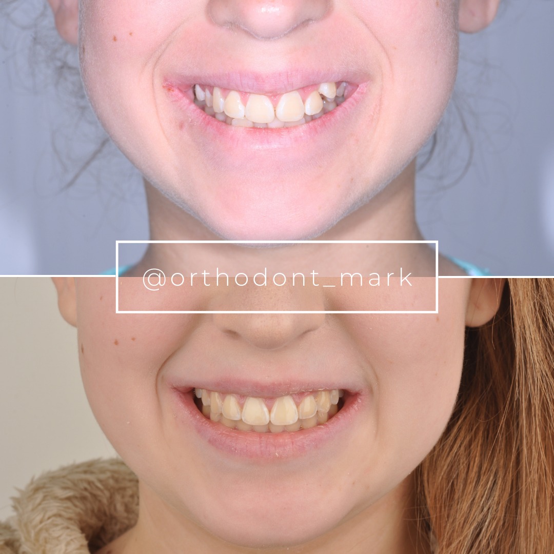
Clinical cases from Dr. Mark Radzhabov's practice
Learn the full MARPE protocol from Dr. Mark Radzhabov
Fundamental course covering CBCT patient selection, miniscrew planning, activation protocols, and 60+ clinical cases. Choose the access level that fits your practice.
Essentials of rapid palatal expansion for practicing orthodontists.
- Core RPE concepts and biomechanics
- 6 structured video lessons
- Clinical decision checklists
- Lifetime access to recordings
Deep-dive into MARPE protocol, diagnostics, and clinical execution.
- 6 modules covering MARPE from A to Z
- CBCT case selection walkthroughs
- Miniscrew placement and activation
- 60+ annotated clinical cases
- Direct Q&A with Dr. Mark Radzhabov
5-element medical consultation framework for dentists and orthodontists.
- Trust-building consultation protocol
- 5 lesson modules
- Templates for treatment plan delivery
- Works with any clinical specialty
Clinical FAQ
How does facial divergence affect optimal screw angulation in MARPE placement?
A hyperdivergent skeletal pattern tips the palatal vault anteriorly, changing the cortical surface angle at each insertion site. Miniscrew trajectory must be calculated relative to the local cortical tangent at each of the four sites rather than a global palatal plane reference to maintain effective anchorage and symmetric force distribution.
What CBCT measurements should guide miniscrew angulation decisions for high-angle MARPE patients?
Reorient the CBCT to the palatal plane, draw cortical surface tangents at each intended insertion site in both coronal and sagittal planes, and measure Hounsfield units for bone density. These three measurements together determine insertion angle, screw length, and safe depth to avoid nasal floor perforation.
How common is asymmetric expansion in MARPE and what is the angulation-related cause?
Yoon et al. (2022) reported asymmetric expansion greater than 1 mm in 47.8% of MARPE patients. Unequal cortical engagement caused by divergent insertion angles is a primary driver. Left and right screws at mismatched trajectories create an asymmetric force couple that translates one palatal half more than the other.
Does suture maturation stage change the angulation strategy for adolescent MARPE cases?
Yes. Govaerts et al. (2023) found midpalatal suture closure in 61% of 15-year-old females. In patients with advanced ossification, even a perfectly angulated screw cannot generate reliable suture separation. Confirming suture patency on CBCT is a prerequisite before finalizing any insertion trajectory.
Why does MARPE produce less buccal dental displacement than RPE, and how does angulation preserve that advantage?
Chun et al. (2022) confirmed that MARPE produced significantly less buccal displacement of anchor teeth than conventional RPE. When screws are misaligned with the midpalatal suture plane, activation force is partially absorbed by the band-screw interface, recreating tooth-borne loading and negating the bone-borne advantage.
What are the most common MARPE complications and how does screw angulation influence their incidence?
Yoon et al. (2022) identified gingival inflammation in 83.9% of patients and pain in 45% as the top complications. Both are worsened by poorly angled screws that generate eccentric perimucosal loading and cortical stress concentration. Individualized trajectory planning reduces the mechanical stimulus driving these responses.
How should the occlusal plane relationship inform miniscrew insertion trajectory in MARPE planning?
The occlusal plane's angular relationship to the palatal plane shifts with facial divergence. In high-angle patients, using the occlusal plane as an indirect reference for insertion trajectory without correcting for the steeper palatal inclination systematically underestimates the apical angulation needed at anterior insertion sites.
What activation monitoring protocol detects asymmetric expansion early enough to allow correction?
Bilateral caliper measurements at each activation visit allow detection of side-to-side discrepancies before they ossify. When asymmetry exceeds 1 mm — the threshold Yoon et al. (2022) used to define clinically significant asymmetry — differential activation with additional quarter-turns on the lagging side can partially compensate.
At what suture maturation stage does MARPE remain viable without surgical assistance in adolescent females?
Govaerts et al. (2023) found pterygomaxillary suture closure reaching 83–100% and transpalatal suture closure reaching 78–85% from age 15 in females aged 13–17. MARPE without surgical assistance is most viable when CBCT confirms a radiolucent midpalatal gap without trabecular bridging, regardless of chronological age.
How does palatal vault geometry in vertical maxillary excess affect nasal floor clearance during MARPE screw insertion?
Vertical maxillary excess is often accompanied by a narrow, highly arched palatal vault with reduced vertical bone height at the insertion sites. Longer screws needed for adequate density at low-HU sites, combined with a more apical insertion angle in high-angle cases, narrow the safety margin before nasal floor perforation — requiring precise pre-surgical depth marking.
Occlusal plane orientation and facial divergence are not peripheral variables in MARPE planning — they directly govern where cortical anchorage is available and how asymmetric the expansion vector becomes. Matching screw angulation to the patient's skeletal pattern, suture maturation stage, and palatal vault geometry converts a procedurally complex appliance into a predictable tool. Dr. Mark Radzhabov encourages colleagues to audit their current MARPE protocols against the angulation principles outlined here and to explore structured MARPE training at ortodontmark.com to refine case selection and insertion technique before tackling hyperdivergent presentations. Key sources: Chun et al. (2022), BMC Oral Health, doi:https://doi.org/10.1186/s12903–022-02138-w. Govaerts et al. (2023), Journal of Orofacial Orthopedics, doi:https://doi.org/10.1007/s00056–023-00487-x. Yoon et al. (2022), retrospective analysis.