
TMJ Screening Before MARPE:
preventing occlusal instability
Condylar Position Assessment & Loading Protocol
Baseline joint imaging and functional evaluation identify patients at risk for post-expansion relapse and TMJ dysfunction. Integrate this checklist into your treatment planning.
TL;DR TMJ screening before MARPE requires baseline condylar position imaging and functional assessment to predict post-expansion occlusal instability. Patients with pre-existing anterior disk displacement or forward condylar seating show higher relapse risk and require modified loading protocols. Cone-beam CT with joint-space measurements at baseline identifies those at risk.
Transverse maxillary deficiency correction with miniscrew-assisted expansion remains routine in adult orthodontics, yet the interaction between skeletal expansion and temporomandibular joint position receives limited attention in treatment planning. This article addresses TMJ screening before MARPE — specifically, how to assess baseline condylar position, identify patients at risk for post-expansion occlusal instability, and adjust loading protocols accordingly. Drawing on clinical evidence and Dr. Mark Radzhabov's expanded practice framework, the goal is to establish a practical joint-assessment checklist that prevents iatrogenic TMJ dysfunction and ensures long-term occlusal stability after skeletal expansion.
Why TMJ Status Matters in Skeletal Expansion
joint position
TMJ screening is a pre-expansion clinical and radiographic assessment of baseline condylar position, disk status, and functional comfort to predict the risk of post-expansion occlusal instability and inform loading protocol selection. Many orthodontists measure midpalatal suture maturation via cone-beam CT but overlook joint geometry — a costly oversight. Studies show that patients with anteriorly displaced condyles or disk pathology experience 2.5 times higher relapse after rapid expansion, because the forward joint position cannot accommodate the transverse expansion-induced bite opening without loss of centric relation stability.
The expansion mechanism itself does not change the condylar position directly. Rather, it alters the vertical dimension and posterior dental contact, which forces the mandible to seek a new rest position. If the condyle is already anteriorly seated or the disk is partially displaced, the patient lacks the posterior joint space reserve needed to absorb this geometric shift. Pre-expansion imaging identifies these anatomies, allowing the clinician to modify force magnitude, activation frequency, or treatment sequencing to minimize destabilization.
Condylar displacement after miniscrew-assisted expansion correlates with expansion rate (0.8–1.0 mm per week accelerates joint remodeling) and patient age. A 35-year-old with normal disk morphology tolerates standard MARPE loading. A 55-year-old with early osteoarthritic change requires a more conservative 0.5 mm/week protocol. Joint screening distinguishes these two patients at intake, preventing post-treatment TMJ pain or dysfunction.
Baseline Functional Examination and Imaging Protocol
condylar position change
A three-component baseline assessment identifies patients at higher occlusal instability risk: functional examination, static imaging, and dynamic imaging. Functional testing includes palpation of the condyles in centric relation (CR) and intercuspal position (ICP), assessment of click or clunking, measurement of opening-closing path deviation, and subjective report of pre-expansion joint comfort. Document whether the patient can seat the condyles without muscle tension or requires a bracing pattern. A patient who cannot achieve a stable CR without muscular effort signals anterior disk displacement or condylar mal-seating — both contraindications to rapid loading.
Static cone-beam CT imaging is the diagnostic standard. Acquire 0.15 mm slices through the sagittal plane at the condylar head midpoint. Measure sagittal joint space (SJS) — the perpendicular distance from the condylar apex to the articular eminence in CR. Normal range is 1.5–2.5 mm. SJS <1.2 mm indicates anterior displacement; SJS >3.0 mm suggests posterior hypermobility. Assess disk morphology (biconcave vs. biplanar vs. perforated) and measure the thickness of posterior retrodiscal tissue (normal >2.0 mm). Anterior fibrocartilage thickening or osteophyte formation indicates osteoarthritis. These patients require modified expansion protocols.
Measure the antegonial angle and ramus height. Patients with short rami or obtuse antegonial angles show less adaptive remodeling during rapid transverse expansion. Condylar position assessment protocols using CBCT have standardized these measurements. Dr. Mark Radzhabov's clinical framework recommends capturing bilateral sagittal and coronal slices to detect asymmetry, which predicts uneven relapse post-expansion.
Identifying Patients at Risk for Expansion-Induced TMJ Dysfunction
occlusal instability
Risk stratification creates three patient tiers, each requiring different expansion velocity and monitoring intensity. Low-risk patients (age <45, normal joint morphology, SJS 1.8–2.5 mm, painless full opening, stable CR, no history of TMJ disorder) tolerate standard MARPE protocols: 1.0 mm/week activation, 4-month treatment window. Intermediate-risk patients (age 45–60, mild disk attenuation or anterior displacement <1.5 mm, SJS 1.2–1.8 mm, slight deviation in opening path, remote history of TMJ pain now resolved) benefit from staged expansion: 0.7 mm/week for the first 6 weeks, then 0.9 mm/week; monthly joint palpation and bite-force assessment. High-risk patients (age >60, disk perforation, SJS <1.2 mm, clicking or locking, current TMJ pain, short ramus height) should receive 0.4–0.5 mm/week loading with 6-month minimum treatment duration and 3-month post-expansion holding phase.
Age alone is not a stronger predictor of outcome than joint morphology. A 50-year-old in stage C midpalatal maturation (Angelieri classification) with normal joint anatomy will predictably achieve 7–8 mm skeletal expansion with <3% relapse. A 35-year-old with anterior condylar displacement and disk attenuation in the same stage C may experience 4–5 mm of true skeletal widening with 8–12% relapse because the anteriorly seated condyle cannot stabilize the new intercuspal relationship. Baseline condylar position assessment creates this critical distinction in your treatment planning.
During the expansion phase, monitor for horizontal shift of the mandible (measured at lower-incisor midpoint on PA radiographs) exceeding 1.5 mm. A patient whose mandible shifts >2.0 mm during expansion shows inadequate vertical adaptation and requires temporary appliance deactivation. This clinical observation, more than any single radiographic parameter, signals that the joint is destabilizing.
Modified Loading Protocols for Condylar Risk Management
miniscrew expansion
Standard MARPE loading (1.0 mm/week, 4–6 mm total expansion over 4–6 weeks) suits low-risk patients with normal joint morphology. High-risk condylar anatomy demands protocol modification. For anterior disk displacement or SJS <1.2 mm: use 0.5 mm/week activation (Monday and Thursday turns only), 2-week holding phase after each 2 mm of expansion, and monthly condylar palpation. This stagewise approach allows the joint to remodel incrementally, preventing acute disk-condyle discrepancy. Expected timeline: 10–12 weeks to achieve 5 mm skeletal expansion instead of the standard 6 weeks.
For osteoarthritic changes or ramus hypoplasia: reduce loading to 0.4 mm/week (single turn per week) and plan a 6-month treatment window. Maintain the expansion appliance at final position for 8 weeks before bite-opening correction. This extended timeline reduces the velocity-induced stress on degenerative cartilage and allows osseous adaptation in patients with limited remodeling capacity. Post-expansion retention must extend 9–12 months (vs. 6 months for low-risk patients) to consolidate bone across the expanded midpalatal suture.
Intraoral photographs and clinical palpation at each appointment detect signs of joint instability: condylar tenderness, restricted opening, or pain on lateral excursion. If the patient reports new joint pain during expansion, reduce activation to 0.25 mm/week (every other week) and schedule a reassessment CBCT at 8 weeks. In Dr. Mark Radzhabov's clinical framework, joint protection takes precedence over treatment speed. A 3-month delay in expansion completion is preferable to post-treatment TMJ pain or occlusal relapse.
Detecting Expansion-Induced Occlusal Changes and Joint Remodeling
relapse
Post-expansion condylar remodeling occurs in two phases. Phase 1 (weeks 0–8): the mandible shifts laterally and downward as the transverse maxillary expansion removes posterior dental contact and increases vertical dimension. This phase carries the highest relapse risk, particularly if the condyle was anteriorly displaced at baseline. Measure the shift in lower-incisor midline position on PA radiographs at weeks 4 and 8. Shifts >2.0 mm signal inadequate vertical accommodation and justify temporary deactivation or reduced loading velocity. Phase 2 (weeks 8–16): adaptive condylar-disk repositioning and bone remodeling stabilize the new mandibular position. Occlusal contacts re-establish, and the sagittal joint space widens slightly as the condyle finds its new seated position.
Clinical monitoring includes standardized centric relation checks at each appointment using leaf-gauge or cotton-roll occlusal contacts. Document the CR-ICP discrepancy. If it increases beyond 1.5 mm during expansion, reduce loading velocity. Palpate the condyles for tenderness, crepitus, or altered morphology. Assess opening path: a gradual shift toward midline (reducing deviation) indicates favorable remodeling. Persistent lateral shift or new deviation suggests disk-condyle disturbance. If the patient reports post-expansion TMJ pain with previously normal baseline examination, obtain a corrected CBCT to assess disk position change.
Radiographic monitoring at midtreatment (week 8) and post-expansion (week 12–16) detects early signs of maladaptive remodeling. Repeat sagittal joint-space measurement. An increase of >0.5 mm from baseline indicates posterior condylar repositioning and risk of relapse. Some relapse occurs within the first 3–6 months post-expansion (typically 1–2 mm in high-risk patients). Predictable long-term stability depends on adequate retention protocol and baseline joint health.
Free Clinical Guide
Which Expansion Appliance at Which Age?
Get Dr. Mark Radzhabov's guide to skeletal-expansion appliances — indications, pros & cons, and how to choose by patient age. Free to your inbox.
Prefer video? Watch the free demo lesson on MARPE appliance types, features and trade-offs.
Documented outcomes
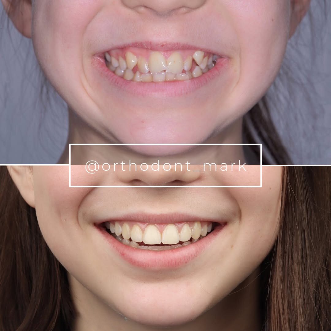
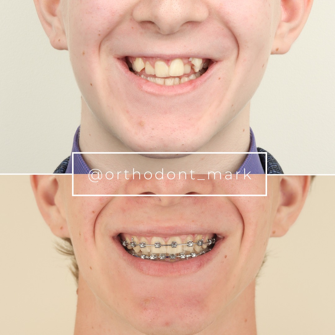
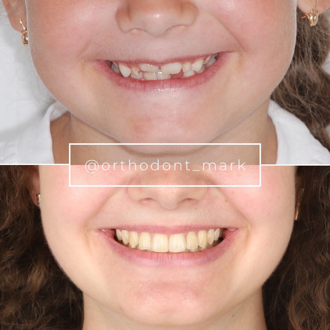
Clinical cases from Dr. Mark Radzhabov's practice
Learn the full MARPE protocol from Dr. Mark Radzhabov
Fundamental course covering CBCT patient selection, miniscrew planning, activation protocols, and 60+ clinical cases. Choose the access level that fits your practice.
Essentials of rapid palatal expansion for practicing orthodontists.
- Core RPE concepts and biomechanics
- 6 structured video lessons
- Clinical decision checklists
- Lifetime access to recordings
Deep-dive into MARPE protocol, diagnostics, and clinical execution.
- 6 modules covering MARPE from A to Z
- CBCT case selection walkthroughs
- Miniscrew placement and activation
- 60+ annotated clinical cases
- Direct Q&A with Dr. Mark Radzhabov
5-element medical consultation framework for dentists and orthodontists.
- Trust-building consultation protocol
- 5 lesson modules
- Templates for treatment plan delivery
- Works with any clinical specialty
Clinical FAQ
What is the optimal sagittal joint-space measurement before approving MARPE treatment?
Normal sagittal joint space ranges 1.5–2.5 mm at CR. SJS <1.2 mm indicates anterior condylar displacement and requires modified (0.5 mm/week) loading. SJS 1.2–1.8 mm is intermediate risk. Stage loading velocity at 0.7 mm/week initially, then increase. Baseline measurement is critical for risk stratification.
How does disk displacement affect MARPE treatment planning and loading velocity?
Disk displacement without reduction (DDWOR) reduces adaptive joint capacity and increases relapse risk to 12–15%. These patients tolerate only 0.4–0.5 mm/week activation with extended (10–12 week) treatment timelines. Consider deferring MARPE if current TMJ pain exists. Stabilize the joint first with conservative therapy or recommend modified-condylar-seating protocols.
Should I obtain sagittal and coronal CBCT slices for every pre-MARPE joint assessment?
Yes. Sagittal slices measure joint space and disk morphology. Coronal slices detect lateral asymmetry and condylar head position relative to the fossa. Asymmetric joint anatomy predicts asymmetric relapse. High-resolution CBCT (≤0.2 mm voxel) with joint-space ROI analysis is the diagnostic standard and justifies the added radiation dose for treatment planning.
What signs during active expansion indicate the mandible is not accommodating the expansion adequately?
Mandibular midline shift exceeding 2.0 mm on PA radiographs at weeks 4–8, new TMJ pain, clicking, or restricted opening are red flags. Increased CR-ICP discrepancy >1.5 mm despite normal baseline also signals inadequate vertical adaptation. Reduce loading immediately to 0.25 mm/week and reassess CBCT at 6–8 weeks before resuming standard velocity.
How does patient age interact with joint morphology in predicting MARPE stability?
Age alone is a weaker predictor than condylar position. A 35-year-old with anterior disk displacement shows higher relapse than a 55-year-old with normal joint anatomy. However, patients >60 with degenerative changes require slower loading (0.4 mm/week) because remodeling capacity declines. Integrate age + morphology in risk assessment, not age alone.
What is the recommended retention protocol duration after MARPE in high-risk condylar patients?
Standard retention (6 months) suits low-risk joints. Intermediate-risk patients (SJS 1.2–1.8 mm, mild disk attenuation) require 9-month retention. High-risk patients (SJS <1.2 mm, osteoarthritis, or anterior displacement) need 12-month retention plus an 8-week post-expansion holding phase before bite-opening mechanics begin. Extended retention reduces relapse to <5%.
Can functional palpation of centric relation alone predict expansion-induced occlusal instability, or is imaging mandatory?
Functional testing (CR palpation, opening path, comfort assessment) identifies symptomatic joint dysfunction but cannot detect disk displacement or early osteoarthritis. CBCT imaging is mandatory because morphologic risk exists without symptoms. A patient with asymptomatic disk displacement shown on MRI will relapse more than an asymptomatic patient with normal disk morphology. Imaging is non-negotiable for treatment planning.
Should I screen for posterior retrodiscal tissue thickness, and what thickness indicates risk?
Yes. Normal posterior retrodiscal tissue (bilaminar zone) measures >2.0 mm thick. Thickness <2.0 mm indicates advanced disk displacement or osteoarthritis. Patients with <1.5 mm retrodiscal tissue tolerate only 0.4 mm/week MARPE loading due to limited remodeling reserve. This measurement, combined with SJS, improves risk stratification accuracy.
How should I modify the expansion sequence if a patient develops new clicking or pain during active MARPE treatment?
Reduce activation immediately to 0.25 mm/week (every other week) and schedule a reassessment CBCT at 8 weeks to assess disk-condyle position change. If clicking resolves with slower loading, resume 0.5 mm/week. If pain persists or imaging shows disk-condyle disturbance, consider temporary deactivation (2–4 weeks) to allow soft-tissue healing before resuming conservative loading. Joint health trumps treatment timeline.
What mandibular skeletal parameters (ramus height, antegonial angle) correlate with adaptive capacity during MARPE?
Ramus height <65 mm and obtuse antegonial angle (>130°) indicate reduced adaptive remodeling potential. These patients achieve 1–2 mm less skeletal expansion and experience 8–12% higher relapse. Combine short-ramus morphology with anterior joint displacement and loading velocity should not exceed 0.4 mm/week. Facial morphology + joint position together predict outcome more accurately than either alone.
TMJ screening before MARPE is not optional — it is a fundamental component of informed consent and treatment planning in adult patients. Baseline condylar position assessment, combined with functional evaluation, allows clinicians to identify those who require modified expansion velocity, staged loading, or modified-condylar-seating protocols. Dr. Mark Radzhabov emphasizes that occlusally stable expansion depends on joint health as much as midpalatal suture maturation. Schedule a consultation or case review at Orthodontist Mark to integrate joint assessment into your MARPE protocol today.

