
RPE Retention Duration:
Ossification
Evidence Translated Into Timelines
Histology and CBCT findings mapped to defensible retention protocols for growing patients.
TL;DR RPE retention duration in growing patients should be guided by histological and CBCT ossification evidence, not arbitrary clinical convention. Caprioglio et al. (2017) document trabecular bone formation within 7 days of expansion, while Ballantia et al. (2009) establish that at least 5 months of retention is necessary to allow adequate mineralization and minimize relapse after RME.
A clinician who releases a growing patient from retention too early risks watching hard-won expansion collapse before the midpalatal suture has consolidated. Translating RPE retention duration evidence into reliable chairside timelines requires understanding what the suture is actually doing at the histological and radiographic levels during those early post-active months. Dr. Mark Radzhabov at ortodontmark.com has compiled the most clinically relevant imaging and histology findings to give orthodontists a defensible, evidence-grounded protocol for deciding when retention can safely end in growing patients.
Why Does RPE Retention Duration Demand a
Biological
Rationale Rather Than a Calendar Rule?
RPE retention duration is the post-active period during which a palatal expander or dedicated retainer maintains the expanded arch width while new bone mineralizes across the opened midpalatal suture. Setting that duration arbitrarily — six weeks because it feels standard, or until the next appointment slot — ignores the incremental tissue events that determine whether expansion holds or collapses. The fundamental challenge is that skeletal expansion creates a fibrous gap filled initially with immature connective tissue, not bone. Until that gap mineralizes sufficiently, any reduction in transverse restraint exposes the patient to relapse driven by the elastic recoil of the surrounding periosteal and mucosal tissues. A retention protocol calibrated to ossification milestones addresses the actual mechanism of failure rather than guessing at a safe interval. Age further modulates the rate and pattern of suture response. Kinzinger et al. (2022) identified that RME response is age-dependent: younger patients tend to express width and height increases posteriorly, whereas older growing patients show a more anteriorly concentrated response. This differential means that the regional distribution of newly formed bone — and therefore the time needed to stabilize it — is not uniform across the mixed-dentition and early-permanent-dentition age groups. Retention planning that ignores this gradient will systematically under-retain older growing patients whose anterior suture activity is highest. For a detailed review of appliance mechanics relevant to these decisions, see our resource on rapid palatal expander mechanics and appliance selection.
What Do Histological Samples Reveal About Early
Suture Healing
After Rapid Palatal Expansion?
The most granular evidence for suture ossification timelines comes from direct histological sampling. Caprioglio et al. (2017) obtained histological samples of the midpalatal suture at 7 days and 30 days after RME in growing patients, providing a rare window into the tissue-level sequence of repair. At 7 days post-expansion, Caprioglio et al. (2017) documented mature bone with small marrow spaces and trabecular bone formation already present within the suture gap. This finding is clinically significant: it confirms that the osteogenic response is rapid and that the suture is biologically active far earlier than many practitioners assume. However, the presence of early trabeculae does not indicate structural consolidation — the tissue at this stage remains mechanically vulnerable. By 30 days, Caprioglio et al. (2017) reported increased newly-formed bone trabeculae with perpendicular orientation to the suture axis, a geometric arrangement associated with greater resistance to transverse loading. Critically, the fibrous component of the bone tissue in the suture decreases between day 7 and day 30, marking a shift from a mixed fibro-osseous matrix toward a more mineralized architecture. This trajectory confirms that active remodeling continues throughout at least the first month, reinforcing why retainer removal well before suture consolidation is a predictable source of relapse.
How Does CBCT Mineralization Data Establish a
Minimum Retention
Threshold for Growing Patients?
Histological findings describe suture biology. CBCT data translate that biology into a clinically actionable timeframe. Ballantia et al. (2009) evaluated 17 growing patients with a mean age of 11.2 years who underwent RME, with CT scans acquired at baseline, end of active phase, and after a 6-month retention period. The expansion protocol achieved 7 mm of transverse opening. From this CBCT evidence, Ballantia et al. (2009) concluded that at least 5 months of retention is necessary to allow adequate mineralization of the midpalatal suture and minimize relapse after RME. That figure is not a comfortable buffer above the minimum — it represents the evidence-derived floor below which mineralization is incomplete and relapse risk is elevated. Protocols that terminate retention at two or three months deny the suture the consolidation period the imaging data demand. The 6-month scanning interval used by Ballantia et al. (2009) also informs how clinicians should sequence radiographic assessment in retention. A CBCT acquired before the 5-month threshold may show incomplete sutural fill, providing a direct imaging criterion to extend retention rather than relying on a fixed endpoint. This approach converts subjective clinical judgment into an objective mineralization check. Orthodontists building or refining their retention protocols around this evidence will find structured RPE training for evidence-based retention protocols a useful resource for applying these thresholds in practice.
How Should Age-Dependent Suture Response Shape
Retention Decisions
Across the Mixed and Early Permanent Dentition?
The Alt-RAMEC protocol adds a further variable worth integrating into retention planning. avu (2019) describes Alt-RAMEC as a 9-week protocol involving initial 7 mm expansion at 1 mm per day, followed by alternating weekly expansion and constriction cycles. The repeated mechanical cycling mobilizes the suture more thoroughly than conventional RME, and that greater initial disruption likely demands at minimum the same 5-month retention floor documented for standard RME — if not a longer consolidation period, given the more extensive fibrous mobilization. Kinzinger et al. (2022) remind clinicians that the regional distribution of the skeletal response shifts with age: younger patients consolidate posteriorly, older growing patients anteriorly. A patient at the upper boundary of the growing category may therefore require particular vigilance over the anterior suture region, where perpendicular trabecular organization — as documented by Caprioglio et al. (2017) at 30 days — will be most critical to the long-term stability of the expanded arch. Practical retention workflows should incorporate three elements drawn from the combined evidence: a minimum retention duration grounded in the 5-month threshold of Ballantia et al. (2009). A CBCT check before appliance removal to confirm sutural fill rather than assuming it. And age-adjusted expectations regarding which suture region is most vulnerable. For complex cases involving MARPE and skeletal expansion case planning, the same ossification principles apply, though the force delivery pathway differs fundamentally from tooth-borne RPE.
Free Clinical Guide
Which Expansion Appliance at Which Age?
Get Dr. Mark Radzhabov's guide to skeletal-expansion appliances — indications, pros & cons, and how to choose by patient age. Free to your inbox.
Prefer video? Watch the free demo lesson on MARPE appliance types, features and trade-offs.
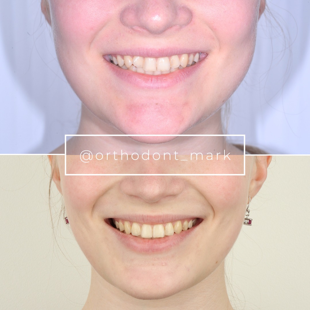
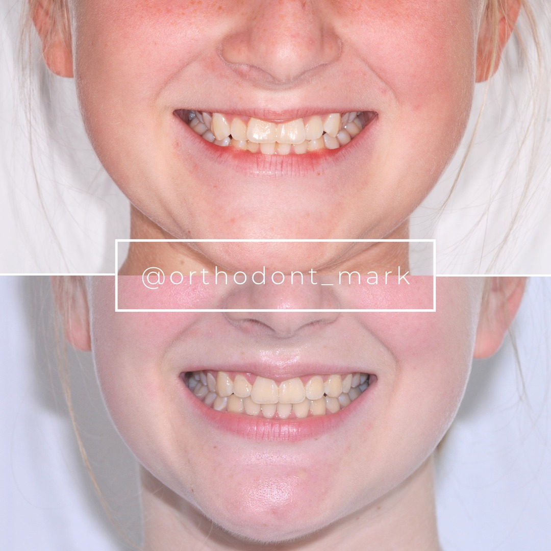
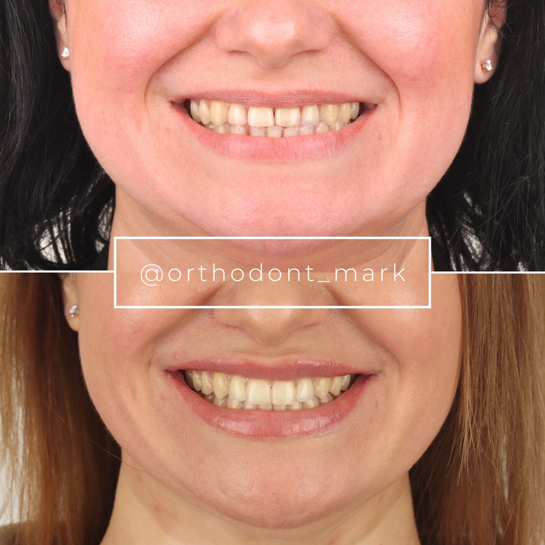
Documented outcomes
Clinical cases from Dr. Mark Radzhabov's practice
Learn the full MARPE protocol from Dr. Mark Radzhabov
Fundamental course covering CBCT patient selection, miniscrew planning, activation protocols, and 60+ clinical cases. Choose the access level that fits your practice.
Essentials of rapid palatal expansion for practicing orthodontists.
- Core RPE concepts and biomechanics
- 6 structured video lessons
- Clinical decision checklists
- Lifetime access to recordings
Deep-dive into MARPE protocol, diagnostics, and clinical execution.
- 6 modules covering MARPE from A to Z
- CBCT case selection walkthroughs
- Miniscrew placement and activation
- 60+ annotated clinical cases
- Direct Q&A with Dr. Mark Radzhabov
5-element medical consultation framework for dentists and orthodontists.
- Trust-building consultation protocol
- 5 lesson modules
- Templates for treatment plan delivery
- Works with any clinical specialty
Clinical FAQ
What is the evidence-based minimum RPE retention duration in growing patients?
Ballantia et al. (2009) established that at least 5 months of retention is necessary to allow adequate midpalatal suture mineralization and minimize relapse after RME in growing patients. This figure derives from CBCT assessment, not clinical convention, and represents a floor rather than a target.
At what timepoint does histology confirm trabecular bone formation after rapid palatal expansion?
Caprioglio et al. (2017) found mature bone with small marrow spaces and trabecular bone formation already present at 7 days post-RME in growing patients. By 30 days, trabeculae displayed perpendicular orientation to the suture axis and the fibrous component had decreased, indicating advancing consolidation.
How does CBCT suture mineralization guide the decision to remove a palatal retainer?
Using CBCT at baseline, post-active phase, and after a retention interval — as in Ballantia et al. (2009) — provides direct imaging evidence of sutural fill. Removal before adequate mineralization is visible on CBCT increases relapse risk regardless of elapsed calendar time.
Does age within the growing dentition affect which suture region requires the longest retention?
Kinzinger et al. (2022) show that younger growing patients express more posterior width and height response, while older growing patients show anterior predominance. Clinicians should therefore monitor anterior suture consolidation most closely before releasing older growing patients from retention.
How does the Alt-RAMEC protocol affect the minimum retention duration needed after expansion?
avu (2019) describes Alt-RAMEC as a 9-week protocol with 7 mm initial expansion at 1 mm per day and alternating weekly cycles. Its greater suture mobilization suggests the 5-month retention floor established for standard RME applies at minimum. Greater disruption may warrant extended monitoring before appliance removal.
What imaging criterion best confirms suture ossification is adequate before ending retention?
Perpendicularly oriented trabeculae and a reduced fibrous component — documented by Caprioglio et al. (2017) at 30 days — represent histological markers of advancing consolidation. On CBCT, increasing radiodensity across the sutural gap at the 5-month interval is the most accessible surrogate for this tissue state.
Why is 6 months of retention the scanning endpoint chosen in the Ballantia et al. CBCT study?
Ballantia et al. (2009) scanned 17 growing patients after 6 months of retention to assess mineralization following 7 mm of RME. Six months captured suture status after the evidence-derived 5-month minimum, allowing the investigators to characterize the degree of consolidation achievable within a clinically relevant timeframe.
What is the relationship between the fibrous suture component and relapse risk after RME?
Caprioglio et al. (2017) show that the fibrous component of suture bone tissue decreases between day 7 and day 30 post-expansion. While fibrous tissue predominates, transverse elastic recoil from periosteal and mucosal structures can overcome it. Reduced fibrous content signals greater structural resistance to that relapse force.
How should retention duration differ between younger and older growing patients after palatal expansion?
Kinzinger et al. (2022) identify age-dependent differences in RME response distribution. Older growing patients who express predominantly anterior skeletal change may need extended retention with specific anterior-suture CBCT assessment, since anterior consolidation is the critical determinant of stability in that subgroup.
Is there histological evidence that expansion stability mechanisms begin before clinical signs of consolidation appear?
Yes. Caprioglio et al. (2017) sampled suture tissue at 7 days post-RME and confirmed mature bone with small marrow spaces and trabeculae already present — well before any clinical or radiographic consolidation would typically be assessed. This underlines why retention must be maintained even when the arch appears clinically stable.
The evidence converges on a single clinical mandate: do not shorten retention duration simply because expansion looks stable at the appliance level. Histological remodeling continues well beyond the active phase, and CBCT mineralization data confirm that the suture requires months of mechanical support to consolidate. Orthodontists who anchor their retention decisions in ossification milestones — rather than elapsed calendar time alone — deliver more durable outcomes for growing patients. Dr. Mark Radzhabov encourages colleagues to review their current retention protocols against this evidence base and to explore structured RPE training at ortodontmark.com for a deeper clinical framework. Key sources: Ballantia et al. (2009), Angle Orthodontist, doi:10.2319/012008–35.1. Caprioglio et al. (2017), doi:10.3390/ijms18030615.