
MARPE Learning Pathway Orthodontist
Framework
From Simulation to First Cases
An evidence-based progression for orthodontists adopting miniscrew-assisted palatal expansion — appliance biomechanics, CBCT staging, and case selection in one clinical framework.
TL;DR The marpe learning pathway orthodontist teams need moves from bench simulation and biomechanics review through supervised cadaver work to carefully selected first cases. Appliance choice, force magnitude, and CBCT-guided suture staging each influence outcomes. Chun et al. (2022) confirmed MARPE achieved suture separation in 95% of patients versus 90% with RPE.
Adopting MARPE into an established orthodontic practice demands more than purchasing an appliance and scheduling a weekend course. The marpe learning pathway orthodontist educators recommend is deliberately sequenced: biomechanics theory, in-vitro force calibration, simulation drilling, and mentored first cases — each stage building the judgment to avoid complications. Dr. Mark Radzhabov at ortodontmark.com has distilled this progression into an evidence-based framework that helps clinicians move confidently from textbook familiarity to predictable skeletal outcomes, without exposing early patients to an undertrained operator.
Why Does the Sequence of MARPE Skill Acquisition
Actually Matter
Before You See Your First Patient?
A MARPE learning pathway is a sequenced clinical curriculum that takes an orthodontist from biomechanical theory and bench simulation through supervised cadaver drilling to independent case selection and first patient treatment. The rationale for strict sequencing is mechanical: the forces generated by palatal expanders vary substantially across appliance designs, and selecting the wrong device for a given patient architecture can overload the midpalatal suture or fail to open it at all. Camporesi et al. (2013) quantified this in vitro, demonstrating that RME appliances generate expansion forces ranging from 7.54 to 15.8 kg, a nearly twofold spread that has direct implications for which appliance a clinician reaches for at each stage of learning. Equally important is understanding what the target tissue will do under load. Chun et al. (2022) tracked suture separation using CBCT imaging at baseline, immediately after 35 turns of expansion, and again following a 3-month consolidation period — a protocol that reveals not just whether the suture opened but how it consolidated over time. Residents who review this imaging sequence before their first case develop an eye for what a successful split looks like radiographically, and what a failed or asymmetric opening pattern signals clinically. The practical consequence of sequencing is error prevention. An orthodontist who has calibrated force expectations on a bench model, reviewed CBCT staging criteria, and observed a mentor's first activation appointment will recognise the early warning signs — unilateral palatal blanching, screw resistance, patient pain disproportionate to activation stage — that an unprepared clinician might dismiss. Structured MARPE training from simulation to first cases closes this gap deliberately rather than leaving it to trial and error on live patients.
How Should Appliance Biomechanics Guide Your
Early Case Selection
as You Adopt MARPE?
Choosing an expander without understanding its mechanical behaviour is the most common error at the adoption stage. Camporesi et al. (2013) demonstrated that A2620 and Hyrax expanders showed significantly greater rigidity than Palatal Split screws across all activation levels in vitro — meaning that a clinician who defaults to a lower-rigidity screw in a high-resistance adult suture may activate repeatedly without generating adequate separation force. Conversely, deploying a high-rigidity device on a patient with a still-mobile suture risks excessive dental tipping rather than pure skeletal opening. The force range reported by Camporesi et al. (2013) — 7.54 to 15.8 kg across RME appliance types — should anchor the learner's mental model when reviewing a new case. An adolescent patient with a radiographically open suture sits toward the lower end of necessary force. A skeletally maturing patient with early fusion bridging in the posterior third requires a device capable of approaching the upper range. This is why appliance selection in the MARPE skill acquisition sequence comes after, not before, suture staging on CBCT. For clinicians developing their MARPE biomechanics training, the practical exercise is to map each appliance in their inventory to its published force output, then cross-reference with the patient's CBCT suture stage before the consent appointment. This pre-case checklist approach reduces the likelihood that force mismatch will be discovered mid-treatment.
Which Clinical and Imaging Criteria Define a Safe
First MARPE Case
for the Adopting Orthodontist?
Case selection at the learning stage should be deliberately conservative. Chun et al. (2022) confirmed that MARPE achieved midpalatal suture separation in 95% of patients, compared with 90% for conventional RPE — a meaningful difference that reflects the bone-borne force path and the additional anchorage miniscrews provide through the palatal cortex rather than through tooth roots. For a first case, selecting a patient whose suture morphology and skeletal maturity align clearly with the high-probability end of that range reduces the risk of a non-separation outcome during the practitioner's learning period. Beyond suture stage, the learner should screen for adequate palatal bone volume at the planned miniscrew insertion site, confirm that the intermaxillary width discrepancy is primarily skeletal rather than dental, and rule out compromised mucosal thickness that would complicate seating. CBCT is the diagnostic standard at this stage. The sagittal and coronal cuts allow simultaneous assessment of suture fusion pattern, available bone height between root apices and the nasal floor, and any anatomical asymmetry that would alter insertion angulation. Chun et al. (2022) also found that MARPE produced significantly greater maxillary width at the first premolar and molar regions compared to RPE (P < 0.05), reinforcing that the skeletal effect is genuine rather than primarily dental expansion. For the adopting orthodontist, this outcome difference strengthens the case for transitioning to MARPE in adult and late-adolescent patients, provided the case selection criteria and insertion technique have been rehearsed through structured MARPE training before the first appointment.
How Do Simulation Exercises Build the Insertion Precision
MARPE Demands
Before a Live Patient?
Miniscrew placement in the hard palate is technically unforgiving. The insertion corridor between the posterior nasal spine and the midsagittal raphe is narrow, the palatal mucosa is tightly adherent, and even a two-degree deviation in angulation can shift the miniscrew tip toward a root or the nasal floor. Bench simulation on typodont or cadaver models allows the learner to rehearse depth stops, insertion torque feel, and orientation confirmation with a periapical film or CBCT before any of these variables are encountered in a patient setting. An effective simulation exercise sequence moves from acrylic block drilling — where the learner develops consistent hand pressure and angular control — to cadaver or synthetic skull models, where the bone density and mucosal resistance approximate clinical conditions. At each stage, the exercise should replicate the full operative sequence: appliance try-in, pilot hole preparation, miniscrew insertion under controlled torque, and post-insertion angulation check. Skipping any step in simulation tends to reproduce exactly that omission during the first live case. For clinicians enrolling in RPE fundamentals and tooth-borne expansion technique before advancing to MARPE, the bench simulation transfers directly: the force-application concepts, activation schedules, and patient monitoring checkpoints are shared across both appliance families. The step from RPE to MARPE simulation is therefore shorter for orthodontists who have already drilled appliance placement and activation mechanics on models, making the RPE skill base a genuine prerequisite rather than an optional background.
How Should the First Three Cases Be Structured to
Consolidate
MARPE Competence Independently?
The transition from supervised to independent MARPE practice is not a single event. It is a checkpoint-based progression. The first case should be performed with a mentor present or available for real-time consultation. The second case consolidates the insertion and activation protocol under independent hands but with a scheduled case-review call at the 3-month consolidation point — the same imaging milestone Chun et al. (2022) built into their prospective protocol to confirm suture separation and early bone fill. By the third case, the clinician should be performing pre-treatment CBCT interpretation, appliance selection, insertion, activation schedule delivery, and consolidation imaging independently. If any of the three cases produced a complication — root contact, miniscrew mobility, asymmetric opening — that case becomes the most instructive teaching file in the practitioner's library. Detailed records including pre- and post-treatment CBCT and digital casts should be archived from the outset, as this documentation supports both clinical audit and future case presentations. For orthodontists who have not yet taken a palatal expansion course, MARPE biomechanics and skeletal expansion protocols on ortodontmark.com provide the foundational imaging interpretation and case selection framework before the first supervised case begins. Beginning this structured sequence positions the clinician to reach independent competence in fewer total cases, with a lower complication rate, and with the documentation habits already in place to support continuous quality review.
Free Clinical Guide
Which Expansion Appliance at Which Age?
Get Dr. Mark Radzhabov's guide to skeletal-expansion appliances — indications, pros & cons, and how to choose by patient age. Free to your inbox.
Prefer video? Watch the free demo lesson on MARPE appliance types, features and trade-offs.
Documented outcomes
Clinical cases from Dr. Mark Radzhabov's practice
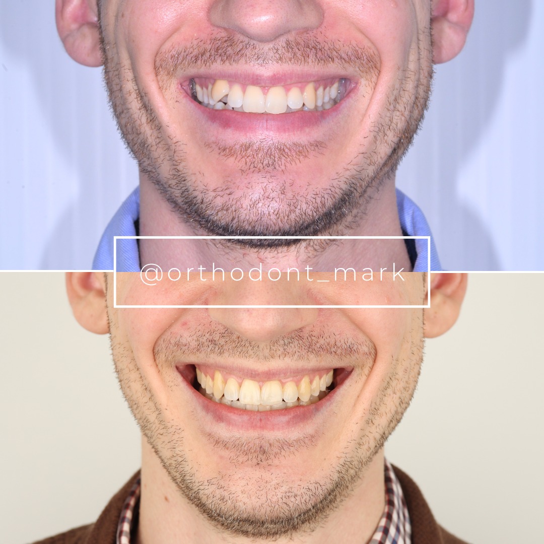
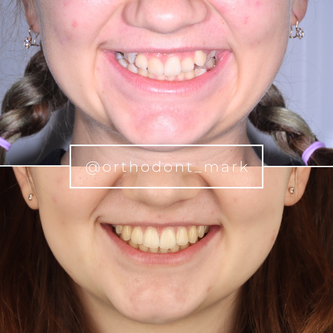
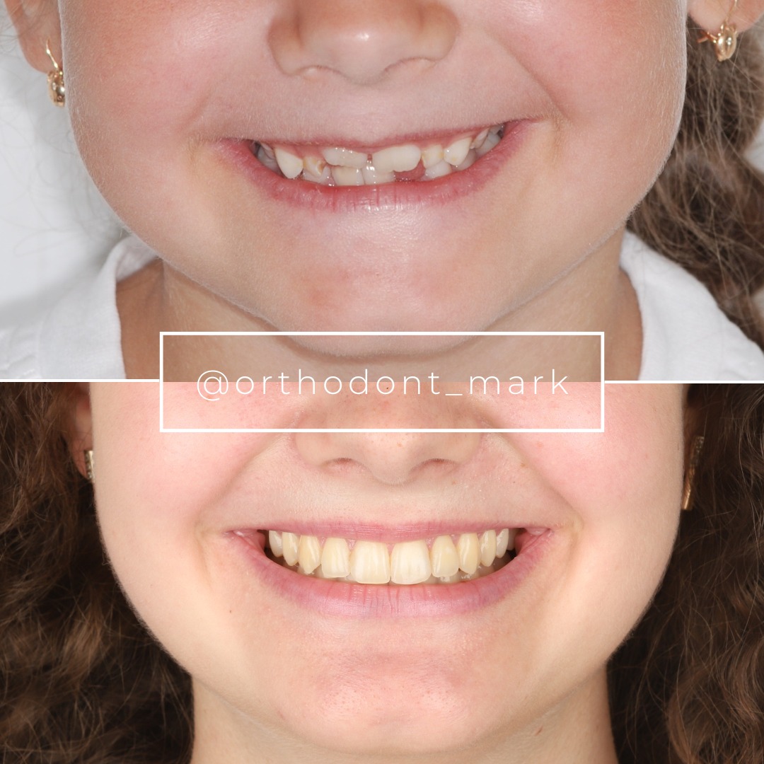
Learn the full MARPE protocol from Dr. Mark Radzhabov
Fundamental course covering CBCT patient selection, miniscrew planning, activation protocols, and 60+ clinical cases. Choose the access level that fits your practice.
Essentials of rapid palatal expansion for practicing orthodontists.
- Core RPE concepts and biomechanics
- 6 structured video lessons
- Clinical decision checklists
- Lifetime access to recordings
Deep-dive into MARPE protocol, diagnostics, and clinical execution.
- 6 modules covering MARPE from A to Z
- CBCT case selection walkthroughs
- Miniscrew placement and activation
- 60+ annotated clinical cases
- Direct Q&A with Dr. Mark Radzhabov
5-element medical consultation framework for dentists and orthodontists.
- Trust-building consultation protocol
- 5 lesson modules
- Templates for treatment plan delivery
- Works with any clinical specialty
Clinical FAQ
What is the recommended sequence for MARPE skill acquisition in orthodontic residency?
The evidence-supported sequence moves from biomechanics review and force calibration through bench simulation, cadaver or synthetic skull drilling, mentored first case, and finally independent practice with structured case review. Each stage develops a specific competency that the next stage depends on.
How does MARPE compare to conventional RPE for suture separation rate?
Chun et al. (2022) reported midpalatal suture separation in 95% of MARPE patients compared with 90% of RPE patients in a prospective randomized trial, with MARPE also producing significantly greater maxillary width at premolar and molar regions (P < 0.05).
Which appliance generates the highest expansion force in vitro?
Camporesi et al. (2013) showed that Hyrax and A2620 expanders generated forces exceeding 20 kg, while Palatal Split screws reached approximately 16 kg. Both Hyrax and A2620 demonstrated significantly greater rigidity than Palatal Split across all activation levels.
At what activation milestone should CBCT be taken during MARPE treatment?
Chun et al. (2022) performed CBCT at baseline, immediately after 35 turns of expansion, and after a 3-month consolidation period. This three-point imaging protocol confirms suture separation and tracks early bone fill during consolidation.
How do I select a safe first MARPE case as an adopting orthodontist?
Prioritise patients with clearly skeletal transverse deficit on CBCT, suture morphology indicating patency rather than advanced fusion, and adequate palatal bone volume at the planned miniscrew site. Avoid cases with anatomical asymmetry or thin mucosal coverage for initial cases.
What is the role of bench simulation in the MARPE learning curve?
Bench simulation on acrylic blocks and typodont models builds consistent insertion angulation and depth-stop technique before live tissue is encountered. Errors in trajectory that would contact roots or the nasal floor in a patient are identified and corrected during the simulation phase.
Why does appliance rigidity matter when adopting MARPE for skeletally mature patients?
Higher suture resistance in maturing patients requires a device capable of sustaining adequate force across repeated activations. Camporesi et al. (2013) showed that lower-rigidity appliances, such as Palatal Split screws, generate substantially less force than Hyrax or A2620 designs, which may be insufficient in high-resistance sutures.
How many supervised cases are needed before independent MARPE practice?
Evidence does not specify a universal number, but a three-case mentored progression — fully supervised, independently executed with mentor review at consolidation, and fully independent with self-audit — is a clinically sound framework. Complication-free completion of all three stages guides the transition decision.
What CBCT findings should a trainee identify before selecting a MARPE patient?
Key findings include midpalatal suture fusion pattern across anterior, middle, and posterior thirds. Vertical bone height between root apices and nasal floor at the insertion corridor. Intermaxillary width discrepancy at the basal bone level. And any palatal vault asymmetry affecting planned miniscrew angulation.
How does RPE training serve as a prerequisite for MARPE adoption?
RPE training establishes the force-application concepts, activation scheduling, and patient monitoring protocols shared by both appliance families. Orthodontists who have completed structured RPE training already possess the foundational mechanics knowledge that MARPE simulation builds directly upon.
Building competence in MARPE is a staged process, not a single course event. Clinicians who invest in simulation before their first live case, calibrate appliance selection against measured force outputs, and interpret CBCT suture staging systematically will experience fewer complications and more predictable skeletal responses. Dr. Mark Radzhabov offers structured MARPE training at ortodontmark.com to guide orthodontists through exactly this progression — from mechanical fundamentals to independent case management. Enroll, review your pending cases, or book a case consultation to define where you stand on the pathway. Key sources: Chun et al. (2022), BMC Oral Health, doi:https://doi.org/10.1186/s12903–022-02138-w. Camporesi et al. (2013), BioMedical Engineering OnLine, doi:http://www.biomedical-engineering-online.com/content/12/1/128.