
Adding MARPE to an Established Practice:
Learning Pathway
Case Volume and When to Seek External Review
A structured clinical guide for orthodontists ready to integrate bone-borne expansion—covering appliance mechanics, imaging criteria, and practice workflow decisions.
TL;DR Adding MARPE to an established practice requires deliberate case selection, appliance-force literacy, and a clear threshold for seeking external case review. CBCT-guided suture assessment and an understanding of force mechanics—documented by Camporesi et al. (2013) and Chun et al. (2022)—form the clinical foundation every orthodontist needs before expanding their caseload.
Integrating bone-borne skeletal expansion into a busy orthodontic schedule is less a purchasing decision than a clinical discipline built on imaging interpretation, appliance biomechanics, and honest case-volume progression. At ortodontmark.com, Dr. Mark Radzhabov argues that the most preventable errors in early MARPE adoption stem not from technique but from gaps in case selection and force management. This article gives practicing orthodontists a structured learning pathway—what to assess before the first case, which clinical signals warrant external review, and how peer-reviewed mechanics data should inform appliance choice from day one.
Why Does Your First MARPE Case Feel Riskier Than It Should?
Adding MARPE to an established practice is the process by which an orthodontist systematically incorporates miniscrew-assisted rapid palatal expansion into their clinical workflow through staged case selection, appliance-force training, and defined thresholds for peer or specialist review. The procedural gap most practitioners feel at the outset is rarely about screw insertion technique—it is about not yet having a reliable mental model for predicting skeletal versus dental response before treatment begins. Age alone does not fully characterize the expansion challenge. A 2022 CBCT and dental cast analysis demonstrated that the midpalatal suture opening pattern shifts from parallel to a triangular orientation in both the cranial and horizontal planes as patients age, with children up to age 10 showing parallel opening and adolescents 12 years and older showing V-shaped, anteriorly dominant patterns. Understanding this geometry helps the clinician anticipate where transverse gain will concentrate—and where it will not. Tooth-borne expansion complicates that prediction further. Bud et al. (2023) identified that palatal expander insertion is an invasive procedure that frequently causes local and regional hyperplasia of the oral mucosa as a secondary traumatic effect, a finding that reinforces why mucosal health and palatal vault morphology belong in the pre-treatment checklist long before MARPE is considered. Documenting baseline tissue status protects the clinician and sets a measurable standard for monitoring throughout the active phase.
Which Appliance Force Profile Matches the Case You Are Planning?
Orthodontists adding MARPE to their armamentarium often select appliances by brand familiarity rather than force output. Camporesi et al. (2013) quantified why that distinction matters: in mechanical testing, Hyrax and A2620 expanders generated forces exceeding 20 kg, while Palatal Split screws generated approximately 16 kg. This is not a trivial spread when you are planning load distribution across miniscrews anchored in palatal cortical bone rather than distributing force through molar bands. RPE appliances produce expansion forces ranging from 7.54 to 15.8 kg (16.6 to 34.8 pounds) according to Camporesi et al. (2013), and the activation increment matters equally. The same study documented that RME screws measured 10 mm in total travel, with each full turn delivering 0.8 mm of expansion across 4 activations of 0.2 mm each. Knowing this arithmetic allows the clinician to map a total expansion prescription against an activation schedule rather than simply instructing the patient on turns per day without a skeletal endpoint in mind. For practitioners building MARPE caseload, matching force output to bone density and suture maturity is the central biomechanical skill. A high-force appliance applied to a mature, partially fused suture without CBCT-confirmed bone density data is a protocol gap, not a technique choice. Referring to a structured MARPE training pathway that addresses these load calculations systematically can shorten the learning curve before a complication makes the lesson expensive.
What Does Suture Response Data Tell You About Protocol Design?
When counseling a patient or designing a protocol, separation rate benchmarks from controlled trials provide a realistic reference for informed consent and clinical expectation-setting. Chun et al. (2022) reported midpalatal suture separation in 90% of RPE patients and 95% of MARPE patients following expansion in a prospective randomized clinical trial—figures that support MARPE as a reliable skeletal intervention when patient selection and imaging are rigorous. The protocol rigor in that trial also deserves clinical attention. Chun et al. (2022) performed CBCT imaging at baseline, immediately after 35 turns of expansion, and after 3 months of consolidation. That three-stage imaging structure—pre-treatment, post-active expansion, post-consolidation—is a model worth replicating in practice, both for tracking skeletal response and for identifying early signs of inadequate suture opening before the consolidation phase locks in a suboptimal result. Critically, Chun et al. (2022) found that MARPE resulted in significantly less buccal displacement of anchor teeth during expansion and consolidation compared to RPE. For orthodontists managing posterior dental crowding, Class II molar relationships, or thin buccal bone on the anchor premolars and molars, this is not a minor biomechanical footnote—it is a primary criterion for appliance selection. Exploring miniscrew-assisted skeletal expansion protocols in depth before the first case allows clinicians to anticipate this advantage and document it in treatment records.
When Should an Orthodontist Seek External Case Review for Expansion?
Knowing when to seek external case review is not a sign of limited competence—it is the structural skill that separates practitioners who grow their MARPE caseload safely from those who plateau after a difficult outcome. The trigger for review should be defined prospectively, not after a complication surfaces. Concrete thresholds include: suture morphology that is difficult to stage confidently on CBCT, bone density readings outside the range of your prior cases, or a patient who has previously undergone tooth-borne expansion with documented buccal bone loss. Mucosal findings also raise the review threshold. Because Bud et al. (2023) confirmed that palatal expanders can induce local and regional mucosal hyperplasia as a secondary traumatic effect, a patient presenting with existing palatal pathology or prior mucosal reactions warrants specialist input before appliance placement. This is especially relevant when planning miniscrew insertion sites close to anatomical structures where hyperplastic tissue would complicate access. The practical workflow recommendation is to build a review checklist into your treatment planning appointment for any expansion case—not just the novel ones. Documenting your decision rationale at each step also creates an audit trail that supports clinical growth. For cases that clearly exceed current case complexity, an external case review consultation through a structured platform is more efficient than trial-and-error adjustment after screw placement.
How Does Expansion Pattern Geometry Affect Your Clinical Protocol?
Expansion Geometry
One of the less-discussed dimensions of building a MARPE caseload is developing an intuitive model of how expansion force distributes across the palate at different skeletal ages. A 2022 CBCT and dental cast analysis established that the midpalatal suture opening pattern shifts from parallel in younger children to a V-shaped, anteriorly dominant pattern in older adolescents—specifically, children up to 10 years showed parallel expansion while those 12 years and older showed anterior-predominant opening geometry. This has direct implications for where you expect to gain transverse width and where relapse risk concentrates. For the orthodontist adding MARPE to an established practice, this geometric shift matters during appliance selection and when setting patient expectations. A case that presents with a wider anterior deficit than posterior will respond differently than one with uniform transverse deficiency—and the activation protocol, consolidation duration, and post-retention monitoring should reflect that. Failing to account for geometry means the total expansion achieved on a dental cast may overstate the posterior skeletal gain that actually occurred. This evidence also clarifies why CBCT is not optional in MARPE case planning—it is the only imaging modality that allows direct visualization of both the suture morphology and the spatial distribution of early opening. When the posterior thirds show minimal separation relative to the anterior, that disparity changes the retention prescription. Orthodontists who review this geometry systematically at the mid-treatment CBCT develop pattern recognition that accelerates their clinical judgment across subsequent cases.
Free Clinical Guide
Which Expansion Appliance at Which Age?
Get Dr. Mark Radzhabov's guide to skeletal-expansion appliances — indications, pros & cons, and how to choose by patient age. Free to your inbox.
Prefer video? Watch the free demo lesson on MARPE appliance types, features and trade-offs.
Documented outcomes
Clinical cases from Dr. Mark Radzhabov's practice
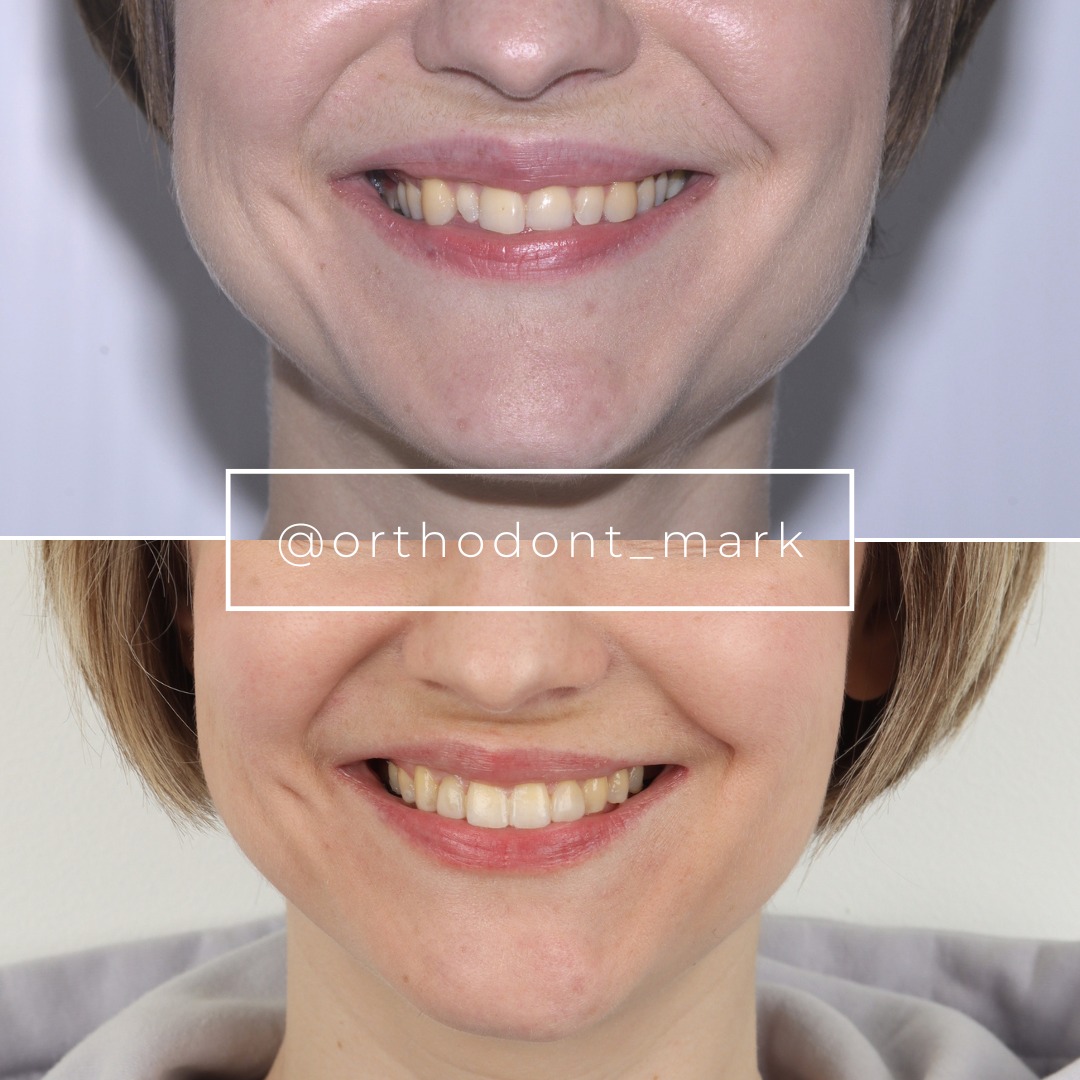

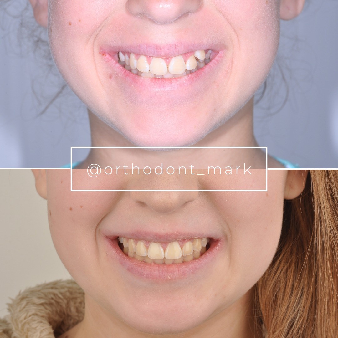
Learn the full MARPE protocol from Dr. Mark Radzhabov
Fundamental course covering CBCT patient selection, miniscrew planning, activation protocols, and 60+ clinical cases. Choose the access level that fits your practice.
Essentials of rapid palatal expansion for practicing orthodontists.
- Core RPE concepts and biomechanics
- 6 structured video lessons
- Clinical decision checklists
- Lifetime access to recordings
Deep-dive into MARPE protocol, diagnostics, and clinical execution.
- 6 modules covering MARPE from A to Z
- CBCT case selection walkthroughs
- Miniscrew placement and activation
- 60+ annotated clinical cases
- Direct Q&A with Dr. Mark Radzhabov
5-element medical consultation framework for dentists and orthodontists.
- Trust-building consultation protocol
- 5 lesson modules
- Templates for treatment plan delivery
- Works with any clinical specialty
Clinical FAQ
What imaging criteria should I use when deciding between MARPE and RPE for a new patient?
CBCT assessment of suture morphology, bone density at planned miniscrew sites, and buccal bone thickness at anchor teeth are the primary imaging criteria. Chun et al. (2022) documented significantly less buccal tooth displacement with MARPE, making it preferable where posterior buccal bone is thin.
How does appliance force output differ between common palatal expander designs?
Camporesi et al. (2013) found that Hyrax and A2620 expanders generated forces exceeding 20 kg in mechanical testing, while Palatal Split screws generated approximately 16 kg. RPE appliances overall produced forces ranging from 7.54 to 15.8 kg, depending on design.
What is the activation increment per turn for standard RME screws?
Camporesi et al. (2013) documented that RME screws deliver 0.8 mm of expansion per full turn across 4 activations of 0.2 mm each. Knowing this arithmetic allows precise mapping of a total expansion prescription against a structured activation schedule.
How should I structure CBCT imaging through the MARPE treatment timeline?
Chun et al. (2022) used a three-stage protocol: baseline, immediately after 35 turns of expansion, and after 3 months of consolidation. This structure captures both the active suture response and the early consolidation phase, allowing mid-course correction before retention.
What mucosal complication should I monitor during palatal expansion cases?
Bud et al. (2023) identified local and regional mucosal hyperplasia as a frequent secondary traumatic effect of palatal expander insertion. Serial photographic documentation of palatal tissue at each activation review appointment is the simplest way to detect this early.
How does expansion geometry differ across skeletal age groups in RME patients?
A 2022 CBCT and dental cast analysis found parallel suture opening in children up to 10 years and V-shaped, anteriorly dominant opening in adolescents 12 years and older. This geometric shift affects where transverse gain concentrates and should inform retention protocol design.
When is it appropriate to seek external case review for a complex MARPE case?
Seek external review when suture staging is ambiguous on CBCT, when bone density falls outside your experience range, when the patient has prior mucosal pathology, or when buccal bone loss from previous tooth-borne expansion complicates miniscrew site planning.
Does MARPE reduce dental side effects compared to conventional RPE?
Yes. Chun et al. (2022) confirmed in a prospective randomized trial that MARPE produced significantly less buccal displacement of anchor teeth during both the active expansion and consolidation phases compared to tooth-borne RPE.
What is the suture separation rate I can quote in consent for MARPE vs. RPE?
Chun et al. (2022) reported midpalatal suture separation in 95% of MARPE patients and 90% of RPE patients. These figures come from a prospective randomized clinical trial and are appropriate to reference during patient consent discussions.
How do I build MARPE clinical skills systematically without overextending on complex cases?
Progress through case complexity by first mastering CBCT suture staging, then appliance force selection based on bone density, then miniscrew site planning. Define your external review threshold before your first case, and use structured mentorship such as Dr. Mark Radzhabov's MARPE training pathway.
Building a MARPE caseload responsibly means pairing each new case type with the evidence behind it—suture mechanics, dental side-effect profiles, and age-related expansion geometry. When a case sits outside your current experience, external case review is a professional asset, not a limitation. Dr. Mark Radzhabov offers structured clinical consultation through ortodontmark.com for orthodontists navigating complex expansion decisions. The literature reviewed here confirms that rigorous imaging and appliance-force literacy produce better skeletal outcomes than volume alone. Key sources: Chun et al. (2022), BMC Oral Health, doi:https://doi.org/10.1186/s12903–022-02138-w. Camporesi et al. (2013), BioMedical Engineering OnLine, doi:http://www.biomedical-engineering-online.com/content/12/1/128.

