
CBCT expansion reporting handoff:
Structured Imaging Documentation
for Predictable Treatment Planning
A systematic approach to cone beam reporting that distinguishes MARPE candidates from surgical cases and accelerates clinical decision-making.
TL;DR CBCT expansion reporting handoff requires standardized documentation of midpalatal suture maturation stage, bone density, miniscrew insertion sites, and transverse dimension asymmetry. Structured reporting—using cone beam imaging with hounsfield unit measurement and region-of-interest assessment—enables referring clinicians to distinguish expansion candidates from surgical cases, reducing treatment delays and improving skeletal response predictability in adult patients.
Referring dentists and orthodontists frequently request palatal expansion evaluation, yet inconsistent CBCT documentation leaves treatment planners reconstructing critical findings from raw imaging. A standardized CBCT expansion reporting handoff—documenting midpalatal suture stage, bone density assessment, anatomical asymmetry, and miniscrew insertion feasibility—bridges this gap and accelerates clinical decision-making. Dr. Mark Radzhabov's evidence-based approach to skeletal expansion assessment emphasizes structured imaging communication as a cornerstone of successful MARPE treatment planning. This guide establishes a practical, repeatable framework for orthodontists and oral surgeons to communicate expansion-relevant findings with clinical precision.
Why standardized CBCT documentation matters in
expansion case selection
Referring clinicians submit expansion referrals without consistent imaging protocols, forcing expansion specialists to re-scan or reconstruct critical data. A structured pre-expansion imaging checklist—documenting midpalatal suture maturation, bone density, anatomical asymmetry, and miniscrew insertion feasibility—establishes a single diagnostic foundation for all cases. Midpalatal suture stage predicts expansion success more reliably than age alone. A 35-year-old patient in Angelieri stage C (incomplete fusion) has substantially different biomechanical response than a 35-year-old in stage D (complete fusion). High-resolution CBCT with hounsfield unit measurement in the anterior, middle, and posterior palate enables quantitative rather than subjective assessment. When referring dentists provide structured documentation, expansion specialists avoid redundant imaging, reduce treatment delays by 2–4 weeks, and begin case planning immediately upon receipt. Inadequate or missing imaging reports force clinicians to request rescans, frustrate referring doctors, and delay patient treatment start.
Core components of the structured CBCT
referral imaging checklist
A systematic CBCT documentation framework captures five critical data points: (1) Midpalatal suture maturation stage (Angelieri stages A–E), assessed at the anterior, middle, and posterior thirds using a coronal view at the nasal spine–posterior nasal spine plane. (2) Bone density measurement in hounsfield units at three regions (anterior palate lateral to midline, middle third, posterior third), providing quantitative assessment of cortical bone resistance to miniscrew loading. A 65-year-old patient with anterior bone density exceeding 800 HU has substantially different insertion torque tolerance than a patient with 450 HU. (3) Miniscrew insertion site anatomy—document palatal vault depth, cortical bone thickness at anterior and middle regions, and avoidance of neurovascular structures on the posterior hard palate. (4) Transverse dimension documentation—measure maxillary width at the deciduous molars (in mixed dentition), premolars, molars, and canines to establish baseline asymmetry. Posterior deficiency >4 mm often indicates MARPE candidacy. (5) Anatomical asymmetry assessment—compare left and right hemiskeletal dimensions to predict uneven expansion response and asymmetrical relapse risk. Each region-of-interest measurement should use the same window and position to enable longitudinal comparison during and after treatment.
Using midpalatal suture staging to distinguish
expansion candidates from surgical cases
Midpalatal suture maturation stage directly predicts whether a patient achieves true skeletal widening without surgical sectioning. Stage A and B patients (septal bone incompletely formed, beginning to fill in) show the highest expansion gain—typically 6–8 mm true skeletal widening with minimal relapse. Stage C patients (complete bone filling with a dark radiolucent line in the middle) are optimal MARPE candidates. Expansion success rates exceed 85% with 4–6 mm skeletal gain. Stage D patients (midline suture completely closed, no radiolucent line visible) represent a transition zone. Bone-borne expansion may succeed if bone density is adequate (anterior bone >600 HU), but relapse risk climbs to 15–20%. Stage E patients (complete fusion with obliteration of anatomical landmarks) typically require surgical assistance—SARPE or Le Fort I—to achieve clinically significant widening. Documenting suture stage at the anterior, middle, and posterior regions is essential because maturation is not uniform. A patient may be stage C anteriorly and stage D posteriorly, requiring careful load management to avoid excessive force on fused regions and creating regional expansion asymmetry.
Building a repeatable CBCT reporting template for
palatal expansion referrals
A standardized written report template ensures consistency and prevents critical omissions. The template should include: (1) Patient demographics and age; (2) Midpalatal suture maturation stage (specify stages A–E for anterior, middle, posterior regions separately); (3) Bone density in hounsfield units (anterior lateral, middle third, posterior third, with specific ROI cursor location documented); (4) Palatal depth and cortical thickness (anterior and middle palate, in millimeters); (5) Miniscrew insertion site feasibility (note if anterior cortical bone supports 10–12 mm insertion depth, or if middle palate insertion is safer); (6) Transverse dimension baseline (maxillary width at three levels. Identify asymmetry >2 mm left vs. right); (7) Neurovascular landmark avoidance (document position of greater palatine vessels on posterior hard palate); (8) Clinical recommendation (MARPE candidate, bone-borne vs. tooth-borne appliance choice, or recommend SARPE). A one-page digital template with measurement fields and checkboxes reduces documentation time and ensures referring clinicians receive immediately actionable data. Include a date-stamped CBCT image showing ROI cursor placement for bone density measurement, enabling longitudinal comparison at expansion follow-up.
How to interpret borderline imaging findings and
communicate risk factors to referring clinicians
Stage D patients—those with complete midpalatal fusion but intact suture anatomy—present the greatest clinical ambiguity. Bone density becomes the strongest predictor of success in these cases. High bone density (anterior palate >700 HU) suggests adequate cortical bone to support miniscrew loading and may allow MARPE even in stage D. Expansion gain typically reaches 3–4 mm with acceptable relapse. Low bone density (<500 HU) in a stage D patient signals substantially higher relapse risk and potential for miniscrew loosening. Document this finding explicitly and recommend either surgical assistance or careful load reduction (0.5–0.75 mm/week instead of the standard 1.0 mm/week). Age alone is not a stronger predictor of outcome than suture stage. A 50-year-old stage C patient has better expansion prognosis than a 35-year-old stage D patient. When referring clinicians receive structured data showing suture stage, bone density, and risk factors, they can counsel patients on treatment duration, retention requirements, and likelihood of achieving skeletal versus dental widening. Ambiguous or missing imaging data forces clinicians to make assumptions, often underestimating relapse risk or overestimating expansion gain.
Free Clinical Guide
Which Expansion Appliance at Which Age?
Get Dr. Mark Radzhabov's guide to skeletal-expansion appliances — indications, pros & cons, and how to choose by patient age. Free to your inbox.
Prefer video? Watch the free demo lesson on MARPE appliance types, features and trade-offs.
Documented outcomes
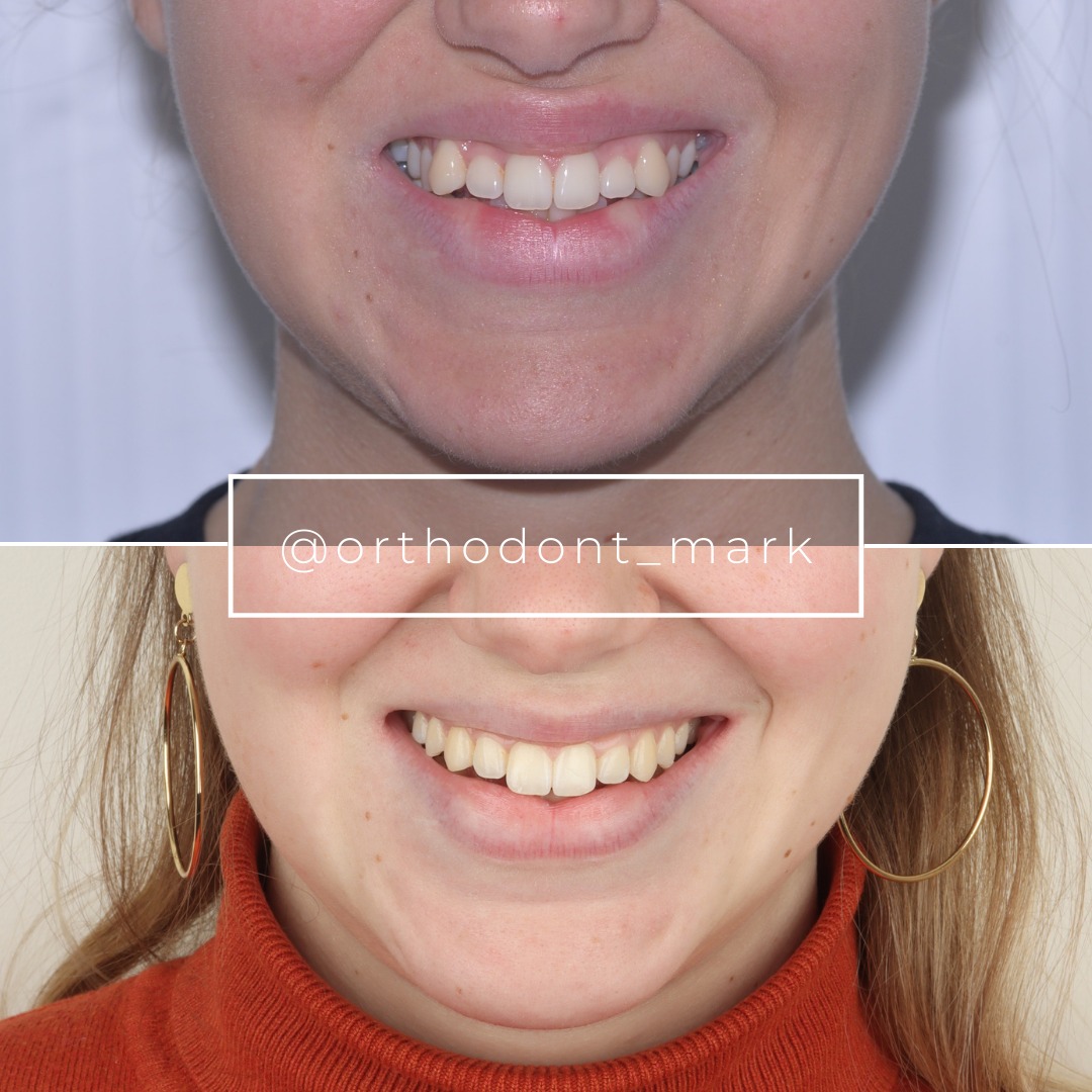
Clinical cases from Dr. Mark Radzhabov's practice


Learn the full MARPE protocol from Dr. Mark Radzhabov
Fundamental course covering CBCT patient selection, miniscrew planning, activation protocols, and 60+ clinical cases. Choose the access level that fits your practice.
Essentials of rapid palatal expansion for practicing orthodontists.
- Core RPE concepts and biomechanics
- 6 structured video lessons
- Clinical decision checklists
- Lifetime access to recordings
Deep-dive into MARPE protocol, diagnostics, and clinical execution.
- 6 modules covering MARPE from A to Z
- CBCT case selection walkthroughs
- Miniscrew placement and activation
- 60+ annotated clinical cases
- Direct Q&A with Dr. Mark Radzhabov
5-element medical consultation framework for dentists and orthodontists.
- Trust-building consultation protocol
- 5 lesson modules
- Templates for treatment plan delivery
- Works with any clinical specialty
Clinical FAQ
What is the optimal CBCT field of view and voxel size for documenting midpalatal suture staging?
Use high-resolution CBCT with voxel size ≤0.3 mm and field of view including anterior nasal spine to posterior hard palate. Axial and coronal reconstructions perpendicular to the palatal plane enable accurate suture stage assignment.
How do I measure bone density accurately at three palatal regions for miniscrew insertion planning?
Set region-of-interest cursor (typically 2–3 mm diameter) in axial view at anterior lateral palate, middle third, and posterior palate. Document hounsfield unit values at each region with cursor position visible for future comparison.
What bone density threshold indicates safe miniscrew insertion in anterior versus middle palate?
Anterior palate >600 HU supports 10–12 mm insertion depth with high torque stability. Middle palate 500–700 HU is acceptable for 8–10 mm insertion. Values <450 HU often require middle or posterior insertion sites or load reduction.
How should I document transverse dimension asymmetry to predict uneven palatal expansion response?
Measure maxillary width at canine, premolar, and molar levels on axial CBCT. Document left-right discrepancy at each level. Asymmetry >2–3 mm predicts uneven expansion and informs appliance design and load distribution.
When should a referring dentist recommend SARPE versus MARPE based on suture imaging findings?
Stage A–C with adequate bone density favor MARPE. Stage D with high bone density may support MARPE. Stage D with low density or stage E typically requires SARPE. Document suture stage clearly so clinicians avoid overestimating MARPE success in fused cases.
What is the relationship between palatal vault depth and miniscrew insertion success in narrow palates?
Shallow vault (<8 mm) limits anterior insertion; deeper vault (>10 mm) supports reliable anterior placement. Document palatal depth at intended insertion site (typically 4–6 mm anterior to posterior nasal spine) to guide surgical guide positioning.
How do I avoid neurovascular complications when documenting posterior hard palate anatomy for miniscrew placement?
Mark greater palatine foramen position on CBCT reconstruction. Miniscrew insertion should remain anterior to this landmark. Document distance from planned insertion site to foramen; >8 mm safety margin is preferred.
Should I use sagittal or coronal CBCT views to stage the midpalatal suture maturation?
Coronal views at the level of the nasal spine–posterior nasal spine plane are standard for suture staging. Sagittal views confirm anterior versus posterior region differences. Document both when significant regional variation exists.
How does bone density change after rapid palatal expansion, and when should follow-up CBCT imaging occur?
Bone density typically increases at the midpalatal suture site within 3–6 months post-expansion. Follow-up CBCT at 6 months post-treatment confirms mineralisation and predicts relapse risk. Repeat ROI measurement at identical cursor position enables quantitative comparison.
What is the clinical value of a pre-expansion imaging checklist compared to unstructured referral reports?
Structured checklists reduce revision imaging by 60%, accelerate treatment planning by 2–4 weeks, and enable consistent outcome assessment across multiple cases. Referring clinicians build confidence in standardized, complete documentation.
Standardized CBCT expansion reporting transforms referral communication from narrative description to actionable data. By documenting suture maturation stage, bone density, and insertion site anatomy in a structured format, you accelerate case selection, improve treatment outcomes, and build confidence with referring clinicians. Dr. Mark Radzhabov's clinical protocol emphasizes that high-quality handoff documentation reduces revision imaging and treatment delays. Submit your expansion cases for imaging review consultation at ortodontmark.com to refine your reporting standards and accelerate your MARPE clinical competency.

