
Transverse Consultation Workflow:
Two-Visit
Records, CBCT, and Appliance Sequencing
A practical framework for ordering CBCT imaging and staging suture maturation before committing to MARPE or RPE—built for practicing orthodontists.
TL;DR A structured transverse consultation workflow sequences diagnostic records and CBCT imaging before appliance selection, not after. Suture maturation staging, cast morphology analysis, and skeletal versus dental expansion data each inform a distinct visit. Splitting the consultation into two purposeful appointments reduces chairtime waste and sharpens appliance decisions for MARPE or RPE cases.
When a transverse consultation workflow collapses into a single appointment, clinicians are forced to discuss MARPE or RPE options before holding the diagnostic records that actually justify those choices. Dr. Mark Radzhabov at ortodontmark.com outlines a two-visit intake model that separates record acquisition from appliance discussion—an approach grounded in peer-reviewed imaging studies on suture maturation and expansion morphology. Orthodontists who read this gain a sequenced protocol that aligns CBCT ordering, cast analysis, and patient staging with the moment of clinical decision-making, reducing both diagnostic error and unnecessary imaging.
Why Does Appliance Discussion Belong at Visit Two,
Not Visit One?
A transverse consultation workflow is a sequenced clinical intake process that separates diagnostic record acquisition—including CBCT imaging and dental cast analysis—from appliance selection discussion, distributing each task across two purpose-defined appointments. The most common workflow failure in transverse case planning is presenting the patient with an appliance option before the suture status is known. Without a CBCT, the clinician is estimating whether the midpalatal suture is patent, partially bridged, or fused—an estimate that directly controls whether the patient is appropriate for a bone-borne device or requires a surgical referral first. Angelieri et al. (2013) established five radiographic maturation stages of the midpalatal suture across a CBCT cross-sectional study of 140 subjects aged 5.6 to 58.4 years—ranging from a straight, uninvolved suture at Stage A to complete anterior maxillary fusion at Stage E. That staging system is the interpretive anchor of Visit 2, and it cannot be applied without a CBCT acquired at Visit 1. Scheduling the imaging before the consultation appointment is the single structural change that converts a one-visit impression into a two-visit diagnostic protocol. Cast morphology adds a second layer of information that is equally visit-sequenced. Kinzinger et al. (2022) analyzed 60 patients stratified by age into three groups and found that expansion morphology differs significantly across children under 10 years, ages 10–12, and ages 12 and older. Those cast differences are visible before any appliance is placed and should be documented at Visit 1 so that the Visit 2 conversation begins with objective expansion morphology data already in hand.
When in the Intake Sequence Should CBCT
Actually Be Ordered?
CBCT should be ordered at or immediately following the initial clinical screening—before any appliance discussion occurs. The rationale is that suture staging drives appliance choice, not the other way around. A clinician who orders imaging after a preliminary appliance conversation must either retract that conversation or rationalize a device choice that may contradict the imaging findings, both of which erode patient confidence and clinical credibility. Angelieri et al. (2013) identified that Stages A and B of midpalatal suture maturation occur typically before age 13, while Stage C appears primarily between ages 11 and 17. Because Stage C encompasses a wide and clinically mixed adolescent population, age alone cannot serve as a proxy for suture status in that bracket—CBCT is the only reliable discriminator. This is precisely the age range where a premature appliance recommendation, made without imaging, is most likely to be wrong. Chun et al. (2022) used CBCT imaging at three time points—baseline, immediately post-expansion, and a 3-month consolidation interval—to document skeletal and dentoalveolar changes following expansion. That temporal imaging structure underscores that the baseline CBCT is not optional documentation. It is the reference frame against which all subsequent skeletal and dental change is measured. Ordering it before Visit 2 positions the clinician to discuss not just appliance type but measurable skeletal outcomes with the patient at the appliance consultation itself.
How Does Pre-Treatment Cast Morphology Shape
Appliance Selection?
Dental cast analysis at Visit 1 is not redundant with CBCT—it captures the dental expression of transverse deficiency independently of skeletal suture status. The two data sources address different clinical questions: the cast documents where the teeth are and how the arch has compensated, while the CBCT documents what the suture will permit. Kinzinger et al. (2022) demonstrated that rapid maxillary expansion produces parallel expansion in children under age 10, but V-shaped, anterior-dominant expansion in adolescents aged 12 and older. That morphological shift has direct implications for appliance design: a patient presenting with a pre-existing V-shaped arch at baseline is already exhibiting the pattern associated with older skeletal age, which should heighten the clinician's index of suspicion for a partially fused or fusing suture and reinforce the need for CBCT staging before commitment to a tooth-borne rapid palatal expander. For cases where cast analysis and clinical age both point toward adolescent or adult suture status, miniscrew-assisted expansion protocol considerations come into focus at Visit 2. Chun et al. (2022) found that miniscrew-assisted RPE produced significantly greater skeletal nasal width gain and greater palatine foramen expansion than conventional tooth-borne RPE (p < 0.05), while also demonstrating significantly reduced buccal displacement of anchor teeth throughout expansion and consolidation (p < 0.05). Presenting both findings at the Visit 2 appliance discussion gives patients and referring clinicians a concrete, evidence-sourced rationale for bone-borne device selection rather than an opinion.
Interpreting Midpalatal Suture Stages at Visit Two:
Decision Thresholds
for Bone-Borne Versus Surgical Referral
The five-stage classification system described by Angelieri et al. (2013) maps suture morphology from a radiographically visible, straight dark line at Stage A through progressive interdigitation and ossification to complete anterior maxillary fusion at Stage E. Each stage carries a different clinical probability of successful non-surgical skeletal opening, which is precisely why the Visit 2 appliance conversation must begin with the stage assignment rather than the patient's chronological age. Stage A and Stage B sutures—appearing typically before age 13—offer the most predictable response to conventional tooth-borne rapid palatal expander force, and this is the population where a tooth-borne rapid palatal expander selection discussion is straightforward. Stage C, which spans ages 11 to 17, represents the ambiguous zone: the suture may still open under bone-borne load, but the probability decreases as interdigitation advances. Presenting the CBCT image directly during Visit 2 and naming the assigned stage converts an ambiguous clinical impression into a documented, patient-visible rationale. Stages D and E shift the conversation toward surgical adjuncts or alternative treatment goals entirely. The staging system established from 140 subjects by Angelieri et al. (2013) provides the reference distribution against which a single patient's CBCT cross-section is read. Clinicians who defer imaging until after the appliance conversation lose the ability to make this assignment prospectively—and risk placing a device into a suture environment that will not respond to the applied load.
At What Point in the Workflow Does MARPE Outperform
Conventional RPE
on Skeletal Outcome Metrics?
When Visit 2 imaging reveals a Stage C suture in a patient whose cast morphology shows anterior-dominant arch narrowing, the appliance discussion pivots to bone-borne options. Chun et al. (2022) documented that suture separation occurred in 90% of RPE patients and 95% of MARPE patients following identical expansion protocols—a difference that, while numerically modest, is accompanied by a clinically significant divergence in skeletal versus dental effect distribution. Miniscrew-assisted expansion protocol advantages become quantifiable at the anchor tooth level. Chun et al. (2022) showed that MARPE produced significantly reduced buccal displacement of anchor teeth compared to tooth-borne RPE across both the active expansion phase and the 3-month consolidation interval (p < 0.05). For patients with compromised periodontal support at the premolars or molars, this distinction is not cosmetic—it is a risk-management criterion that belongs explicitly in the Visit 2 record. The skeletal gain data is equally specific: Chun et al. (2022) reported significantly greater skeletal nasal width gain and greater palatine foramen expansion with MARPE than with conventional RPE. Presenting those paired findings—suture separation probability and skeletal nasal width outcome—at Visit 2 completes the evidentiary case for appliance selection without requiring the clinician to rely on clinical intuition alone. The workflow earns its structure precisely because each data point arrives at the moment it is needed.
Free Clinical Guide
Which Expansion Appliance at Which Age?
Get Dr. Mark Radzhabov's guide to skeletal-expansion appliances — indications, pros & cons, and how to choose by patient age. Free to your inbox.
Prefer video? Watch the free demo lesson on MARPE appliance types, features and trade-offs.
Documented outcomes
Clinical cases from Dr. Mark Radzhabov's practice
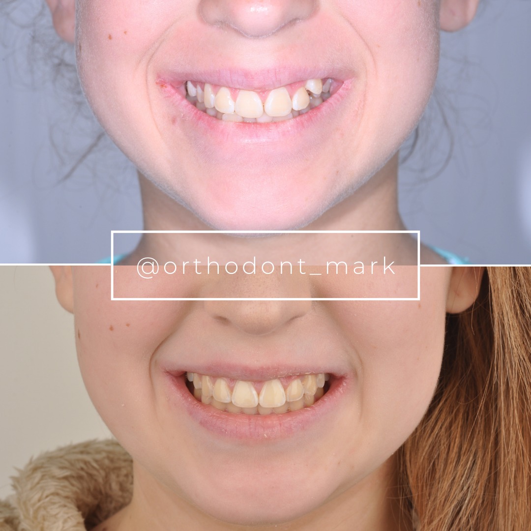

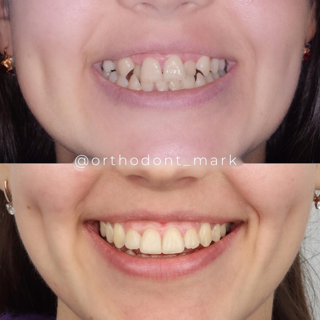
Learn the full MARPE protocol from Dr. Mark Radzhabov
Fundamental course covering CBCT patient selection, miniscrew planning, activation protocols, and 60+ clinical cases. Choose the access level that fits your practice.
Essentials of rapid palatal expansion for practicing orthodontists.
- Core RPE concepts and biomechanics
- 6 structured video lessons
- Clinical decision checklists
- Lifetime access to recordings
Deep-dive into MARPE protocol, diagnostics, and clinical execution.
- 6 modules covering MARPE from A to Z
- CBCT case selection walkthroughs
- Miniscrew placement and activation
- 60+ annotated clinical cases
- Direct Q&A with Dr. Mark Radzhabov
5-element medical consultation framework for dentists and orthodontists.
- Trust-building consultation protocol
- 5 lesson modules
- Templates for treatment plan delivery
- Works with any clinical specialty
Clinical FAQ
Why should CBCT be ordered before the appliance consultation in a transverse expansion workflow?
Midpalatal suture maturation stage determines whether non-surgical expansion is viable. Angelieri et al. (2013) established five distinct radiographic stages across a wide age range. Without that staging data in hand, appliance selection at the consultation lacks a defensible skeletal basis.
What information should be collected at Visit 1 of a two-visit transverse consultation protocol?
Visit 1 should yield full diagnostic records: CBCT, dental casts, photographs, and periodontal screening. No appliance is named. Kinzinger et al. (2022) showed cast morphology differs significantly across age groups, making early cast documentation essential before appliance planning.
How does midpalatal suture maturation staging change the expansion appliance discussion at Visit 2?
Stage assignment from CBCT maps directly to clinical probability of non-surgical suture opening. Angelieri et al. (2013) traced Stages A and B before age 13, Stage C between ages 11–17, and Stages D and E representing progressive fusion—each requiring a different appliance or surgical pathway.
How do dental cast morphology findings from Visit 1 inform the MARPE versus RPE decision at Visit 2?
Kinzinger et al. (2022) demonstrated that expansion produces parallel arch widening under age 10 but V-shaped, anterior-dominant morphology at age 12 and older. A pre-treatment V-shaped arch at baseline suggests more advanced skeletal age and raises the index of suspicion for a partially fused suture.
What CBCT time points are clinically necessary to document skeletal expansion outcomes after MARPE?
Chun et al. (2022) used baseline, immediately post-expansion, and 3-month consolidation CBCTs to track skeletal and dentoalveolar changes. The baseline scan is the reference frame for all subsequent measurement. Acquiring it before treatment starts is non-negotiable for outcome documentation.
How does MARPE compare to conventional RPE on skeletal nasal width gain and anchor tooth displacement?
Chun et al. (2022) found MARPE produced significantly greater skeletal nasal width gain and greater palatine foramen expansion than conventional RPE, while also showing significantly reduced buccal displacement of anchor teeth at p < 0.05 across expansion and consolidation phases.
Is chronological age a reliable substitute for CBCT-based suture staging in transverse case selection?
No. Angelieri et al. (2013) showed Stage C sutures span ages 11–17, a range where suture status varies dramatically between individuals. Age provides a probabilistic estimate. CBCT staging provides direct radiographic evidence of fusion status at the time of treatment planning.
What suture maturation stage justifies proceeding with non-surgical miniscrew-assisted expansion in adolescents?
Stages A, B, and early C—identified on CBCT per Angelieri et al. (2013)—carry the highest probability of successful non-surgical suture opening. Stage D and E findings indicate substantial ossification where bone-borne expansion force alone is unlikely to achieve predictable skeletal separation.
How does expansion morphology differ between children under 10 and adolescents aged 12 and older on dental casts?
Kinzinger et al. (2022) analyzed 60 patients and found that rapid maxillary expansion produces parallel widening in children under 10 years but V-shaped, anterior-dominant expansion in patients aged 12 and older—a morphological distinction detectable on pre-treatment casts before any appliance is placed.
What is the evidence-based rationale for separating records acquisition from appliance selection into two distinct consultation visits?
Suture maturation staging requires CBCT (Angelieri et al., 2013), cast morphology analysis requires physical models (Kinzinger et al., 2022), and outcome benchmarking requires baseline imaging (Chun et al., 2022). Each data source is collected at Visit 1 and interpreted at Visit 2 to avoid premature, unsupported appliance recommendations.
Sequencing records before the appliance conversation is not administrative preference—it is the structural requirement for defensible clinical decisions in transverse cases. Suture maturation data, cast morphology, and skeletal imaging each belong to a defined moment in the workflow, not to the same appointment. Dr. Mark Radzhabov encourages clinicians to audit their current intake process against the two-visit framework described here, then explore structured MARPE training at ortodontmark.com to apply these principles across a full caseload. Key sources: Angelieri et al. (2013), Am J Orthod Dentofacial Orthop, doi:10.1016/j.ajodo.2013.04.022. Chun et al. (2022), BMC Oral Health, doi:10.1186/s12903–022-02138-w. Kinzinger et al. (2022), J Orofac Orthop, doi:10.1007/s00056–022-00429-z.