
MARPE Learning Pathway:
Training,
Mentorship, and First-Case Selection
A practical sequencing guide for orthodontic residents and new operators adding miniscrew-assisted rapid palatal expansion to clinical practice.
TL;DR The MARPE learning pathway sequences hands-on simulation, supervised mentorship, and deliberate first-case selection before independent practice. Appliance force data from Camporesi et al. (2013) and suture separation evidence from Chun et al. (2022) give new operators the biomechanical and imaging benchmarks needed to progress with confidence through each training stage.
Adding miniscrew-assisted rapid palatal expansion to a clinical practice demands more than a weekend course and a supply order. At ortodontmark.com, Dr. Mark Radzhabov argues that the MARPE learning pathway succeeds only when procedural simulation, structured mentorship, and rigorous first-case screening are sequenced in a deliberate order—not assembled ad hoc. This article distills the evidence on appliance biomechanics and suture imaging into a decision-ready progression that helps orthodontic residents and new operators identify exactly where they stand, what they still need, and which patient belongs in chair one.
Why Force Data Must Come First in Any
MARPE
Training Sequence
The MARPE learning pathway is a staged clinical education model that progresses a new operator from mechanical simulation and force-calibration drills through supervised mentorship to independent first-case execution using miniscrew-assisted palatal expansion. Before live placement, the new operator must internalize what the screw actually delivers to the suture—not as an abstract concept but as a measurable range with device-specific variation. Camporesi et al. (2013) quantified this range directly, finding that Hyrax and A2620 expanders generated forces exceeding 20 kg, while Palatal Split screws produced approximately 16 kg under standardized conditions. Each device in that study was subjected to 10 repeated mechanical tests to establish reliable force profiles. For the resident interpreting these figures: the gap between devices is clinically meaningful, and understanding which appliance sits at which end of the force spectrum shapes every subsequent decision about patient selection and activation protocol. Literature values for RME forces span 16.6 to 34.8 pounds (7.54 to 15.8 kg) according to Camporesi et al. (2013), meaning that a new operator who assumes a single average force applies to all screws will systematically misread tissue response. Simulation drills should therefore include force-measurement exercises with each screw type the operator intends to use clinically—not generic torque practice. Appliance familiarity at the bench level converts abstract biomechanics into tactile judgment before the first patient is seated.
Reading Suture Response: What the Evidence Tells
New Operators
to Look For
Hands-on training is incomplete without parallel imaging literacy. The resident needs a published reference point for what successful suture separation looks like on CBCT—and what protocol produced it—so that their post-expansion scans can be interpreted against a defined standard rather than intuition. Chun et al. (2022) supplied that benchmark in a prospective randomized clinical trial: midpalatal suture separation occurred in 90% of RPE patients (18 of 20) and 95% of MARPE patients (19 of 20), with both groups undergoing an identical expansion protocol of 35 activation turns followed by CBCT imaging at baseline, immediately post-expansion, and again at 3 months. The 35-turn protocol and three-timepoint imaging schedule represent a replicable reference that new operators can map directly onto their own post-expansion review workflow. For the resident interpreting these figures: the gap between the two arms is smaller than many expect, but the absolute separation rate in both arms is high enough to justify confidence when the correct patient is selected and the protocol is followed precisely. Where that confidence breaks down is in patient selection—placing a high-maturity suture under a protocol designed for a lower-maturity anatomy. That failure mode is not a technique error. It is a case-screening error, which is why imaging literacy and case selection belong in the same training module, not sequential ones.
How Developmental Suture Patterns Shape
Patient Selection
for the New Operator
Before selecting a first MARPE case, the new operator must understand how midpalatal suture morphology shifts across the developmental window—because the expansion pattern the appliance produces is not fixed. It is a product of the suture anatomy it encounters. A 2022 CBCT and dental cast analysis demonstrated that the expansion mode transitions from a parallel opening pattern to a triangular one with increasing patient age, with progressive reduction in suture opening width in both the cranial and dorsal directions. That geometric shift has direct consequences for case selection: a patient whose suture already shows age-related morphologic change will not distribute force the way a parallel-suture case does, and the new operator who has not screened for this distinction risks misattributing a limited response to appliance failure rather than anatomy. The same analysis evaluated dental cast changes across patients ranging in age from under 10 through 12 years and older, revealing that arch-level measurements alone cannot substitute for suture-level imaging in predicting the expansion pattern. For the resident building a first-case checklist, this means CBCT review of suture morphology—not arch width discrepancy alone—belongs in the screening protocol from case one. Integrating this imaging criterion early, as part of structured MARPE training, prevents the pattern where operators accumulate cases before developing the imaging literacy needed to interpret them correctly.
Free Clinical Guide
Which Expansion Appliance at Which Age?
Get Dr. Mark Radzhabov's guide to skeletal-expansion appliances — indications, pros & cons, and how to choose by patient age. Free to your inbox.
Prefer video? Watch the free demo lesson on MARPE appliance types, features and trade-offs.
Documented outcomes
Clinical cases from Dr. Mark Radzhabov's practice
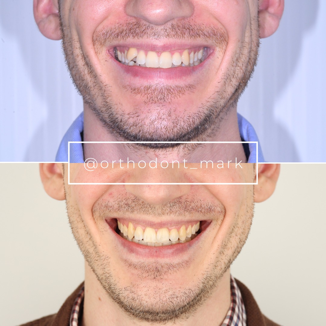
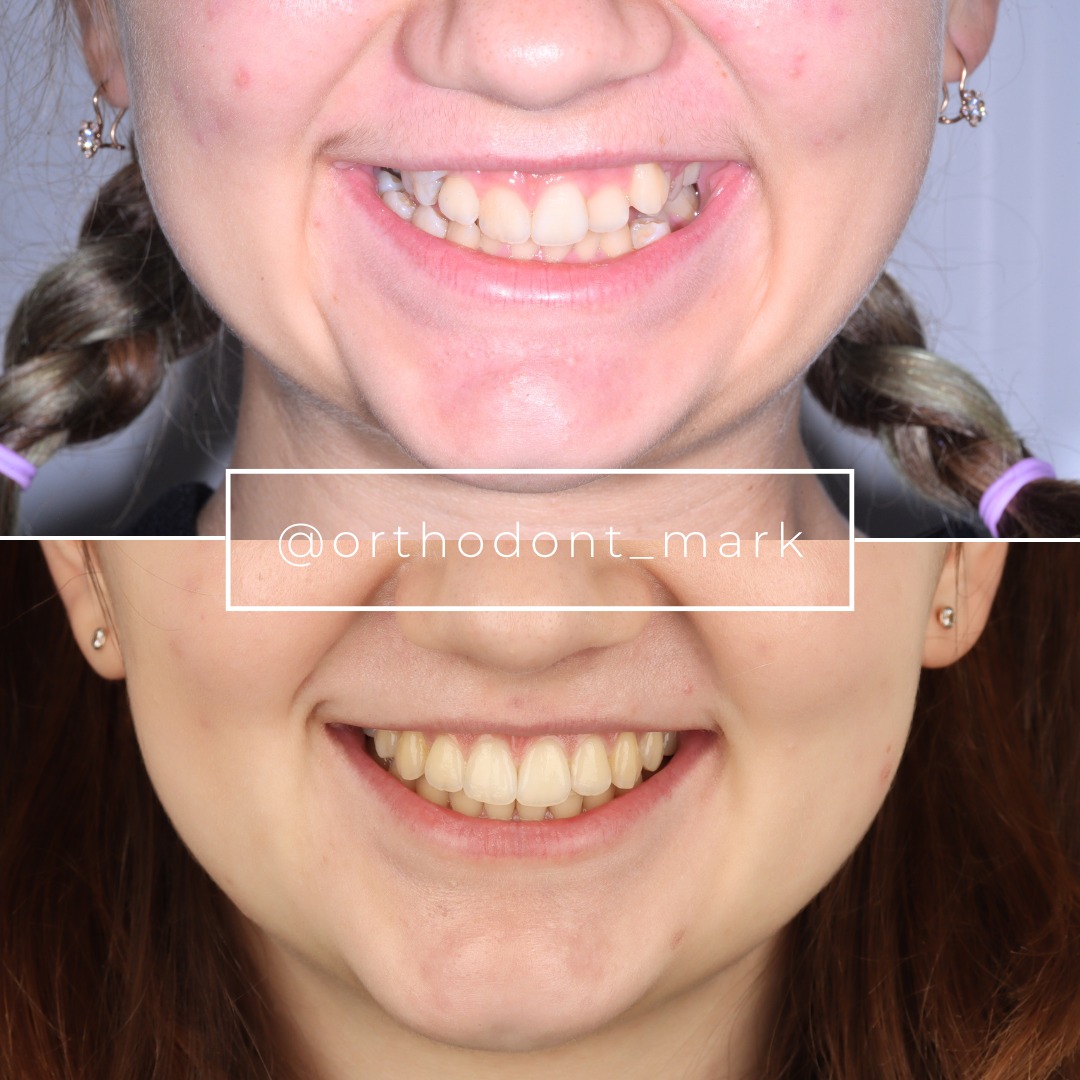
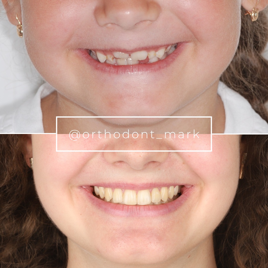
Learn the full MARPE protocol from Dr. Mark Radzhabov
Fundamental course covering CBCT patient selection, miniscrew planning, activation protocols, and 60+ clinical cases. Choose the access level that fits your practice.
Essentials of rapid palatal expansion for practicing orthodontists.
- Core RPE concepts and biomechanics
- 6 structured video lessons
- Clinical decision checklists
- Lifetime access to recordings
Deep-dive into MARPE protocol, diagnostics, and clinical execution.
- 6 modules covering MARPE from A to Z
- CBCT case selection walkthroughs
- Miniscrew placement and activation
- 60+ annotated clinical cases
- Direct Q&A with Dr. Mark Radzhabov
5-element medical consultation framework for dentists and orthodontists.
- Trust-building consultation protocol
- 5 lesson modules
- Templates for treatment plan delivery
- Works with any clinical specialty
Clinical FAQ
What is the MARPE learning pathway and how should it be sequenced for orthodontic residents?
The MARPE learning pathway sequences bench-level force calibration, CBCT imaging literacy, mentored live placement, and post-expansion scan review before independent case management. Establishing biomechanical benchmarks—such as device-specific force ranges from Camporesi et al. (2013)—before the first patient prevents misattributing limited tissue response to technique error.
How do I select the first MARPE patient case as a new operator?
First-case candidates should present with a CBCT-confirmed parallel suture morphology, manageable transverse deficiency, and no significant systemic comorbidities. A 2022 CBCT and dental cast analysis showed that suture opening geometry shifts with age, so imaging review—not arch width alone—drives the screen.
What suture separation rates should a new operator expect when interpreting post-expansion CBCT?
Chun et al. (2022) reported midpalatal suture separation in 90% of RPE patients and 95% of MARPE patients using a standardized 35-turn protocol, with CBCT at baseline, immediately post-expansion, and 3 months. These figures serve as the reference benchmark for new operators reviewing their own scans.
How does MARPE mentorship for orthodontists differ from a standard CE hands-on course?
Mentorship extends beyond placement mechanics to include post-expansion imaging interpretation, complication recognition, and iterative case review. A structured pathway ensures residents develop CBCT reading skills alongside procedural technique rather than acquiring them separately after accumulating early cases.
Which appliance force ranges should inform miniscrew-assisted expansion case planning?
Camporesi et al. (2013) measured Hyrax and A2620 expanders exceeding 20 kg and Palatal Split screws at approximately 16 kg, with literature RME forces ranging from 16.6 to 34.8 pounds (7.54 to 15.8 kg). Device selection should reflect patient anatomy and suture maturity, not default to a single appliance.
Why does suture morphology change with age and how does that affect new operator case screening?
A 2022 CBCT and dental cast analysis documented a shift from parallel to triangular expansion patterns with age, with reduced opening in cranial and dorsal directions. New operators screening cases must account for this geometry change because it alters the force distribution and expected expansion response regardless of appliance choice.
How many activation turns are used in a standardized MARPE protocol for training reference?
Chun et al. (2022) used 35 total activation turns with CBCT imaging at baseline, immediately post-expansion, and at 3 months. This protocol provides new operators with a concrete replicable reference when calibrating their own activation schedules during initial hands-on training.
At what training stage should a new MARPE operator begin independent case management?
Independent management is appropriate after the operator can reliably interpret pre-treatment suture morphology on CBCT, calibrate device-specific force delivery, and review post-expansion scans against published separation benchmarks. Mentorship should continue through at least the first post-expansion imaging review of an independent case.
How does hands-on MARPE training translate force mechanics into clinical decision-making?
Bench drills using the specific screw types intended for clinical use—referencing the force profiles documented by Camporesi et al. (2013)—build tactile judgment before patient contact. That tactile reference then informs real-time decisions about activation rate adjustments when tissue response deviates from expectation.
What CBCT imaging criteria should be included in a MARPE first-case selection checklist?
Minimum criteria include suture morphology classification (parallel versus triangular pattern), bone density assessment at the planned miniscrew insertion sites, and absence of significant suture bridging. A 2022 CBCT and dental cast analysis confirmed that suture opening geometry varies with patient age, making morphologic review non-negotiable.
Acquiring MARPE competency is not a single event—it is a staged progression anchored in biomechanical understanding, imaging literacy, and mentored repetition before independent case load. Clinicians who sequence simulation before live cases, and who screen their first patients against rigorous suture-maturity and force-tolerance criteria, report a markedly smoother transition to independent practice. Dr. Mark Radzhabov has built the structured MARPE training at ortodontmark.com around exactly this logic, offering case-review support and mentorship milestones that match the evidence reviewed here. Key sources: Chun et al. (2022), BMC Oral Health, doi:10.1186/s12903–022-02138-w. Camporesi et al. (2013), BioMedical Engineering OnLine.