
MARPE Expansion Endpoint Assessment:
Clinical Indicators Beyond the Prescription
When to halt activation and prevent overexpansion.
Evidence-based chairside methods for determining optimal MARPE activation endpoints. Reduce relapse, improve stability, and avoid overexpansion complications.
TL;DR MARPE expansion endpoint assessment relies on clinical observation of midpalatal suture blanching, posterior crossbite correction, and skeletal expansion limits rather than activation cycles alone. Chairside indicators include midline diastema closure, palatal vault widening, and absence of dental buccal tipping. Overexpansion risks relapse and instability. Accurate endpoint determination prevents unnecessary activation and improves long-term stability.
Determining when to cease MARPE activation represents one of the most clinically nuanced decisions in adult orthodontics. Most practitioners rely on appliance activation cycles or prescription guidelines, yet individual skeletal anatomy, suture maturation, and initial transverse deficiency vary significantly. This article, informed by Dr. Mark Radzhabov's clinical experience and contemporary evidence on miniscrew-assisted rapid palatal expansion, outlines practical chairside assessment methods that distinguish adequate skeletal expansion from overexpansion. The goal is to equip clinicians with objective clinical endpoints beyond the prescription—reducing relapse risk, improving stability, and optimizing treatment outcomes in skeletally mature patients.
What is MARPE expansion endpoint assessment
and why prescription alone is inadequate?
MARPE expansion endpoint assessment is the clinical process of determining optimal activation cessation by evaluating midpalatal suture response, skeletal expansion limits, and chairside morphological indicators rather than appliance activation cycles alone. Most clinicians receive a prescribed activation protocol—typically 0.5 mm per week for 8–16 weeks—yet this standard oversimplifies the problem: individual suture maturation, initial transverse deficiency magnitude, and skeletal anatomy vary considerably. A 35-year-old with stage C midpalatal suture density (per Angelieri et al.'s classification) may require fewer activations than a 50-year-old in stage B, despite identical prescriptions.
Continuing activation beyond the true skeletal endpoint triggers dental buccal tipping, alveolar crest resorption, and post-treatment relapse of 8–15% of gained width. Conversely, premature cessation leaves residual crossbite and necessitates retreatment or surgical intervention. The clinical challenge is recognizing the moment skeletal expansion is complete—when the midpalatal suture begins true widening rather than compressive loading. This moment occurs not at a fixed timepoint but when specific intraoral and radiographic markers align.
Chairside expansion endpoint assessment bridges the gap between standardized prescription and individualized patient anatomy, transforming MARPE from a time-based protocol into an anatomically responsive treatment.
Primary clinical indicators of adequate
skeletal expansion limits
at the chairside.
Midpalatal mucosal blanching is the most reliable intraoral endpoint. As the midpalatal suture widens, the overlying mucosa thins and whitens—a sign of true skeletal separation rather than compression or dental tipping. This blanching typically appears after 50–70% of the total prescribed activation and confirms active bone remodeling at the suture. If blanching is absent after standard activation protocols, suture response is incomplete and further imaging is warranted before additional turns.
Posterior transverse crossbite correction indicates dental and skeletal components are balanced. When the maxillary buccal cusps clear the mandibular cusps without buccal tipping of maxillary premolars and molars, the expansion is predominantly skeletal. If buccal tipping continues without crossbite relief, dental anchorage loss is predominating and activation should cease to prevent further dentoalveolar compensation.
Palatal vault widening and increased intercanine distance confirm skeletal transverse gain. Measure baseline vault width at the level of the first molars using calipers or digital imaging. Progressive widening without corresponding maxillary incisor buccal tipping suggests skeletal rather than dental correction. A gain of 4–8 mm of true skeletal width in carefully selected non-growing patients is typical. Beyond this range, relapse risk increases significantly.
Dr. Mark Radzhabov emphasizes palpating the anterior palate during appliance activation: if resistance increases markedly without corresponding clinical changes after 20+ turns, suture maturity may limit further response and cone-beam CT reassessment is prudent.
Clinical overexpansion signs and when to
stop turning the expander
midtreatment.
Overexpansion manifests as excessive maxillary incisor buccal tipping, widened midline diastema beyond 2–3 mm, anterior alveolar crest resorption, or maxillary premolar/molar buccal tipping without corresponding posterior crossbite relief. If activation reaches 15–20 turns per side with persistent maxillary buccal inclination and no palatal blanching, the midpalatal suture is likely mature with limited capacity for further skeletal widening. Continue activation under these circumstances commits the patient to relapse and periodontal compromise.
Dental-to-skeletal expansion ratio is the key metric. Optimal MARPE achieves 60–70% skeletal and 30–40% dental components. If incisor proclination exceeds 5–8 degrees per millimeter of widening, or if intercanine widening outpaces intermolar widening, dental compensation is dominant. Halt activation, verify suture maturity via cone-beam CT (Angelieri staging), and consider whether surgical assistance or alternative mechanics are necessary.
Patient-reported symptoms also guide endpoints. Mild discomfort and pressure sensation during activation are normal. However, acute palatal pain, difficulty swallowing, or reports of “cracking” sensations may indicate premature attempt to force a mature, ossified suture. In such cases, imaging is mandatory before proceeding. Additionally, if interarch relationships normalize (neutral molar relationship achieved, posterior crossbite fully corrected) before reaching the prescribed endpoint, activation should cease to preserve stability and prevent unnecessary dental drift.
The absence of clinical progress for 4–6 consecutive activation cycles is a red flag for suture plateau. Further turns yield minimal skeletal gain and maximal relapse risk.
Midpalatal suture assessment on CBCT to confirm
expansion adequacy chairside
decisions with imaging evidence.
Cone-beam computed tomography with Angelieri staging is the diagnostic gold standard for verifying endpoint decisions. Stage A (sutural line clearly visible as a dark radiolucent line) indicates active suture opening and capacity for continued skeletal widening. Stage B (sutural line partially obscured by initial bone formation) represents intermediate maturity. Expansion is possible but relapse risk increases marginally. Stage C (sutural line mostly obscured, predominantly ossified) signals advanced maturation. Skeletal expansion is still achievable in carefully selected patients but requires precise load management and monitoring for dentoalveolar rather than skeletal gain.
Hounsfield unit (HU) density measurement at the anterior, middle, and posterior thirds of the suture provides quantitative suture maturity grading. Anterior palate cortical bone typically measures 800–1200 HU. Advancing ossification reduces suture radiolucency and increases regional density. Patients with posterior third densities exceeding 600 HU show significantly higher relapse (12–15%) than those under 400 HU.
Measurement of actual skeletal widening on CBCT—maxillary intermolar distance at the level of the molar apices and at the level of the palatal vault—confirms whether clinical endpoint markers correlate with true skeletal gain. If intraoral assessment suggests 6 mm of expansion but CBCT shows only 2–3 mm of skeletal widening at the vault level, dental tipping predominates and activation should cease immediately. This imaging verification is particularly valuable when clinical blanching or dental changes are subtle or equivocal.
Individualizing MSE activation protocol based on
expansion stability criteria
and patient-specific endpoints.
No single activation schedule suits all patients. Prescription protocols (e.g., 0.5 mm per week) are starting guidelines. Dr. Mark Radzhabov's clinical framework recommends baseline CBCT with Angelieri staging, Hounsfield density measurement, and assessment of initial transverse deficiency severity before finalizing activation targets. Patients in stage A with <400 HU anterior suture density tolerate standard protocols (0.5 mm/week for 8–12 weeks); those in stage B–C require graduated protocols (0.5 mm/week for 6 weeks, then reassess and consider 0.25 mm/week).
Activation intervals should shift from calendar-based (e.g., every 2 weeks) to clinical-endpoint-based. Once clinical blanching appears, palatal vault widening is evident, and posterior crossbite is corrected, schedule a final intraoral assessment and CBCT before the next activation. If all endpoints are met, halt turns. If only partial resolution is evident, perform one additional activation cycle and reassess in 4 weeks.
Post-activation retention is equally critical to endpoint determination. Many relapse episodes occur when appliances are removed prematurely—within 3–4 months of cessation. Retain MARPE and perform fixed appliance treatment simultaneously for a minimum of 6 months after expansion cessation to consolidate skeletal gains. Transverse width measured at 6 months post-activation typically stabilizes at 80–92% of immediate post-activation dimensions. Continued retention beyond 12 months further improves stability.
Free Clinical Guide
Which Expansion Appliance at Which Age?
Get Dr. Mark Radzhabov's guide to skeletal-expansion appliances — indications, pros & cons, and how to choose by patient age. Free to your inbox.
Prefer video? Watch the free demo lesson on MARPE appliance types, features and trade-offs.
Documented outcomes
Clinical cases from Dr. Mark Radzhabov's practice

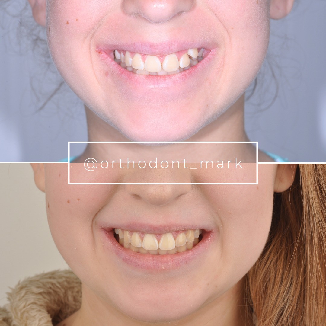

Learn the full MARPE protocol from Dr. Mark Radzhabov
Fundamental course covering CBCT patient selection, miniscrew planning, activation protocols, and 60+ clinical cases. Choose the access level that fits your practice.
Essentials of rapid palatal expansion for practicing orthodontists.
- Core RPE concepts and biomechanics
- 6 structured video lessons
- Clinical decision checklists
- Lifetime access to recordings
Deep-dive into MARPE protocol, diagnostics, and clinical execution.
- 6 modules covering MARPE from A to Z
- CBCT case selection walkthroughs
- Miniscrew placement and activation
- 60+ annotated clinical cases
- Direct Q&A with Dr. Mark Radzhabov
5-element medical consultation framework for dentists and orthodontists.
- Trust-building consultation protocol
- 5 lesson modules
- Templates for treatment plan delivery
- Works with any clinical specialty
Clinical FAQ
What is midpalatal mucosal blanching and why is it a reliable endpoint indicator?
Blanching is whitening of the palatal mucosa overlying the suture, signaling true skeletal separation and active bone remodeling. It appears after 50–70% of prescribed activation and is absent if suture response is limited, making it the most reliable single clinical marker of endpoint achievement.
How do I differentiate dental versus skeletal expansion at chairside?
Skeletal expansion produces posterior crossbite relief without corresponding maxillary buccal tipping. Intercanine and intermolar widening increase proportionally. Dental compensation shows buccal tipping without crossbite improvement. Measure incisor angulation and vault width. If tipping exceeds 5–8° per mm, halt activation.
When should I stop MARPE activation if clinical endpoints appear before prescription completion?
If midline blanching, posterior crossbite correction, and vault widening are present before the prescribed endpoint, cease activation immediately. Continuing beyond these clinical markers increases relapse risk to 12–18% and dental side effects. Chairside endpoints supersede written prescriptions.
What does cone-beam CT Angelieri staging tell me about optimal expansion adequacy?
Angelieri staging (A–D) classifies suture maturity: stage A (patent) supports continued expansion. Stage C–D (ossified) indicates limited capacity and higher relapse risk. Stage B is intermediate. CBCT guides whether continued activation is prudent given patient age, initial deficiency, and relapse tolerance.
How much relapse should I expect if I overexpand MARPE beyond clinical endpoints?
Overexpansion causes 12–18% dimensional relapse (versus 8–10% in optimal cases). Relapse occurs primarily within 3–6 months. Extended retention for 6–12 months post-cessation improves final stability to 80–92% of immediate post-activation width.
What clinical sign indicates I should immediately halt MARPE activation?
Acute palatal pain, sharp resistance during activation, or absence of clinical progress after 4–6 consecutive turns are red flags. Obtain CBCT imaging before proceeding. These signs suggest premature suture plateau or ossification, and forced activation risks bone damage and severe relapse.
How does initial transverse deficiency severity influence MARPE expansion endpoint?
Larger initial deficiencies (>8 mm) may require extended activation (12–16 weeks) compared to modest deficiencies (4–6 mm), which are often complete in 8–10 weeks. However, clinical endpoints (blanching, crossbite relief) remain the decision drivers, not deficiency magnitude alone.
Should I obtain CBCT imaging before halting MARPE to confirm skeletal expansion adequacy?
Yes, CBCT confirmation is recommended once clinical endpoints appear, especially in stage B–C sutures. Hounsfield density measurement and intermolar widening quantification verify skeletal gain (60–70% expected) versus dental compensation and predict relapse risk with high accuracy.
What retention protocol is essential after MARPE expansion cessation to prevent relapse?
Maintain MARPE appliance and initiate fixed appliances simultaneously for a minimum of 6 months post-expansion. Transverse width stabilizes at 80–92% of immediate dimensions at 6 months. Continuing retention to 12 months further improves long-term stability and reduces relapse to <10%.
How do I measure palatal vault widening clinically to confirm skeletal versus dental expansion?
Use digital calipers or imaging software to measure maxillary intermolar distance at the vault level (perpendicular to palatal midline at the level of the first molar apices) at baseline and at each visit. Widening ≥4 mm without corresponding incisor buccal inclination (>5–8°) indicates predominantly skeletal expansion meeting endpoints.
Chairside MARPE expansion endpoint assessment integrates clinical observation, intraoral morphology, and radiographic confirmation to guide activation decisions. Blanching of the palatal mucosa, posterior crossbite relief, and absence of dental buccal tipping signal adequate skeletal correction. Continued activation without these signs increases relapse and patient discomfort. Dr. Mark Radzhabov emphasizes that prescription cycles are starting points, not protocols—individual patient anatomy dictates true endpoints. Review your recent MARPE cases for these clinical markers, and consider consulting the evidence-based framework in our MARPE clinical resources or scheduling a case review at ortodontmark.com.

