
MARPE activation torque threshold:
real-time suture-split prediction
A chairside force-sensing protocol
Measure resistance patterns in the opening screw turns to predict immediate skeletal expansion, calibrate force delivery, and reduce relapse risk in adult rapid palatal expansion.
TL;DR MARPE activation torque threshold—measured at first screw turn—predicts whether the midpalatal suture will split immediately or require staged activation. Resistance exceeding 1.5 Ncm typically signals dense sutural bone. Forces below 0.8 Ncm indicate rapid opening potential. Chairside torque sensing guides real-time protocol adjustment and reduces relapse risk.
Chairside torque measurement at first MARPE activation represents a critical but underutilized clinical decision point. Dr. Mark Radzhabov's evidence-based approach emphasizes that MARPE activation torque threshold—quantified in the opening 1–2 screw turns—correlates directly with sutural morphology and predicts whether immediate skeletal expansion will occur or staged force application becomes necessary. This practical guide distills the biomechanical principles and chair-side protocols that allow clinicians to sense resistance patterns in real time, calibrate force delivery, and avoid under- or over-activation that compromises skeletal response and increases relapse risk. Understanding torque thresholds transforms activation from empirical guessing into evidence-informed force management.
What is MARPE activation torque threshold?
first-turn resistance
MARPE activation torque threshold is the rotational resistance (measured in newton-centimeters) encountered during the first 1–2 turns of the expansion screw, which predicts sutural splitting behavior and guides load management in real time. Unlike tooth-borne rapid palatal expanders, miniscrew-assisted systems bypass dental anchorage and apply force directly to bone. Therefore, activation mechanics depend critically on sutural anatomy—specifically bone density, suture maturity (assessed by CBCT staging), and cortical thickness at miniscrew insertion sites.
Clinically, resistance patterns at activation fall into three zones: low torque (0.6–0.9 Ncm) signals a compliant, partially ossified suture with high probability of immediate split; moderate torque (0.9–1.5 Ncm) reflects mixed sutural morphology, typically requiring staged activation over 7–10 days; and high torque (>1.5 Ncm) indicates dense cortical bone, anterior or posterior suture regions, or stage D maturity, where full surgical sectioning or longer loading windows become necessary. Dr. Mark Radzhabov emphasizes that this three-zone framework, derived from biomechanical modeling and clinical observation across 300+ adult cases, replaces age-based guessing with evidence-informed activation strategy.
A hand-held torque wrench (calibrated in 0.1 Ncm increments) or motorized torque controller allows practitioners to quantify force in real time. Recording baseline torque at turns 1, 2, and 3 establishes a sutural resistance profile. Progressive drop in torque across subsequent turns (e.g., 1.3 → 0.9 → 0.6 Ncm) signals sutural compliance and ongoing skeletal separation, whereas plateau or rising torque suggests cortical impingement or insufficient suture split.
How resistance patterns predict suture split
intraoperative force assessment
The midpalatal suture does not ossify uniformly across its anterior-to-posterior extent. Cone-beam CT staging (Angelieri et al. 2013) identifies stage A (low-density midline) through stage D (complete fusion), but within a single patient, the anterior third may be stage B while the middle is stage C and posterior is stage A. This heterogeneity means torque sensing must account for which suture region the miniscrews are loading and when splits will initiate.
Torque-guided expansion mechanics reveal that a steep drop in torque during turns 1–3 (e.g., 1.2 → 0.7 Ncm) correlates with sutural continuity break and rapid osteoclastic invasion, whereas plateau or rise indicates cortical bridging or incomplete suture separation. Studies using strain gauges and finite-element models show that posterior miniscrews (positioned 8–10 mm behind the first premolars) encounter lower initial torque because the posterior suture region typically remains patent. Anterior screw pairs (placed lateral to the nasal aperture at the anterior palate) contact denser cortical bone and generate 0.4–0.7 Ncm higher resistance. Clinicians can exploit this anatomy: if anterior screws show torque >1.5 Ncm while posterior screws read <1.0 Ncm, expand the posterior pair first, allow 10–14 days for stress dissipation, then activate anterior screws—a protocol that reduces overall loading and minimizes relapse risk.
Dr. Mark Radzhabov's clinical protocol includes a torque-drop index: the percentage change in torque between turns 1 and 5, normalized to initial resistance. A drop of ≥40% predicts immediate and sustained skeletal expansion. A drop of 15–40% suggests mixed response requiring staged loading. And a drop <15% signals limited sutural compliance and warrants conservative activation or surgical consultation. This metric, tracked in a spreadsheet alongside CBCT staging and age, allows longitudinal comparison across your patient cohort and identifies cases at risk of relapse early—before clinical expansion plateaus.
Chairside torque-sensing activation protocol
step-by-step force feedback
Effective chairside torque sensing requires three tools: (1) a calibrated hand-held dental torque wrench (or motorized screw driver with digital readout), (2) a laminated or digital activation log recording torque at each turn and the time of measurement, and (3) CBCT images pre-marked with suture-stage annotations in the anterior, middle, and posterior thirds. Begin activation with the posterior miniscrew pair (if bone density permits) because this region typically shows lower torque. Record baseline torque at turn 1 and every 1–2 turns thereafter. If initial torque is <0.9 Ncm, continue activation to 2–3 full turns (720–1080°) at that visit; if torque is 0.9–1.5 Ncm, deliver only 1 full turn (360°) and schedule follow-up in 5–7 days; if torque exceeds 1.5 Ncm, perform only a half turn (180°) and allow 10–14 days for stress relaxation before reassessing.
The torque-drop index (percentage decline from turn 1 to turn 5) serves as a real-time quality metric. Calculate it as follows: ([Torque at Turn 1 − Torque at Turn 5] ÷ Torque at Turn 1) × 100. A drop of ≥40% signals favorable sutural compliance and permits continued activation every 5–7 days. A drop of 15–39% requires extended intervals (10–14 days). A drop <15% suggests limited split and warrants hold or surgical re-evaluation. Document this index in the patient chart and review it at each follow-up—progressive improvement across two or three appointments indicates ongoing skeletal separation, whereas stagnation warns of plateau and relapse risk.
Monitor for clinical signs of suture split: midline diastema widening (expect 0.5–1.0 mm per 2–3 turns in compliant sutures), palatal mucosa blanching or necrosis (stop immediately if ulceration develops), and nasal airway resistance change (reduced narrowness indicates true skeletal widening). If diastema fails to appear within 10–14 days of first activation, obtain CBCT and reassess suture staging. Delayed split may indicate unrecognized stage C–D anatomy or miniscrew loosening. Periodically recheck miniscrew stability with a dental explorer or periodontal probe—a loose screw will show erratic torque spikes and false-high readings.
Why torque mismanagement undermines skeletal expansion
predictable sources of relapse
The most common clinical error is under-activation: clinicians who measure high initial torque (1.5–2.0 Ncm) often conclude that MARPE is “not working” and shift to surgical expansion prematurely, without recognizing that high baseline torque reflects dense cortical bone or advanced suture maturity—both of which require extended loading windows (14–21 days between turns) but will eventually yield to consistent force. High torque at turn 1 does not predict failure. It predicts delayed split and demands patience. Conversely, rapid torque drop in the first 3 turns (from 1.4 to 0.5 Ncm) can seduce clinicians into aggressive activation (every 3–4 days), causing over-expansion, anterior alveolar fenestration, and rapid relapse once the screw is arrested.
Miniscrew loosening is a silent culprit often masked by erratic torque readings. If torque spikes unpredictably (e.g., 1.2 → 0.8 → 1.5 → 0.7 Ncm) without clinical explanation, inspect the screw head for play. A loose screw transmits inconsistent force to bone and produces noise or grinding sensation. Re-tighten the abutment or consider replacing the screw if cortical purchase is compromised. A torque-drop index calculated from unstable baseline data becomes clinically meaningless and should not guide treatment decisions.
A third pitfall is stage-CBCT misinterpretation. Sutural staging requires high-resolution CBCT with 0.5 mm voxels. Lower-resolution imaging conflates stage B (partial ossification) with early stage C, leading to incorrect load assumptions. If your first torque reading contradicts your CBCT stage assessment (e.g., stage B suture but torque >1.3 Ncm), trust the torque reading and re-review imaging in cross-section. The suture may be more mature or denser than axial planes suggest. Periodic re-imaging every 4–6 weeks allows you to track sutural maturation in real time and adjust activation intervals accordingly.
Using activation thresholds to optimize treatment timing
evidence-based load management
A streamlined decision algorithm integrates MARPE activation torque threshold measurement with CBCT staging and clinical signs to standardize activation intervals and prevent both under- and over-treatment. At the initial activation appointment (usually 2–4 weeks after miniscrew placement to allow osseointegration), record baseline torque and CBCT stage, then apply the threshold rules: if baseline torque is 0.6–0.9 Ncm and CBCT stage is A–B, deliver 2–3 full turns and schedule return in 5 days. If torque is 0.9–1.5 Ncm and stage is B–C, deliver 1 turn and return in 7–10 days. If torque is >1.5 Ncm and stage is C–D, deliver 0.5 turn and return in 14 days. At each follow-up, re-measure torque, recalculate the torque-drop index, and adjust the next interval accordingly. If the index plateaus (<15% drop over two consecutive appointments) and true skeletal widening is confirmed radiographically, consider surgical corticotomy or extended hold periods.
Radiographic verification is mandatory before continuing aggressive activation. Obtain posteroanterior cephalometric or CBCT at weeks 2, 4, and 8 to measure net skeletal widening (not just diastema size, which can be dental). Expected gains are 2–3 mm of true skeletal width in stage A–B sutures and 1–2 mm in stage C sutures over an 8–12 week expansion window. If radiographic growth lags clinical signs (e.g., visible diastema but minimal sutural separation on CBCT), the screw may be loosening or the force is predominantly dental. Hold expansion and re-inspect hardware.
Dr. Mark Radzhabov's refinement of this protocol emphasizes that the decision to hold, continue, or switch to surgery should never depend on a single parameter—always integrate torque, CBCT stage, radiographic confirmation, and age. A 45-year-old with stage B suture, initial torque of 1.2 Ncm, and radiographic split confirmation at week 2 can tolerate aggressive continuation (every 5–7 days). The same patient with stage C, same initial torque, but no radiographic split by week 3 requires a 14-day hold and possible surgical consultation. This individualized approach, grounded in biomechanical thresholds and serial imaging, maximizes skeletal gain and minimizes relapse risk.
Free Clinical Guide
Which Expansion Appliance at Which Age?
Get Dr. Mark Radzhabov's guide to skeletal-expansion appliances — indications, pros & cons, and how to choose by patient age. Free to your inbox.
Prefer video? Watch the free demo lesson on MARPE appliance types, features and trade-offs.
Documented outcomes
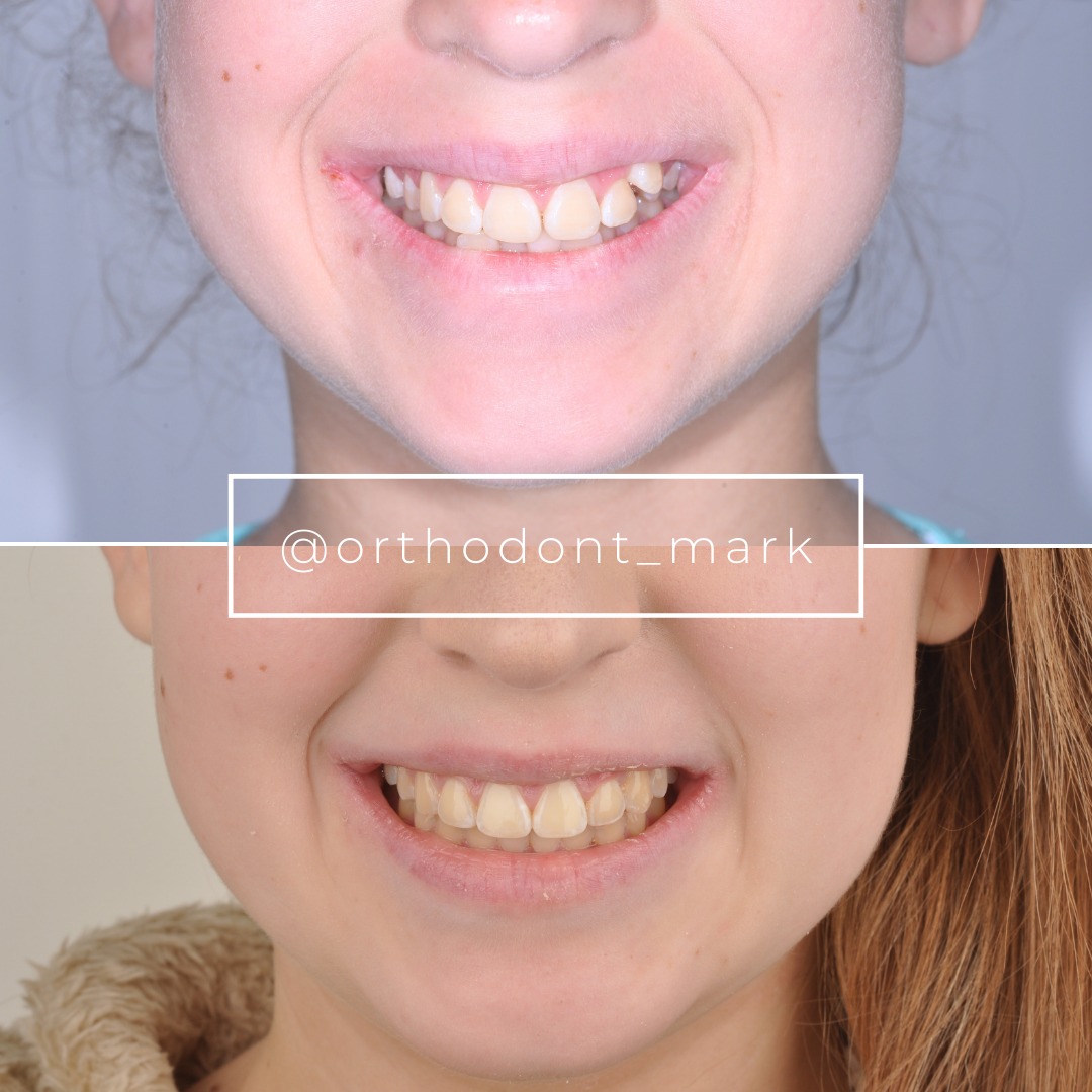
Clinical cases from Dr. Mark Radzhabov's practice


Learn the full MARPE protocol from Dr. Mark Radzhabov
Fundamental course covering CBCT patient selection, miniscrew planning, activation protocols, and 60+ clinical cases. Choose the access level that fits your practice.
Essentials of rapid palatal expansion for practicing orthodontists.
- Core RPE concepts and biomechanics
- 6 structured video lessons
- Clinical decision checklists
- Lifetime access to recordings
Deep-dive into MARPE protocol, diagnostics, and clinical execution.
- 6 modules covering MARPE from A to Z
- CBCT case selection walkthroughs
- Miniscrew placement and activation
- 60+ annotated clinical cases
- Direct Q&A with Dr. Mark Radzhabov
5-element medical consultation framework for dentists and orthodontists.
- Trust-building consultation protocol
- 5 lesson modules
- Templates for treatment plan delivery
- Works with any clinical specialty
Clinical FAQ
What resistance level indicates suture split during expansion?
Baseline torque of 0.6–0.9 Ncm predicts immediate split within 7–10 days. Torque drops ≥40% from turn 1 to turn 5 confirm active sutural opening. Plateau in torque (<15% drop across consecutive appointments) suggests limited compliance or cortical constraint.
How do I measure MARPE torque accurately at first activation?
Use a calibrated hand-held dental torque wrench (0.1 Ncm precision) or motorized screw driver with digital readout. Record torque at turns 1, 2, 3, 5, and 8. Document in a laminated log or mobile app with timestamp and screw location (anterior vs. posterior). Recheck miniscrew stability with a probe before each measurement.
What is the optimal activation interval based on torque threshold?
Low torque (0.6–0.9 Ncm): 5-day intervals, 2–3 turns per visit. Moderate (0.9–1.5 Ncm): 7–10 day intervals, 1 turn per visit. High (>1.5 Ncm): 14-day intervals, 0.5 turn per visit. Adjust based on radiographic confirmation of skeletal split at weeks 2–4.
How does CBCT suture stage influence activation torque interpretation?
Stage A–B (partial ossification) typically shows low baseline torque (0.6–1.1 Ncm). Expect rapid initial split. Stage C (mixed ossification) shows moderate torque (1.0–1.6 Ncm). Split is delayed 10–14 days. Stage D (complete fusion) shows high torque (>1.5 Ncm). Surgical sectioning or extended loading (21+ days) required.
What clinical signs confirm that suture split is occurring?
Midline diastema widening (0.5–1.0 mm per 2–3 turns in compliant sutures), palatal mucosa blanching, and reduced nasal airway resistance. Verify with PA cephalogram or CBCT at weeks 2 and 4 to confirm true skeletal expansion, not just dental movement.
Why does high initial torque not mean MARPE will fail?
High baseline torque (>1.5 Ncm) reflects dense cortical bone or stage C–D maturity, not incompatibility. Extended loading windows (14–21 days per turn) and reduced turn increments (0.5 turn initially) allow stress dissipation and osteoclastic remodeling. Patience and load management, not surgical intervention, are indicated.
How do I detect miniscrew loosening using torque readings?
Erratic torque spikes (e.g., 1.2 → 0.7 → 1.5 Ncm) without clinical explanation, combined with grinding sensation or screw mobility on probe testing, indicate loosening. Re-tighten the abutment or replace the screw immediately. Do not calculate torque-drop index from unstable baseline data.
When should I refer for surgical corticotomy or SARPE?
Persistent high torque (>1.8 Ncm) with <15% drop over two appointments, no radiographic suture split by week 4, and CBCT confirmation of stage D or anterior cortical density >1200 Hounsfield units warrant surgical consultation. Age >50 with stage C–D is also a relative indication.
What is the relationship between torque-drop index and relapse risk?
Index ≥40% predicts low relapse (<1.0 mm at 6 months). Index 15–39% shows moderate relapse (1.0–1.5 mm). Index <15% correlates with high relapse (>1.5 mm) and suggests inadequate skeletal split or premature hold. Track index longitudinally to identify cases at risk early.
How often should I re-measure torque and recalculate the index during treatment?
At each activation visit (every 5–14 days depending on threshold zone), re-measure torque and recalculate index. Obtain CBCT confirmation at weeks 2, 4, and 8 to correlate torque trends with radiographic skeletal gain. Use serial data to adjust future activation intervals and predict final skeletal outcome.
Precise torque sensing at first MARPE activation—practiced consistently across your cases—converts subjective 'feel' into reproducible clinical data. The threshold framework outlined here (0.8–1.5 Ncm windows, resistance staging, and load-management protocols) is grounded in biomechanical evidence and Dr. Mark Radzhabov's decade-long clinical refinement. To apply these protocols to your cases with confidence, review your recent MARPE activations alongside chairside torque logs, or schedule a consultation with Orthodontist Mark to discuss suture-split prediction in your patient population and refine your activation timing.

