MSE anchor screw angulation protocols:
perpendicular vs angled
placement for bicortical engagement
A CBCT-grounded comparison of insertion trajectories, skeletal force distribution, and documented clinical outcomes to sharpen your MSE protocol decisions.
TL;DR MSE anchor screw angulation protocols—perpendicular versus angled placement—directly determine the quality of bicortical engagement and the skeletal versus dental distribution of expansion forces. Paredes et al. (2020) documented skeletal expansion reaching 96.58% of total displacement in a cohort of 39 patients, with dental tipping held to 4.23%. Insertion site selection and trajectory are therefore non-trivial protocol decisions.
Anchor screw angulation in MSE therapy is among the least standardized yet most mechanically consequential decisions a clinician makes before activating a palatal expander. Whether a perpendicular or angled insertion trajectory achieves reliable bicortical purchase depends on palatal bone availability, screw length, and the displacement mechanics the appliance must generate. At ortodontmark.com, Dr. Mark Radzhabov distills the published CBCT evidence on MSE anchor screw positioning to give practicing orthodontists a decision-ready framework for choosing and documenting their insertion protocol before the first activation turn.
Why does insertion trajectory matter for
bicortical purchase
in MSE therapy?
MSE anchor screw angulation protocol is the standardized specification of insertion trajectory—perpendicular or angled relative to the palatal plane—used to achieve predictable bicortical engagement of miniscrews during palatal expansion. The choice of trajectory is not cosmetic: it governs whether the screw tip engages the nasal-floor cortex or terminates within cancellous bone, a difference that determines primary stability under cyclic activation loads. Giudice et al. (2020) established that MSE miniscrews are placed in the anterior palate because that region offers adequate bone availability and carries low risk to critical anatomical structures such as the nasopalatine canal and greater palatine vessels. This anatomical preference constrains the angulation window: the palatal vault geometry in the anterior zone means that a strictly perpendicular trajectory may encounter thinner cortical bone coronally, while a mesially or distally angled path can reach the denser cortical layer closer to the nasal floor. Conventional MARPE placement, as described by Giudice et al. (2020), positions two miniscrews in the anterior palate posterior to the second palatal rugae, with the expander body retained by screws or cementation caps. This spatial arrangement defines the starting angulation envelope: screws inserted too steeply risk nasal mucosal perforation, while an excessively oblique angle reduces the effective depth of cortical engagement and increases the lever arm that translates activation force into bending rather than separation. Documenting the chosen trajectory on the pre-treatment CBCT slice is therefore a baseline requirement for meaningful protocol comparison across cases.
How does screw angulation influence
skeletal versus dental
displacement during MSE expansion?
The downstream effect of palatal screw insertion angle becomes measurable when CBCT landmarks are used to decompose total transverse displacement into its skeletal, alveolar, and dental components. Paredes et al. (2020) quantified this in 39 patients treated with MSE: angular measurements relative to the rotational fulcrum showed 96.58% to 95.44% of displacement was skeletal, with alveolar bone bending contributing only 0.34% to 0.33% and dental tipping accounting for 3.08% to 4.23% (Paredes et al., 2020). The rotational fulcrum itself is not a fixed anatomical point. Paredes et al. (2020) located it at the most external and inferior point of the zygomatic process, or slightly above the interfrontal distance in the coronal plane, with 20 of 39 subjects demonstrating a rotational fulcrum at an interfrontal distance measuring 101.6 ± 4.7 mm. This means the lever geometry the appliance works against is patient-specific, and the screw angulation that maximizes bicortical engagement in one patient may not map identically to the next. For the clinician, these figures reframe angulation as a force-routing decision. A trajectory that achieves deep cortical anchorage directs activation load across the midpalatal suture with minimal deflection into alveolar bending—consistent with the near-exclusive skeletal displacement proportions Paredes et al. (2020) documented. Angled placements that intentionally orient the screw toward the nasal-floor cortex can improve lever efficiency when the anterior palatal vault is shallow, whereas a perpendicular path may be adequate when bone depth is generous enough to guarantee bicortical purchase without redirecting force toward the zygomatic fulcrum.
What does the evidence say about
complete suture separation
across MSE and BAME appliances?
Sarraj et al. (2021) compared MSE and BAME appliances in a CBCT study and found that both appliances achieved complete midpalatal suture separation in 100% of patients. While this result applies across appliance designs, it does not decouple screw angulation from outcome—it confirms that when insertion protocols are executed correctly within each appliance system, full suture separation is achievable. The clinical implication is that suture separation alone is an insufficient benchmark for protocol quality. The distribution of displacement between skeletal, alveolar, and dental structures—documented most precisely by Paredes et al. (2020)—is the more discriminating metric. Two cases can both achieve complete separation while differing substantially in how much of that separation reflects true skeletal widening versus dental compensation. Angulation protocol is one of the variables that shifts that ratio. For orthodontists auditing their own insertion technique, the combination of findings across these studies supports a pre-treatment workflow that uses CBCT to measure anterior palatal bone depth at the planned insertion coordinates, select screw length to ensure cortical engagement at the nasal floor, and document the chosen trajectory angle for comparison across cases. Reviewing MSE anchor screw positioning against outcomes in your own records, referencing the skeletal displacement proportions reported by Paredes et al. (2020), provides a practical internal quality loop that published case series cannot substitute for.
Free Clinical Guide
Which Expansion Appliance at Which Age?
Get Dr. Mark Radzhabov's guide to skeletal-expansion appliances — indications, pros & cons, and how to choose by patient age. Free to your inbox.
Prefer video? Watch the free demo lesson on MARPE appliance types, features and trade-offs.
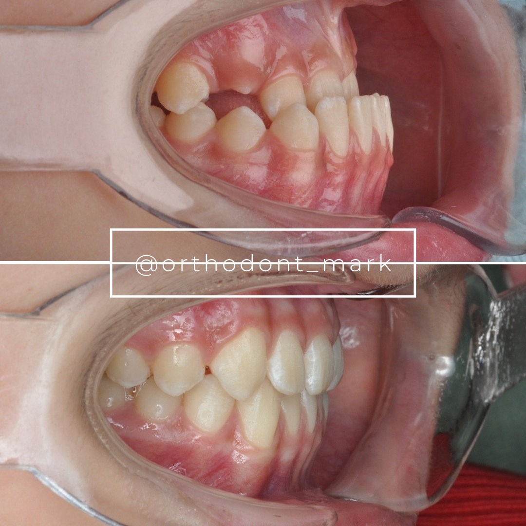
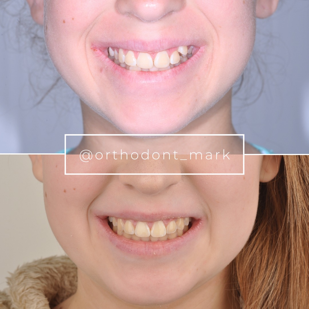
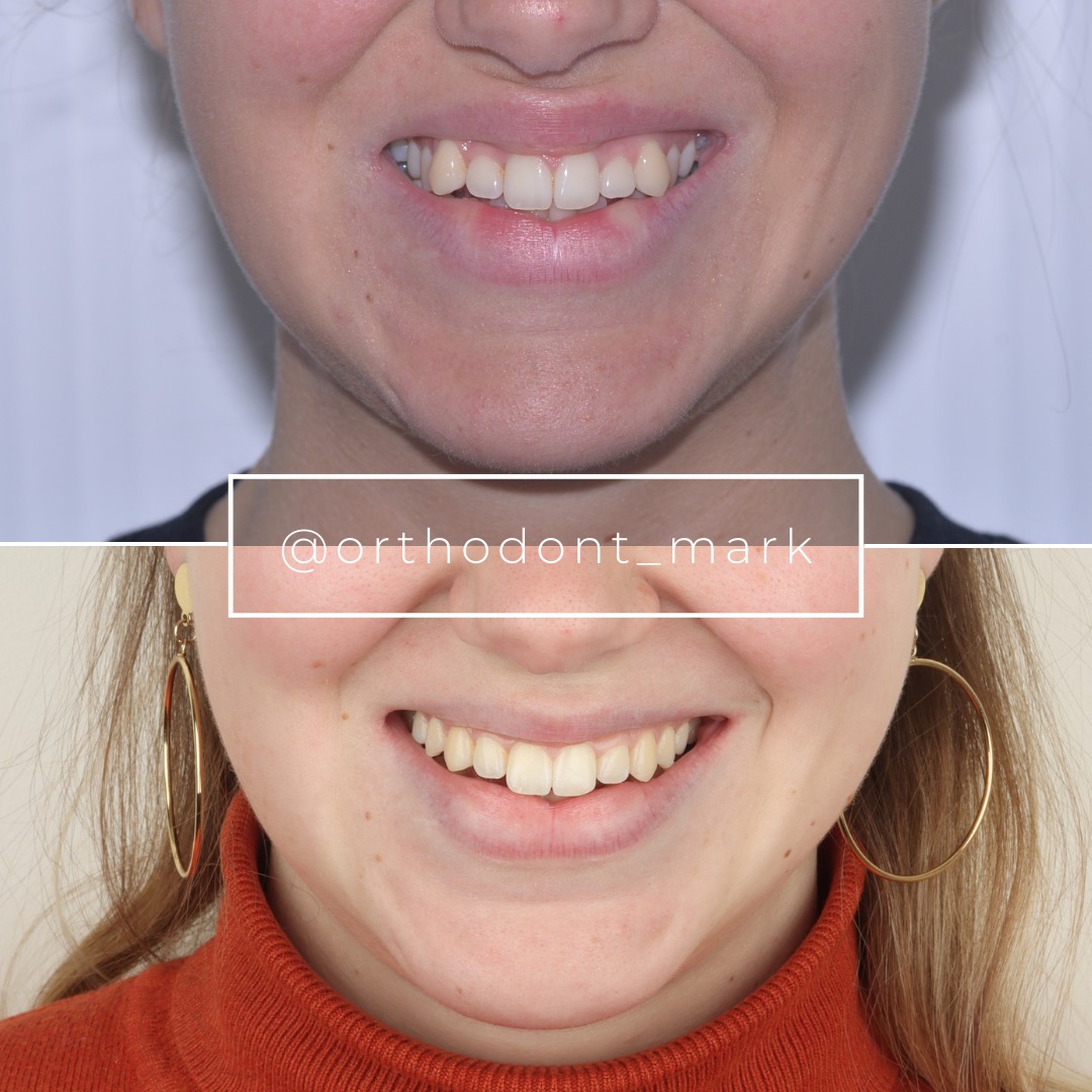
Documented outcomes
Clinical cases from Dr. Mark Radzhabov's practice
Learn the full MARPE protocol from Dr. Mark Radzhabov
Fundamental course covering CBCT patient selection, miniscrew planning, activation protocols, and 60+ clinical cases. Choose the access level that fits your practice.
Essentials of rapid palatal expansion for practicing orthodontists.
- Core RPE concepts and biomechanics
- 6 structured video lessons
- Clinical decision checklists
- Lifetime access to recordings
Deep-dive into MARPE protocol, diagnostics, and clinical execution.
- 6 modules covering MARPE from A to Z
- CBCT case selection walkthroughs
- Miniscrew placement and activation
- 60+ annotated clinical cases
- Direct Q&A with Dr. Mark Radzhabov
5-element medical consultation framework for dentists and orthodontists.
- Trust-building consultation protocol
- 5 lesson modules
- Templates for treatment plan delivery
- Works with any clinical specialty
Clinical FAQ
What is the clinical rationale for perpendicular MSE screw placement versus an angled insertion trajectory?
Perpendicular placement suits cases where anterior palatal bone depth is sufficient for bicortical purchase without redirection. An angled trajectory is favored when the vault geometry requires orienting the screw tip toward the nasal-floor cortex to guarantee cortical engagement and minimize alveolar bending.
How do I confirm bicortical engagement after MSE screw insertion?
Post-insertion CBCT allows direct visualization of screw tip position relative to the nasal-floor cortex. Clinically, firm primary stability with no detectable micro-movement under digital loading pressure is a preliminary indicator, but imaging confirmation is the definitive standard.
Which CBCT landmarks should I use to plan MSE anchor screw angulation?
Plan on a sagittal CBCT slice at the intended insertion coordinate posterior to the second palatal rugae, as described by Giudice et al. (2020). Measure cortical bone thickness at the proposed entry point and nasal-floor depth to determine the angulation needed for bicortical purchase.
Does screw angulation protocol affect the skeletal-to-dental displacement ratio in MSE?
Yes. Paredes et al. (2020) documented that optimized MSE anchorage produced skeletal displacement of 96.58% to 95.44% of total expansion in 39 patients, with dental tipping limited to 3.08% to 4.23%. Trajectories that compromise cortical anchorage shift this ratio toward dental effects.
Where is the MSE rotational fulcrum located and how does it affect angulation planning?
Paredes et al. (2020) located the fulcrum at the most external and inferior point of the zygomatic process or slightly above the interfrontal distance, measuring 101.6 ± 4.7 mm in 20 of 39 patients. This patient-specific location means no single angulation rule covers every case.
Can both MSE and BAME appliances achieve complete midpalatal suture separation regardless of screw angulation?
Sarraj et al. (2021) found complete suture separation in all patients across both MSE and BAME appliances. However, suture separation alone does not capture the skeletal versus dental distribution of that displacement—angulation protocol influences that more sensitive outcome metric.
What anterior palate anatomy makes angled MSE screw placement preferable to perpendicular insertion?
A shallow anterior palatal vault reduces the available perpendicular depth for cortical engagement. In these cases, a distally or mesially angled trajectory allows the screw to reach the denser cortical layer near the nasal floor, as Giudice et al. (2020) identify that zone as the preferred insertion region.
How should I document MSE insertion trajectory for protocol comparison across cases?
Record the angulation relative to the palatal plane on the pre-treatment CBCT sagittal slice at the screw coordinate. Pairing this with post-treatment displacement analysis—referencing the skeletal and dental ratios established by Paredes et al. (2020)—enables meaningful within-practice protocol audit.
Does posterior-to-second-rugae placement constrain the angulation options for MSE miniscrews?
Yes. Giudice et al. (2020) describe the second palatal rugae as the posterior boundary for conventional MARPE placement. This zone's vault curvature limits the safe perpendicular depth and often makes a slight angled trajectory necessary to direct the screw tip toward adequate cortical bone.
What is the minimum information a clinician should record to compare perpendicular versus angled MSE screw placement outcomes?
At minimum: pre-treatment CBCT sagittal slice showing planned angulation, screw length, anterior palatal bone depth at insertion, and post-treatment CBCT with displacement decomposed into skeletal, alveolar, and dental components using the methodology described in Paredes et al. (2020).
Anchor screw angulation is not a detail to be resolved chairside by feel—it is a protocol commitment that determines whether the MSE appliance distributes load skeletally or deflects it into dental tipping and alveolar bending. The evidence from Paredes et al. (2020) and Giudice et al. (2020) points to the anterior palate as the preferred insertion zone, with trajectory tailored to available cortical depth. Clinicians who want to audit and strengthen their own insertion protocol are encouraged to review documented case workflows at ortodontmark.com or register for structured MARPE training with Dr. Mark Radzhabov. Key sources: Paredes et al. (2020), Progress in Orthodontics, doi:10.1186/s40510–020-00320-w. Giudice et al. (2020), Materials, doi:10.3390/ma13081815. Sarraj et al. (2021), APOS Trends in Orthodontics, doi:10.25259/APOS_160_2020.