
Suture Split Verification CBCT Landmarks:
Anterior-Posterior Confirmation
Distinguishing True Skeletal Expansion from Dentoalveolar Bending
Post-activation CBCT analysis protocol to confirm midpalatal suture opening and prevent relapse-prone dentoalveolar compensation.
TL;DR Suture split verification CBCT landmarks distinguish true skeletal expansion from dentoalveolar bending by assessing anterior-posterior opening at specific anatomical zones. Key markers include parallel widening at the midpalatal suture with minimal alveolar plate angulation, increased nasal cavity width, and posterior palatal vault elevation. Accurate verification requires high-resolution imaging and region-of-interest measurement.
Post-activation imaging remains one of the most critical yet underutilized steps in miniscrew-assisted rapid palatal expansion (MARPE) protocol. After 4–6 weeks of loading, many clinicians activate appliances without radiographic confirmation that the midpalatal suture has actually split. Dentoalveolar bending—lateral movement of tooth-bearing structures without true suture separation—mimics skeletal expansion on 2D radiographs but produces unstable, relapse-prone results. High-resolution cone-beam computed tomography (CBCT) with anatomical landmark identification now allows clinicians to verify genuine anterior-posterior split versus compensation, distinguishing responders from non-responders before committing to months of force application. This guide reviews the specific CBCT landmarks and measurement protocol that Dr. Mark Radzhabov and leading centers use to confirm skeletal versus dental effects in real clinical time.
What Are Suture Split Verification CBCT Landmarks?
Suture split verification CBCT landmarks are specific anatomical measurements and radiographic signs used to confirm that true midpalatal suture separation has occurred, distinguishing skeletal expansion from dentoalveolar bending or root movement. After 4–6 weeks of MARPE activation, post-activation CBCT imaging reveals whether force has widened the midpalatal suture itself or simply tipped teeth laterally and bent alveolar bone—a critical distinction that predicts stability and relapse risk.
The anterior-posterior split confirmation process requires measurement at three distinct anatomical zones: the anterior palate (anterior to the incisive foramen), the middle third (transitional zone near the palatal vault apex), and the posterior palate (posterior to the palatal vault). True skeletal expansion produces nearly parallel widening across all three zones with minimal angulation of the alveolar plates, whereas dentoalveolar bending concentrates opening at the alveolar crest with convergent plates and root movement.
High-resolution CBCT with voxel size ≤0.3 mm and reformatted coronal and parasagittal views allows measurement accuracy within 0.5 mm. Clinicians place region-of-interest (ROI) cursors at the midline radiolucent line (the unfused suture interface) and compare widening from baseline to post-activation scans, ensuring that opening is uniform and centrally located rather than lateralized toward teeth or alveolar structures.
The Three Anatomical Zones:
Anterior, Middle, Posterior
Mapping Expansion Across the Palate
The anterior palate zone extends from the midline behind the central incisors to just anterior to the incisive foramen. At this region, healthy MARPE response shows 2–3 mm of midpalatal widening within 4–6 weeks, with no lateral tilt of the incisor teeth and no angulation of the interdental alveolar crest. Dentoalveolar bending produces convergent plates (wider at the midline, narrower at the alveolar crest) and incisor tipping.
The middle palatal zone (transitional region near the hard palate vault) serves as the inflection point where skeletal and dentoalveolar forces compete. In successful expansion, this zone shows 1.5–2.5 mm of widening with the suture opening as a clean radiolucent line running through the center of the palatal vault. If widening is asymmetric or offset from the anatomical midline, uneven miniscrew loading or early bony bridging is suspected.
The posterior palate zone (soft palate junction and posterior hard palate) typically shows slightly less opening (1.0–2.0 mm) than the anterior region due to anatomical tapering. Posterior opening that is significantly less than anterior opening (difference >2 mm) suggests early fusion posteriorly or inadequate posterior force transmission, which may require load redistribution or protocol adjustment.
Post-Activation CBCT Verification Technique
Measurement Protocol
The post-activation CBCT verification protocol begins with baseline (pre-activation) and post-activation scans acquired in the same head position and with identical voxel size and field-of-view parameters. Reformatted coronal slices perpendicular to the sagittal plane allow direct visualization of the midpalatal suture midline and bilateral alveolar plate positions. The clinician places a horizontal reference line through the nasal floor to standardize measurement orientation.
At each of the three anatomical zones, the ROI cursor measures the distance between the most medial margins of the right and left palatal shelves at the suture interface (the radiolucent line separating them). Record baseline and post-activation distances at each level. True skeletal opening is calculated as the difference between post-activation and baseline measurements at the suture center, not at the alveolar margin. Root-to-midpalatal distance measurements rule out dentoalveolar compensation: in pure dentoalveolar bending, roots move laterally while the suture itself remains nearly closed.
Dr. Mark Radzhabov's clinical framework recommends acquiring parasagittal slices (angled 5–10° off true sagittal) along the midline to assess palatal vault height change. True skeletal expansion elevates the hard palate vault (increasing palatal height) and widens the nasal cavity width by 1–2 mm, whereas dentoalveolar bending shows little change in vault height and minimal nasal widening. Compare pre- and post-activation nasal cavity widths at the level of the anterior nasal spine to corroborate suture opening.
Interpreting Results: Skeletal Responders vs. Dentoalveolar Compensation
Decision Algorithm
After post-activation CBCT measurement, clinicians classify outcomes into three categories. Skeletal responders show ≥2 mm opening at anterior and middle zones with parallel alveolar plates and minimal root movement—these patients continue MARPE activation until the desired transverse width is achieved, typically requiring an additional 4–12 weeks depending on the initial deficiency. Retention follows standard post-MARPE protocol (fixed retention 6–12 months, followed by removable retention).
Partial responders display 1.0–1.5 mm of true suture opening but with simultaneous root tipping or early dentoalveolar bending compensating for inadequate skeletal response. These patients benefit from load reduction (decrease activation frequency or force level) combined with continued CBCT monitoring every 3–4 weeks to prevent excessive dental side effects while allowing suture maturation. Some practitioners also increase the inter-activation interval to 2–3 weeks to allow bone remodeling.
Non-responders or minimal responders (suture opening <1.0 mm with >50% of expansion occurring as dentoalveolar bending) signal either patient age/bone density factors incompatible with non-surgical expansion or inadequate miniscrew anchorage. At this decision point, clinicians must discuss SARPE (surgically assisted rapid palatal expansion) versus conservative retention and future orthognathic options with the patient. Continuing MARPE in non-responders risks excessive root angulation, dehiscence, and relapse without true skeletal gain.
Avoiding Misinterpretation:
What to Watch For
Mistaking tipped roots for suture opening is the most common interpretation error. On coronal CBCT, roots tipped laterally can create the visual impression of midpalatal widening even when the suture itself remains closed. The key check: measure widening at the hard palate midline (suture interface), not at the alveolar crest or root apex. If alveolar crest widening significantly exceeds midpalatal widening, dentoalveolar bending is dominant.
A second pitfall involves comparing imaging at different head positions or voxel resolutions. Baseline and post-activation CBCT must use identical acquisition parameters; even slight changes in head rotation or voxel size introduce ±0.5–1.0 mm measurement error. Always reformat both scans with the same coronal slice orientation perpendicular to the sagittal plane. If the CBCT software lacks multiplanar reconstruction capability, measurement reliability falls below clinical usefulness.
Asymmetric opening or midline offset can indicate miniscrew failure, uneven load application, or early bony bridging in specific regions. If left-right widening differs by >1.0 mm at any anatomical zone, acquire additional parasagittal slices to assess bone density, suture maturity, and miniscrew position. Some clinicians obtain supplemental imaging 2–3 weeks after initial post-activation CBCT if asymmetry is detected, allowing time to adjust loading before relapse mechanisms activate.
Free Clinical Guide
Which Expansion Appliance at Which Age?
Get Dr. Mark Radzhabov's guide to skeletal-expansion appliances — indications, pros & cons, and how to choose by patient age. Free to your inbox.
Prefer video? Watch the free demo lesson on MARPE appliance types, features and trade-offs.
Documented outcomes
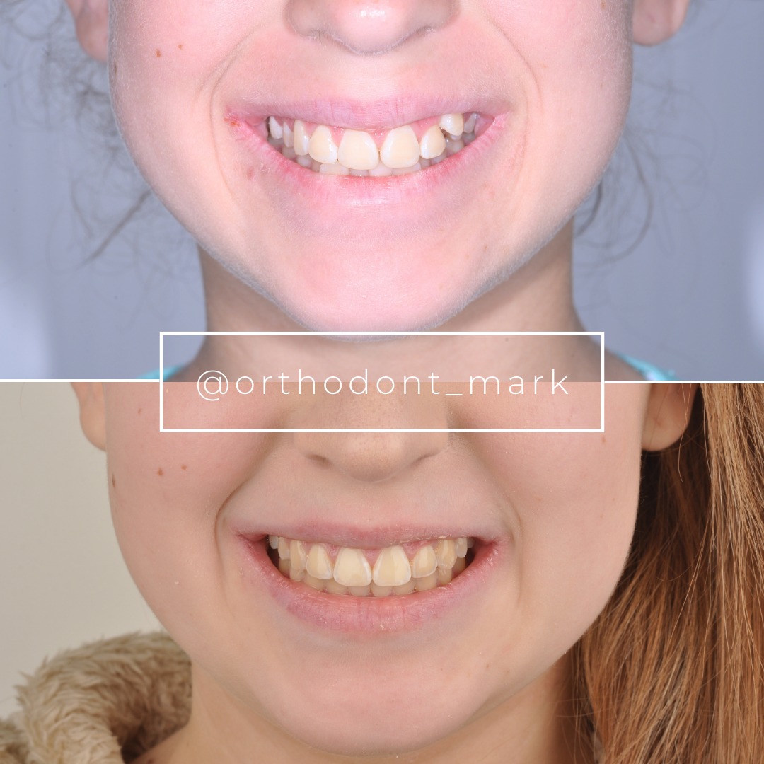
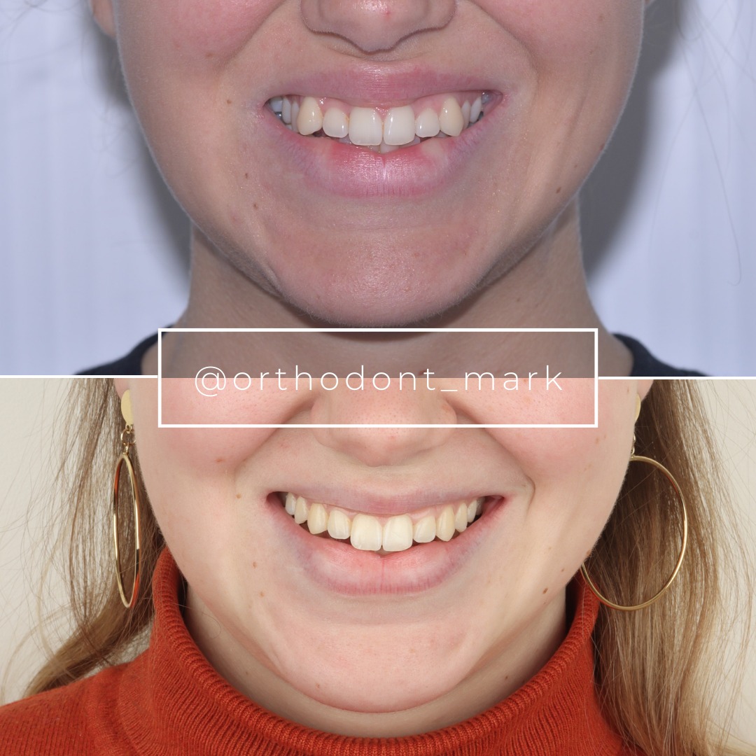
Clinical cases from Dr. Mark Radzhabov's practice

Learn the full MARPE protocol from Dr. Mark Radzhabov
Fundamental course covering CBCT patient selection, miniscrew planning, activation protocols, and 60+ clinical cases. Choose the access level that fits your practice.
Essentials of rapid palatal expansion for practicing orthodontists.
- Core RPE concepts and biomechanics
- 6 structured video lessons
- Clinical decision checklists
- Lifetime access to recordings
Deep-dive into MARPE protocol, diagnostics, and clinical execution.
- 6 modules covering MARPE from A to Z
- CBCT case selection walkthroughs
- Miniscrew placement and activation
- 60+ annotated clinical cases
- Direct Q&A with Dr. Mark Radzhabov
5-element medical consultation framework for dentists and orthodontists.
- Trust-building consultation protocol
- 5 lesson modules
- Templates for treatment plan delivery
- Works with any clinical specialty
Clinical FAQ
How do I differentiate true midpalatal suture opening from dentoalveolar bending on CBCT?
Measure widening at the hard palate midline (suture center) on coronal CBCT, not at the alveolar crest. True opening shows parallel alveolar plates and minimal root tipping. Bending shows convergent plates and lateral root movement with minimal suture change.
What is the target amount of anterior palate widening at 4–6 weeks post-MARPE activation?
Healthy skeletal responders show 2–3 mm of midpalatal widening at the anterior palate zone within 4–6 weeks. Widening <1.0 mm signals inadequate skeletal response and dentoalveolar compensation dominance.
Should anterior-posterior split confirmation be performed on all MARPE patients?
Yes. Post-activation CBCT verification within 4–6 weeks is essential to distinguish responders from non-responders and prevent relapse-prone dentoalveolar bending compensation. Without verification, clinicians cannot reliably guide further activation or protocol adjustment.
What CBCT voxel size and parameters are required for accurate suture split verification?
Voxel size ≤0.3 mm with high-resolution reconstruction is optimal. Baseline and post-activation scans must use identical acquisition parameters, FOV, and head position to ensure measurement accuracy within ±0.5 mm.
If a patient shows asymmetric midpalatal opening (left-right difference >1.0 mm), what does this indicate?
Asymmetric opening suggests miniscrew failure, uneven load application, or early bony bridging in specific regions. Acquire parasagittal slices to assess bone density and miniscrew position. Consider load redistribution or reactivation protocol review.
How does nasal cavity widening help confirm skeletal expansion?
True skeletal expansion widens the nasal cavity width by 1–2 mm and elevates the palatal vault height on parasagittal CBCT. Minimal nasal widening with suture opening suggests predominant dentoalveolar compensation rather than skeletal response.
What should I do if post-activation CBCT shows <1.0 mm true suture opening with dentoalveolar bending?
Discontinue MARPE to prevent excessive root angulation, dehiscence, and relapse. Discuss SARPE (surgically assisted rapid palatal expansion) or conservative retention with orthognathic options for future management.
Can I use 2D radiographs instead of CBCT for suture split verification?
No. Panoramic and lateral cephalometric radiographs cannot reliably distinguish true suture opening from dentoalveolar bending or assess three-dimensional expansion patterns. CBCT with multiplanar reformatting is the diagnostic gold standard.
How frequently should I obtain post-activation CBCT scans during MARPE treatment?
Initial post-activation CBCT at 4–6 weeks is essential for classification. Partial responders benefit from follow-up imaging every 3–4 weeks if load reduction or protocol adjustment occurs. Skeletal responders require CBCT only at final expansion endpoint.
Does the midpalatal suture maturation stage (Angelieri classification) affect suture split verification interpretation?
Yes. Stage A (immature, completely radiolucent) and Stage B (mostly radiolucent) patients typically show more reliable skeletal response than Stage C and D (dense or fused). Stage classification guides load selection and helps predict whether dentoalveolar compensation is patient-biology-related or protocol-related.
Accurate suture split verification CBCT landmarks transform post-activation decision-making: they separate patients who are achieving true skeletal expansion from those experiencing pure dentoalveolar bending, allowing immediate protocol adjustments before relapse or excessive root movement occurs. The anterior-posterior split confirmation protocol—measuring widening across three anatomical zones, comparing nasal cavity expansion, and assessing palatal vault height—requires only 2–3 minutes of CBCT interpretation but prevents months of inefficient treatment. Schedule a consultation or case review through Orthodontist Mark's clinical platform to integrate this verification protocol into your practice, or join our evidence-based expansion course. Dr. Mark Radzhabov's framework has become the standard in centers pursuing skeletal expansion verification worldwide.

