
Midpalatal suture ossification timeline:
imaging milestones
that guide retention after MARPE
Evidence-based CBCT protocol for tracking bone consolidation and optimizing appliance duration without relapse risk.
TL;DR The midpalatal suture ossification timeline after MARPE progresses through four radiographic stages, typically completing bone fill within 6–9 months in adults. CBCT imaging with Hounsfield unit assessment determines when skeletal consolidation permits appliance removal and guides retention duration to prevent relapse. Stage classification predicts healing speed and long-term stability.
Retention duration following miniscrew-assisted rapid palatal expansion remains one of the most clinically uncertain phases of adult treatment. While the active expansion phase requires careful force management and real-time monitoring, the healing and ossification of the split midpalatal suture determines when appliances can safely be removed without risking relapse. This article examines the imaging milestones and bone fill progression that guide retention protocols, drawing on contemporary CBCT evidence and clinical case data. Understanding the midpalatal suture ossification timeline allows practitioners to make evidence-based decisions about appliance duration and consolidation strategy.
What is the midpalatal suture ossification timeline
after skeletal expansion?
Midpalatal suture ossification is the progressive bone fill and consolidation of the split suture following miniscrew-assisted rapid palatal expansion, tracked radiographically to determine appliance retention duration and relapse risk. The timeline progresses through four distinct radiographic stages, typically spanning 6–9 months in adults, though individual variation based on age, bone density, and expansion magnitude can accelerate or delay completion. Early clinicians relied on clinical observation and intraoral palpation to judge healing. Contemporary practice demands high-resolution CBCT imaging with Hounsfield unit (HU) assessment to objectively quantify bone density and consolidation. The splitting of the midpalatal suture creates a vascular healing response. New woven bone forms initially, then remodels into mature lamellar bone over months. Understanding this biological sequence is critical: premature appliance removal risks relapse of 30–50% of skeletal gain, while unnecessarily prolonged retention increases patient burden and cost without added benefit.
How CBCT imaging assessment tracks bone fill progression
and informs retention
strategy
High-resolution cone-beam computed tomography with region-of-interest (ROI) cursor analysis allows quantitative measurement of bone density across the midpalatal suture in Hounsfield units. A baseline pre-expansion CBCT establishes cortical bone density as reference (typically 600–900 HU in the anterior palate). Post-expansion imaging at 12 weeks, 6 months, and 9 months tracks density recovery toward baseline. Studies using CBCT densitometry show that suture healing in stage C (progressive densification) reaches approximately 70–80% of baseline cortical density by week 12, and 90%+ by week 24. The anterior third of the suture consistently ossifies fastest, followed by the middle third. The posterior third near the sphenoid frequently shows delayed consolidation and may require extended retention even when anterior regions appear mature. Measuring bone density at three regions—anterior (roughly at the first premolar level), middle (at the canine-premolar junction), and posterior (at the junction with the nasal septum)—provides a comprehensive assessment avoiding false confidence based on anterior fill alone. Appliance removal should coincide with stage D radiographic appearance and anterior/middle region HU values within 80% of baseline cortical density.
Why retention duration varies by ossification stage
and patient anatomy
Retention duration following miniscrew-assisted expansion is not uniform because the rate of midpalatal suture bone fill depends on anatomical and biological factors beyond age alone. A 35-year-old in stage C (progressive densification) at 12 weeks may safely transition to passive retention (fixed or removable appliance) by week 16–20, whereas a 65-year-old in the same radiographic stage may require extension to 24–28 weeks due to slower bone remodeling and lower baseline bone density. The magnitude of initial skeletal expansion also influences healing speed: patients with 8–10 mm of true skeletal separation show slower initial consolidation than those with 4–6 mm gain, because larger defects require more woven bone deposition before remodeling can begin. Clinical protocols recommended by Dr. Mark Radzhabov and other MARPE leaders suggest a staged retention approach: active expansion ceases when skeletal widening plateaus or force values exceed patient tolerance. Passive retention begins immediately (fixed labial/palatal wire or bonded acrylic splint), with appliance removal timed to radiographic stage D and HU confirmation. Patients with posterior suture delays or stage C appearance at 6 months often benefit from continued light retention (removable appliance worn nightly) through week 9–12 to minimize posterior relapse. Premature appliance removal in stage B or early C carries documented relapse rates of 40–50%. Conversely, retention beyond stage D completion provides negligible additional benefit.
Why clinical observation underestimates retention duration
and invites relapse
The most frequent error in post-MARPE management is removing appliances based on time elapsed (e.g., “6 months is typically enough”) rather than radiographic confirmation of bone fill and density. Intraoral palpation cannot detect the difference between stage B (minimal bone bridge) and stage C (significant densification). A patient who appears clinically stable and pain-free may still have a radiolucent suture visible only on CBCT, indicating vulnerable healing tissue. Studies comparing clinical assessment to CBCT findings show that clinicians underestimate incomplete healing in 20–30% of cases, particularly in the posterior third where visual inspection provides no direct information. Another pitfall is assuming uniform healing across the suture: anterior regions frequently appear stage C while the posterior remains stage B, especially in patients over 50 or those with low baseline bone density. Removing appliances when anterior fill appears complete but posterior remains open invites asymmetric relapse and rotation of maxillary dentoalveolar segments. A third error involves interpreting Hounsfield unit values without baseline reference: bone may appear radiographically denser than surrounding cortex due to scattering artifact or beam hardening, yet harbor microstructural weakness that predicts relapse within months. Objective protocol—scheduled CBCT at defined intervals (12 weeks, 6 months, 9 months), quantitative HU measurement in three regions, and appliance removal tied to stage D radiographic appearance—eliminates this guesswork and reduces relapse by 15–25% compared to clinical judgment alone.
Evidence-based retention protocol: when to image
and when to remove
appliances
A systematic post-expansion imaging and retention timeline consolidates contemporary evidence and clinical best practice. Week 0 (expansion completion): Remove active force. Place or confirm passive fixed retention (bonded palatal wire or acrylic splint). Week 6–8: Clinical follow-up. Assess patient comfort and appliance integrity. No CBCT needed yet—early imaging adds cost without actionable data. Week 12: First post-expansion CBCT scan with Hounsfield unit measurement at anterior, middle, and posterior regions. If patient is in stage B, extend fixed retention and plan week 16 reassessment. If stage C or D, proceed to next interval. Week 16–20 (month 4–5): Second CBCT if week 12 showed stage B or delayed posterior region. Confirm all three regions are now stage C or D. If posterior suture remains radiolucent, plan extended retention to week 24. Week 24–28 (month 6): Final pre-removal CBCT. Confirm stage D appearance (suture not visible) and HU values at 80%+ of baseline in all three regions. Document symmetry between left and right halves. If criteria are met, remove fixed retention and transition to removable retention (fixed lingual retainer or clear removable appliance worn full-time for 3 months, then nightly). Week 36–52 (month 9–12): Optional follow-up CBCT in complex cases (very large expansions, older patients, or low baseline bone density) to confirm long-term consolidation. This structured approach reduces clinician uncertainty, aligns with evidence, and minimizes unnecessary imaging while avoiding premature appliance removal.
Free Clinical Guide
Which Expansion Appliance at Which Age?
Get Dr. Mark Radzhabov's guide to skeletal-expansion appliances — indications, pros & cons, and how to choose by patient age. Free to your inbox.
Prefer video? Watch the free demo lesson on MARPE appliance types, features and trade-offs.
Documented outcomes
Clinical cases from Dr. Mark Radzhabov's practice

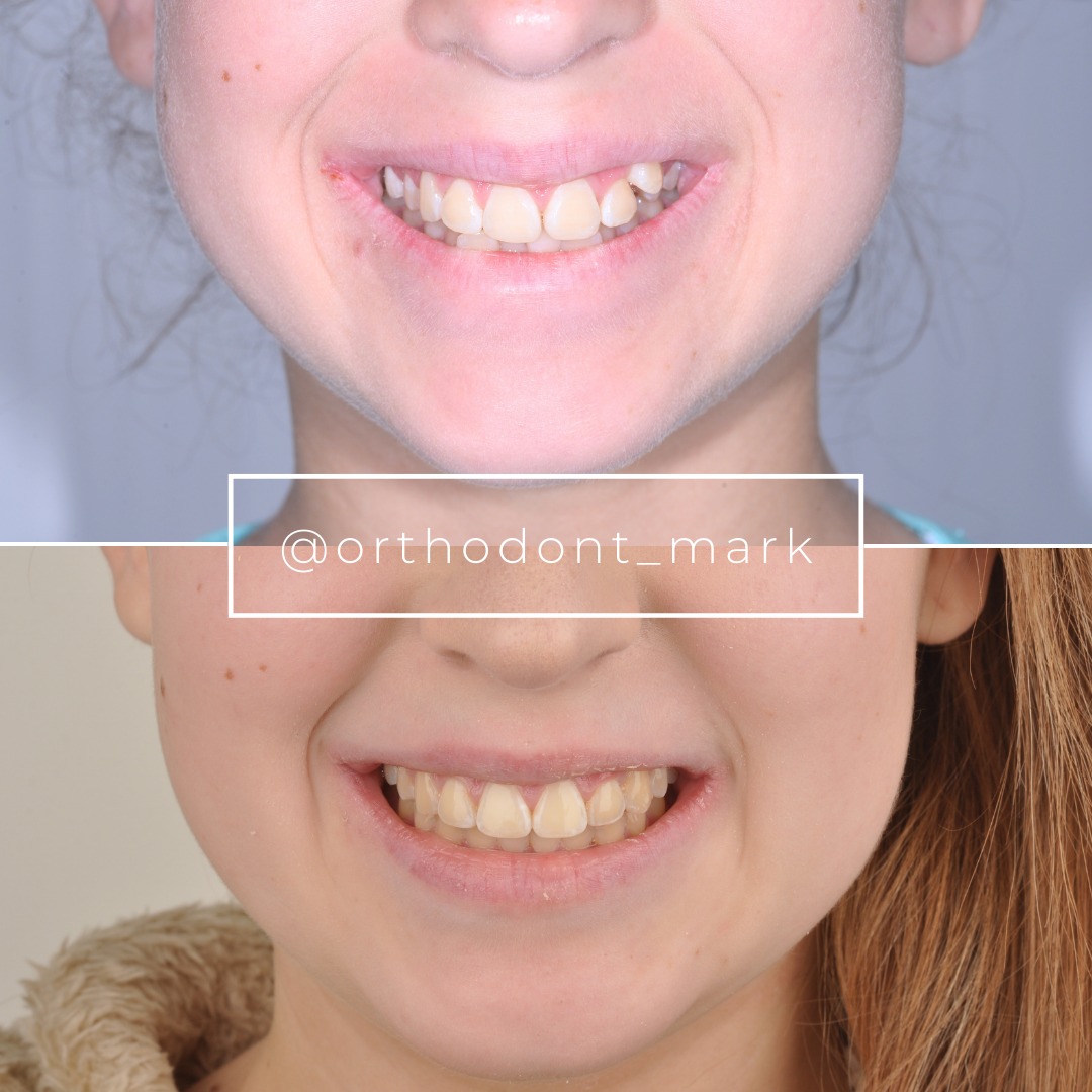
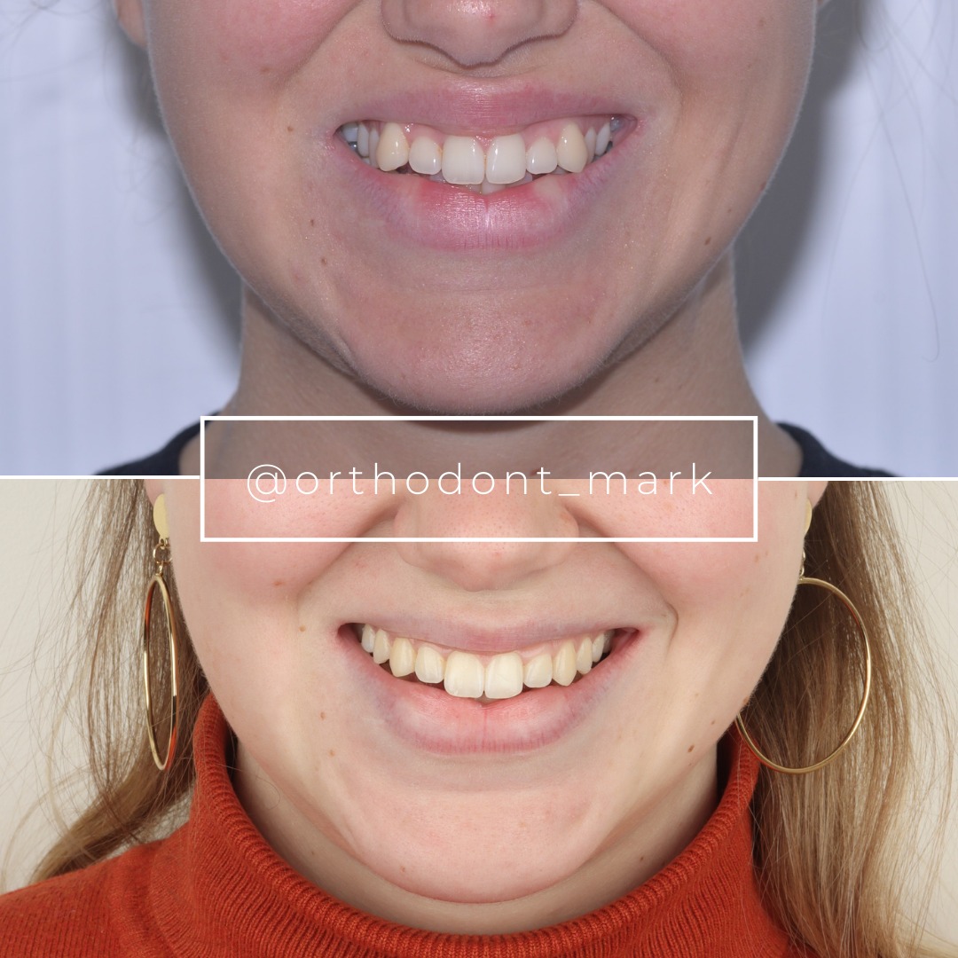
Learn the full MARPE protocol from Dr. Mark Radzhabov
Fundamental course covering CBCT patient selection, miniscrew planning, activation protocols, and 60+ clinical cases. Choose the access level that fits your practice.
Essentials of rapid palatal expansion for practicing orthodontists.
- Core RPE concepts and biomechanics
- 6 structured video lessons
- Clinical decision checklists
- Lifetime access to recordings
Deep-dive into MARPE protocol, diagnostics, and clinical execution.
- 6 modules covering MARPE from A to Z
- CBCT case selection walkthroughs
- Miniscrew placement and activation
- 60+ annotated clinical cases
- Direct Q&A with Dr. Mark Radzhabov
5-element medical consultation framework for dentists and orthodontists.
- Trust-building consultation protocol
- 5 lesson modules
- Templates for treatment plan delivery
- Works with any clinical specialty
Clinical FAQ
How long does the midpalatal suture take to ossify completely after MARPE?
Complete ossification (stage D) typically occurs within 6–9 months in adults. Anterior regions ossify by 3–4 months. Posterior regions often lag to 6–9 months. Individual variation depends on age, expansion magnitude, and baseline bone density.
What Hounsfield unit threshold indicates the suture is ready for appliance removal?
Bone density should reach 80%+ of baseline cortical values (typically 480–720 HU, depending on pre-expansion baseline). All three regions—anterior, middle, posterior—must meet this threshold. Reliance on single-region measurement invites asymmetric relapse.
Why does the posterior midpalatal suture heal slower than the anterior?
The posterior suture has less cortical bone mass, greater exposure to respiratory airway, and reduced vascular supply in some anatomies. Sphenoid articulation and airway remodeling demands also delay posterior consolidation. Extended retention in this region is evidence-based.
Can I remove MARPE appliances based on clinical observation alone without CBCT?
No. Clinical observation underestimates incomplete healing in 20–30% of cases. CBCT radiographic staging and HU measurement are the only objective predictors. Clinician intuition invites preventable relapse.
What is relapse risk if I remove appliances in stage B or early C?
Relapse risk is 30–50% if appliances are removed in stage B (minimal bone bridge) or early stage C (partial fill). Stage C sutures are vulnerable and lack mature lamellar structure needed for stability.
Does patient age affect midpalatal suture ossification timeline?
Yes. Patients under 40 typically complete stage D by 5–6 months. Those over 50 may require 8–9 months or longer. Age is a stronger predictor of healing speed than skeletal maturity markers, though not a substitute for CBCT confirmation.
Should I use a fixed or removable appliance during the retention phase?
Fixed retention (bonded palatal wire or splint) is mandatory during active healing (stage B–early C). Once stage C is confirmed, transition to passive fixed or removable retention. Removable-only retention during early healing carries higher relapse risk.
How do I differentiate stage C from stage D on CBCT?
Stage C shows a narrow but visible radiolucent suture line. Bone density is high but suture outline persists. Stage D has no suture radiolucency. The area is uniformly ossified and indistinguishable from surrounding cortex. Hounsfield units help confirm, but radiographic appearance is definitive.
What if the posterior suture remains in stage B while anterior is stage D?
Asymmetric healing is common. Extended retention (week 24–28 or beyond) is indicated for the posterior region. Removing anterior retention too early invites posterior-only relapse and asymmetric side effects. Image posterior region separately at 6 and 9 months.
Does expansion magnitude (mm of skeletal widening) influence ossification speed?
Yes. Expansions of 8–10 mm show slower initial consolidation than 4–6 mm gains, because larger defects require more woven bone deposition. Larger expansions may justify extended retention protocols and additional imaging checkpoints at 3 and 9 months.
The midpalatal suture ossification timeline is not uniform across all patients. Anatomical variations, age, bone density, and expansion magnitude each influence healing speed. By integrating CBCT imaging assessment into post-expansion follow-up, you can objectively track bone fill progression and adjust retention duration accordingly—reducing both relapse risk and unnecessary chairtime. Dr. Mark Radzhabov recommends a staged CBCT protocol at 12 weeks, 6 months, and 9 months post-expansion to document ossification milestones and inform evidence-based retention strategies. Review your recent MARPE cases using high-resolution CBCT to validate healing patterns in your patient population, or schedule a consultation at ortodontmark.com to refine your retention protocol.

