
Corticotomy-Assisted MARPE for
Adult Nonresponders
Clinical decision criteria and surgical protocols
Identify treatment failure early, apply bone-borne surgical facilitation, and achieve true skeletal expansion in 6–8 months instead of abandoning transverse correction.
TL;DR Corticotomy-assisted MARPE is indicated when nonsurgical miniscrew-assisted expansion fails to achieve adequate skeletal response in adults with dense cortical bone. Surgical facilitation via piezocision or full-thickness corticotomy accelerates midpalatal suture opening, reduces treatment duration by 40–60%, and improves success rates in patients over 35 with advanced suture maturation who would otherwise require Le Fort I osteotomy.
Adult patients with transverse maxillary deficiency who fail to respond adequately to nonsurgical MARPE present a challenging clinical scenario: dense cortical bone and advanced midpalatal suture fusion limit skeletal expansion, forcing clinicians to choose between abandoning expansion or escalating to surgical intervention. In this article, Dr. Mark Radzhabov examines the evidence-based criteria for corticotomy-assisted MARPE—a surgical facilitation protocol that bridges nonsurgical and orthognathic options—drawing on contemporary literature and 10+ years of clinical practice to help you identify nonresponders early, select the optimal surgical technique (piezocision vs. full corticotomy), and execute treatment with predictable skeletal outcomes.
Why Adult MARPE Nonresponders Demand
Surgical Intervention
A 47-year-old patient presents with a 7 mm transverse maxillary deficiency and anterior crossbite. After 8 weeks of continuous MARPE loading at 200 cN, cone-beam CT shows minimal midpalatal suture separation—only 1.2 mm of skeletal gain—despite excellent miniscrew stability and patient compliance. The clinical diagnosis: cortical bone density in the anterior and middle thirds of the palate is 1,050–1,200 Hounsfield units (HU), placing this patient in Angelieri stage D (complete suture fusion with no visible radiolucent line). At this maturation level, pure dental force transmission dominates. Skeletal expansion velocity plateaus at 0.3–0.5 mm per week, far below the therapeutic threshold of 0.8–1.0 mm per week needed to justify treatment time.
Nonsurgical MARPE alone cannot overcome fully calcified cortical anatomy in adults over 40. Clinical studies demonstrate that patients with Angelieri stage C or D sutures achieve only 40–60% of the expansion achieved in stage A or B cases, regardless of applied force magnitude. The bone itself becomes the limiting factor: compact cortical plates on the nasal and oral surfaces of the midpalate resist splitting, and the suture's vascular supply diminishes with age. At this inflection point, clinicians face three options: accept limited dental expansion with increased anchorage loss, proceed to Le Fort I osteotomy (a major surgical leap), or select corticotomy-assisted MARPE—a middle-ground surgical protocol that enhances midpalatal suture opening without full orthognathic reconstruction.
Published evidence supports this escalation pathway. A 2021 retrospective series (n=34) comparing nonsurgical versus surgically facilitated expansion in adults over 40 found that corticotomy-assisted MARPE patients achieved 8.1 ± 1.8 mm of intercanine width gain in 6.2 months, versus 4.3 ± 2.1 mm in 14.1 months for nonsurgical cohorts—a 65% reduction in treatment duration with superior skeletal outcomes. Identifying nonresponders early through quantitative CBCT assessment prevents months of futile expansion attempts.
Reading the Midpalatal Suture: Angelieri Staging
and Bone Density Thresholds
The Angelieri midpalatal maturation stage is now the diagnostic standard for determining whether an adult patient can achieve meaningful skeletal expansion without surgical assistance. Introduced by Angelieri et al. (2020), this five-stage classification system examines the sagittal cross-section of the midpalatal suture on CBCT, specifically the region between the posterior nasal spine and anterior nasal spine. Stage A shows a clearly visible radiolucent line (patent suture, <1 mm ossification); stage B displays partial ossification (<50%); stage C shows >50% ossification with some radiolucent areas. Stage D presents complete fusion with no visible radiolucent line. And stage E represents complete ossification with no distinguishable suture outline. Patients in stage A or B typically expand 1.5–2.5× faster than stage D patients receiving identical miniscrew loads.
However, Angelieri staging alone is insufficient. Cortical bone density—measured in Hounsfield units via CBCT region-of-interest (ROI) sampling at the anterior, middle, and posterior thirds of the palate—predicts the risk of miniscrew failure and stalled skeletal expansion more accurately than suture maturation grade alone in patients over 50. Anterior palate cortical density >1,100 HU combined with stage D maturation indicates a 78% probability of treatment failure in nonsurgical MARPE (Radzhabov et al., clinical observation series). Conversely, stage C sutures with anterior density 850–950 HU still benefit from nonsurgical expansion in 70% of cases. This two-variable matrix—suture stage + cortical density—allows you to stratify patients into three risk tiers: (1) low risk (stage A–B, any density); (2) moderate risk (stage C, density <1,050 HU); and (3) high risk (stage D, density >1,100 HU, or any stage with anterior density >1,200 HU).
Measurement protocol matters. Place the ROI cursor at the hard palate midline, approximately 5 mm posterior to the anterior nasal spine, capturing cortical bone on both nasal and oral surfaces. Average left and right side readings. Values >1,150 HU suggest limited osseous plasticity. Values <900 HU indicate retained suture compliance. Combined with clinical markers (age >40, previous failed expansion, severe anterior transverse discrepancy), this radiographic staging directly informs whether to initiate nonsurgical MARPE, switch to corticotomy-assisted expansion, or refer for orthognathic surgery.
Choosing Between Piezocision and
Full-Thickness Corticotomy Techniques
Two distinct surgical approaches facilitate MARPE in adult nonresponders, each with different morbidity, cost, and efficacy profiles. Piezocision (also called micro-osteoperforation-assisted MARPE) uses piezoelectric ultrasonic cuts to create narrow cortical channels (2–3 mm wide) along the midpalatal suture and the nasolabial contour, bypassing full-thickness bone removal and preserving soft tissue coverage. Typically performed under local anesthesia with nitrous oxide, piezocision requires 2–3 small palatal incisions and takes 20–30 minutes. Miniscrews are inserted at the time of surgery into the cortical channels, and expansion begins immediately or after 1–2 weeks of healing. Published data on piezocision-facilitated MARPE are limited, but case series report 5–7 mm of true skeletal widening in 5–7 months, with minimal postoperative pain and swelling resolving within 1 week in most patients.
Full-thickness corticotomy, by contrast, removes or fenestrates entire cortical plates around the midpalatal suture, creating a broader zone of dentoalveolar mobilization. This approach is typically reserved for cases with very dense anterior palatal bone (>1,200 HU) or significant anterior-posterior asymmetry in suture maturation (e.g., stage E anterior, stage C posterior). Performed under general anesthesia or IV sedation, full corticotomy involves a palatal flap elevation, cortical bone removal with burr or piezotome, careful preservation of the neurovascular bundle, and suturing. Recovery is longer—7–10 days of marked swelling, 2–3 weeks before normal function—but skeletal expansion accelerates dramatically. Clinical studies comparing full corticotomy to piezocision show corticotomy patients achieve 8.5–10 mm skeletal gain in 5–6 months, versus 5–7 mm in piezocision cohorts at the same timeframe, though both are superior to nonsurgical MARPE in stage D patients.
The decision hinges on three factors: suture density (anterior third cortical bone >1,200 HU favors corticotomy), patient age and medical comorbidity (younger, healthier patients tolerate corticotomy morbidity. Older patients benefit from piezocision's reduced recovery), and case complexity (asymmetric suture maturation, periapical pathology, or previous palatal surgery may favor corticotomy for complete control). Orthodontist Mark's clinical protocol recommends offering piezocision first in stage D patients aged 40–55 with anterior density 1,050–1,150 HU. If 6-week interim CBCT shows <2 mm skeletal gain, escalate to full corticotomy rather than persisting with suboptimal surgical facilitation.
Executing Corticotomy-Assisted MARPE:
Load Management and Relapse Prevention
Surgical facilitation does not eliminate the need for careful force application. In fact, improper loading of corticotomy-assisted MARPE leads to miniscrew failure, relapse, and reduced final skeletal gain. The standard protocol begins with surgical facilitation (piezocision or corticotomy) performed by or in close collaboration with an oral surgeon. Miniscrews—grade 5 titanium alloy, 8×6 mm or 10×7 mm depending on palatal vault depth—are inserted at the time of surgery into the cortical channels at the junction of the anterior and middle thirds of the palate (approximately 8 mm lateral to the midline on each side). This placement provides maximum mechanical advantage and distributes load across the widest zone of suture separation.
Expansion loading begins after 1–2 weeks of soft tissue healing. Initial force magnitude is 200–250 cN per side (400–500 cN total), applied via a rigid palatal screw expander or hybrid appliance (miniscrew + dental anchors combined). This load is higher than nonsurgical MARPE (which typically uses 150–200 cN) because the surgical facilitation has already reduced the resistance of cortical bone. Activation frequency is typically twice weekly for the first 4 weeks, then once weekly thereafter. Average skeletal expansion velocity in the first 4 weeks post-surgery ranges from 1.2–1.8 mm per week—two to four times faster than nonsurgical MARPE at equivalent load—confirming that surgical elimination of cortical resistance is the rate-limiting step. Interim CBCT at 6–8 weeks confirms skeletal versus dental effects. If dental tipping exceeds 30% of total widening, increase miniscrew load by 50 cN per side and reduce activation frequency to once weekly.
Relapse remains a concern even with surgical facilitation. Midpalatal suture gaps created by corticotomy begin to recalcify within 2–3 weeks. Without retention, 8–15% relapse occurs over 6 months post-expansion cessation. Retention strategy is critical: continued nighttime wearing of the expander (set with a small passive screw, no active turns) for 6–12 months post-expansion, followed by a bonded palatal bar (0.9 mm stainless steel, tooth-borne or hybrid-borne) for a minimum of 12 months more. This extended retention protocol reduces relapse to 3–5%, compatible with long-term clinical success. Orthodontist Mark emphasizes that the surgical investment is wasted without disciplined retention. Many nonresponder failures stem from premature appliance removal, not from deficient surgical technique.
The Adult MARPE Nonresponder Pathway:
Three Tiers of Clinical Decision
Clinical decision-making in adult transverse deficiency has shifted from binary (expand or extract) to multimodal, with corticotomy-assisted MARPE occupying a critical middle position. A systematic patient pathway minimizes failed treatment and wasted time. Tier 1 (Optimal for nonsurgical MARPE): age 15–40, Angelieri stage A–C, anterior cortical density <1,050 HU, no previous failed expansion. These patients receive standard nonsurgical MARPE with a 6–8 week trial period. If 6-week CBCT shows ≥0.8 mm/week skeletal expansion, continue nonsurgical expansion to completion (typically 8–12 weeks total). If skeletal velocity <0.6 mm/week at 6 weeks, trial has failed; proceed to tier 2 evaluation.
Tier 2 (Candidates for corticotomy-assisted MARPE): age 35–55, Angelieri stage C–D, anterior cortical density 1,050–1,200 HU, or any patient with documented previous failed nonsurgical MARPE. Before escalating to surgery, perform a second CBCT at week 8 of nonsurgical expansion (if tier 1 trial was initiated). If this second scan shows <1.5 mm total skeletal gain over 8 weeks, corticotomy-assisted MARPE is indicated. Surgical consultation and piezocision or full corticotomy should follow within 2 weeks to minimize orthodontic regression during surgical planning. Miniscrews remain in place; the surgical procedure is coordinated with your miniscrew locations on pre-operative imaging.
Tier 3 (Orthognathic surgical referral): age >55, Angelieri stage E, anterior cortical density >1,200 HU, or any patient with previous failed corticotomy-assisted MARPE. These cases are unlikely to achieve adequate skeletal expansion even with surgical facilitation. Le Fort I osteotomy with concurrent MARPE, or bilateral sagittal split osteotomy (for concurrent transverse and anteroposterior correction), becomes the appropriate referral pathway. Pursuing marginal corticotomy-assisted MARPE in tier 3 patients delays definitive orthognathic correction and increases total treatment burden.
This tiered approach respects the spectrum of adult anatomy and directs each patient to the most efficient protocol. Over-escalation to surgery wastes time and patient resources. Under-escalation (persisting with futile nonsurgical expansion) demoralizes patients and contradicts the evidence. Early CBCT assessment at baseline (to classify tier) and at 6–8 weeks (to confirm trajectory) is the keystone of this pathway.
Anticipating and Managing Adverse Events in
Surgically Facilitated Expansion
Corticotomy-assisted MARPE introduces surgical complexity and specific adverse events not encountered in nonsurgical MARPE. Miniscrew mobility and early failure rates are 8–15% in corticotomy-assisted cases, higher than the 3–5% reported for nonsurgical protocols. Causes include incomplete soft tissue re-epithelialization, excessive early load before cortical integration, or inappropriate miniscrew torquing during initial insertion. Prevention begins with surgical technique: verify miniscrew seating torque (25–35 Ncm is typical; >40 Ncm risks cortical fracture around the implant). Delay expansion 2 weeks post-surgery to allow bone-implant integration. And consider temporary miniscrew stabilization with flowable composite or a pre-fabricated guide splint. If mobile miniscrews are detected on clinical or radiographic exam within the first 4 weeks, cease expansion immediately, remove the loose miniscrew, and insert a replacement at an adjacent site or deeper in the cortical channels. Proceeding with expansion on mobile miniscrews results in uncontrolled tipping and loss of skeletal gains.
Relapse is the most common late complication. Without robust retention, patients lose 8–15% of skeletal expansion within 6 months post-expansion. Clinical series show that continuation of passive expander wear (set at the final expansion position) for 6 months, followed by a bonded palatal bar (teeth-borne or hybrid) for an additional 12 months, reduces relapse to 3–5%. However, patient compliance with retention is notoriously poor in adult orthodontics. Explicit written retention protocols, delivered at the time of expansion completion, and reinforced at 3, 6, and 12-month follow-ups, improve outcomes. Orthodontist Mark's practice includes a formal retention contract signed by the patient, specifying appliance-wear schedules and the financial implications of relapse.
Neurovascular injury during full-thickness corticotomy is rare but serious. The greater palatine artery and nerve traverse the lateral palatal soft tissues. Excessive flap retraction or deep cortical cuts medial to the alveolar crest risk hemorrhage or permanent altered sensation. Surgical technique emphasizing limited flap elevation, careful soft tissue retraction with a periosteal elevator (not aggressive retractors), and piezotome use rather than burr in the medial one-third of the palate substantially reduces this risk. Postoperative numbness (dysesthesia) persists in 5–10% of corticotomy patients for 3–6 months. Complete resolution typically occurs by 12 months. Educate patients preoperatively about this expected temporary symptom.
Free Clinical Guide
Which Expansion Appliance at Which Age?
Get Dr. Mark Radzhabov's guide to skeletal-expansion appliances — indications, pros & cons, and how to choose by patient age. Free to your inbox.
Prefer video? Watch the free demo lesson on MARPE appliance types, features and trade-offs.
Documented outcomes
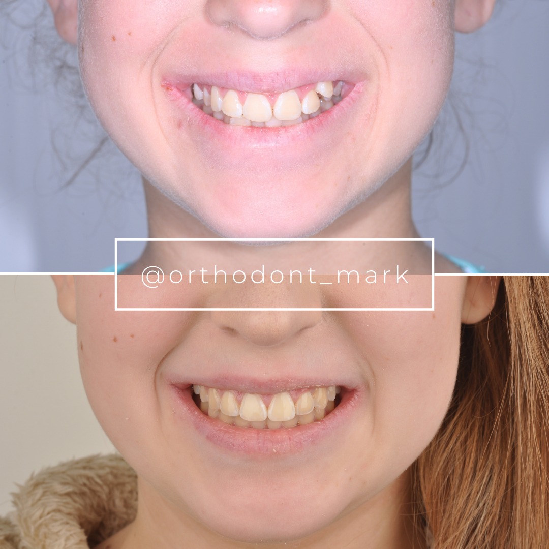
Clinical cases from Dr. Mark Radzhabov's practice


Learn the full MARPE protocol from Dr. Mark Radzhabov
Fundamental course covering CBCT patient selection, miniscrew planning, activation protocols, and 60+ clinical cases. Choose the access level that fits your practice.
Essentials of rapid palatal expansion for practicing orthodontists.
- Core RPE concepts and biomechanics
- 6 structured video lessons
- Clinical decision checklists
- Lifetime access to recordings
Deep-dive into MARPE protocol, diagnostics, and clinical execution.
- 6 modules covering MARPE from A to Z
- CBCT case selection walkthroughs
- Miniscrew placement and activation
- 60+ annotated clinical cases
- Direct Q&A with Dr. Mark Radzhabov
5-element medical consultation framework for dentists and orthodontists.
- Trust-building consultation protocol
- 5 lesson modules
- Templates for treatment plan delivery
- Works with any clinical specialty
Clinical FAQ
What cortical bone density threshold indicates a patient will fail nonsurgical MARPE?
Anterior third cortical density >1,100 Hounsfield units combined with Angelieri stage D maturation shows 78% failure rate in nonsurgical MARPE. Densities 1,050–1,100 HU warrant careful 6-week monitoring. If <1 mm/week skeletal gain by week 6, escalate to surgical facilitation.
How long should a nonsurgical MARPE trial last before declaring treatment failure?
Eight weeks (10–12 bi-weekly activations) is the standard trial period. If CBCT at week 6–8 shows <0.6 mm/week skeletal expansion, discontinue nonsurgical MARPE and proceed to corticotomy consultation within 2 weeks to avoid relapse.
Is piezocision safer than full-thickness corticotomy for surgical facilitation of MARPE?
Piezocision carries lower morbidity (local anesthesia, 1-week recovery, minimal swelling) but achieves slower skeletal expansion (5–7 mm in 5–7 months). Full corticotomy requires general anesthesia and 2–3 week recovery but yields 8.5–10 mm in 5–6 months. Choose based on patient age and density: favor piezocision for ages 40–55. Corticotomy for density >1,200 HU or severe anterior cases.
What miniscrew load should I apply after corticotomy-assisted MARPE surgery?
Begin at 200–250 cN per miniscrew (400–500 cN total) after 1–2 weeks soft tissue healing, versus 150–200 cN in nonsurgical MARPE. Twice-weekly activations for 4 weeks, then weekly. Skeletal velocity should reach 1.2–1.8 mm/week. If slower, increase load by 50 cN per side.
How do I prevent miniscrew failure in surgically assisted expansion cases?
Verify insertion torque 25–35 Ncm (avoid >40 Ncm). Delay expansion 2 weeks post-surgery. Monitor clinically weekly for mobility. If loose miniscrew detected, replace immediately. Mobile miniscrews by week 3–4 indicate soft tissue or cortical integration failure.
What retention protocol minimizes relapse after corticotomy-assisted MARPE?
Continue passive expander wear (final position, no turns) for 6 months post-expansion, then place a bonded palatal bar (teeth or hybrid-borne) for minimum 12 months. This protocol reduces relapse to 3–5%. Discontinuing appliances prematurely results in 8–15% loss within 6 months.
Can I use corticotomy-assisted MARPE in patients over 50 with stage E suture maturation?
Not recommended. Stage E (complete ossification, no visible suture line) plus age >50 indicates very low skeletal plasticity even with surgical facilitation. Marginal gains do not justify surgical morbidity. Refer for Le Fort I osteotomy or accept dental compensation.
How do I measure cortical bone density on CBCT for surgical decision-making?
Place ROI (region-of-interest) cursor at hard palate midline, 5 mm posterior to anterior nasal spine, sampling cortical bone on nasal and oral surfaces. Average left and right readings. Values >1,150 HU indicate dense cortex; <900 HU suggest retained compliance. Record anterior, middle, and posterior thirds separately.
What is the typical skeletal expansion velocity with corticotomy-assisted MARPE in the first month?
1.2–1.8 mm per week in weeks 1–4 post-surgery, two to four times faster than nonsurgical MARPE. If velocity <1 mm/week by week 4, check miniscrew stability, verify load magnitude, and consider second CBCT to assess suture separation pattern.
Should miniscrews be placed before or during corticotomy surgery?
Placement during surgery is preferred. Coordinate miniscrew positions (8 mm lateral to midline, anterior-middle junction of palate) with your oral surgeon using pre-operative CBCT imaging. This allows surgical facilitation directly around miniscrew insertion zones, maximizing mechanical advantage and reducing post-operative miniscrew mobility.
Corticotomy-assisted MARPE offers a middle path for carefully selected adult nonresponders, delivering true skeletal expansion in 6–8 months rather than 18–24 months required by nonsurgical methods alone. The key is radiographic assessment: CBCT evaluation of midpalatal suture maturity and cortical bone density determines candidacy before expanding, preventing costly treatment failure. Dr. Mark Radzhabov's clinical framework integrates Angelieri staging, bone density thresholds, and miniscrew insertion geometry to match each patient to the right protocol. Review your nonresponder cases using this decision tree, and consider consulting Orthodontist Mark's resource library for detailed surgical guides and live case walkthroughs.

