Expansion Relapse Mechanism:
Dental Rebound
vs Sutural Reclosure — What the Evidence Shows
A peer-reviewed breakdown of which tissue process drives post-expansion instability and how protocol design can address each mechanism.
TL;DR The expansion relapse mechanism involves two distinct processes: dental rebound from elastic periodontal and alveolar forces, and sutural reclosure as new bone fills the widened midpalatal gap. Chun et al. (2022) confirm that both RPE and MARPE exhibit skeletal relapse during consolidation, though MARPE demonstrates less decrease across skeletal, dentoalveolar, and periodontal variables than RPE.
When a patient presents with measurable transverse relapse weeks after appliance removal, the clinical question is not simply whether relapse occurred — it is which tissue mechanism is responsible. The expansion relapse mechanism differs fundamentally depending on whether the force was transmitted through teeth or directly to bone. Dr. Mark Radzhabov at ortodontmark.com examines the peer-reviewed evidence distinguishing dental rebound from sutural reclosure, so clinicians can design retention protocols that target the correct biological process and improve long-term skeletal stability.
Why Conflating Dental and Skeletal Relapse Leads to
Inadequate Retention
Expansion relapse mechanism is the biological process by which gained transverse width is lost after appliance removal, attributable to either elastic recoil of dentally displaced tissues or active osseous infilling at the midpalatal suture. These two processes operate on different timescales and respond to different clinical interventions, yet they are frequently treated as interchangeable in practice — a conflation that undermines retention protocol design. Dental rebound arises from the viscoelastic properties of the periodontal ligament, the gingival fiber network, and the alveolar bone housing the posterior teeth. When a tooth-borne expander transmits force through the premolar and molar roots, the buccal periodontium is placed under chronic tension while the palatal cortex is compressed. Lim et al. (2017) documented this asymmetry directly: after MARPE, alveolar bone thickness decreases on the buccal aspect but increases on the palatal aspect, confirming that even bone-borne approaches generate a measurable dentoalveolar gradient that can contribute to elastic recoil once force is removed. Sutural reclosure is a separate, slower process governed by intramembranous ossification at the freshly separated midpalatal suture. New woven bone deposits into the fibrous mid-sutural zone during the consolidation phase, progressively narrowing the interdigitated gap. Chun et al. (2022) confirm that both RPE and MARPE demonstrate skeletal relapse during the consolidation period after expansion, establishing that sutural reclosure is a universal phenomenon regardless of appliance type — not a failure mode exclusive to tooth-borne devices. Understanding this distinction reframes retention entirely. A protocol built solely around passive dental retention addresses gingival and periodontal recoil but does not arrest active bone deposition at the suture. Clinicians managing post-MARPE stability therefore need to account for both processes, particularly in the first months of consolidation when ossification is most active.
Does Appliance Design Modulate the Rate of
Post-Expansion Skeletal Loss?
The clinical relevance of appliance selection becomes apparent when comparing skeletal behavior during the consolidation window. Chun et al. (2022) conducted a prospective randomized clinical trial and reported that midpalatal suture separation occurred in 90% of RPE patients and 95% of MARPE patients, establishing that both devices reliably open the suture. The more instructive finding is what happens next: MARPE demonstrates less decrease in skeletal, dentoalveolar, and periodontal variables during consolidation compared to RPE, indicating that direct skeletal anchorage attenuates the magnitude of relapse across all three tissue categories. This difference likely reflects the force pathway. Bone-borne MARPE miniscrews inserted into the palatal cortex transmit expansion load directly to the maxillary basal bone, minimizing the elastic strain stored in dental and periodontal structures. A tooth-borne rapid palatal expander, by contrast, couples sutural separation to buccal tipping of the anchor teeth, loading the periodontium and creating a larger elastic rebound vector when the appliance is removed. The result is a higher dentoalveolar relapse contribution in tooth-borne cases, even when sutural separation rates are comparable. For clinicians interpreting post-expansion CBCT data, this evidence supports using both skeletal and dentoalveolar width measurements at the consolidation scan — not arch width alone — as the stability benchmark. A patient whose dental arch appears stable but whose basal skeletal width has decreased substantially may be experiencing differential relapse, with sutural reclosure outpacing the apparent dental endpoint. Tracking the midpalatal suture zone across sequential imaging allows the clinician to distinguish active ossification from stable mineralization and time appliance removal accordingly.
How Surgical Release Alters the Sutural
Reclosure Trajectory
When non-surgical expansion is insufficient, surgically assisted protocols introduce a third variable into the relapse equation. Sant'Ana et al. (2016) demonstrated in a comparative clinical trial that SARME with midpalatal split achieved statistically greater efficacy (p = 0.00) than SARME without midpalatal split, confirming that direct surgical disruption of the suture produces superior separation that is not simply a function of increased force magnitude. From a relapse standpoint, the more complete initial separation achieved with midpalatal splitting may alter the ossification geometry during consolidation. When the interdigitated sutural margins are fully disarticulated, the fibrous mid-sutural zone is wider and the advancing ossification fronts must travel a longer distance to achieve reclosure. This theoretically extends the window during which retention is most critical and may shift the relapse timeline relative to non-surgically assisted cases. Clinicians should therefore not apply the same consolidation duration used for non-surgical MARPE when managing post-SARME retention. For MARPE skeletal expansion protocols where surgical assistance is not indicated, the lesson from the surgical literature is directional: maximizing the quality of initial suture opening — confirmed by the characteristic midpalatal diastema and radiolucent sutural gap on periapical imaging — reduces the proportion of total expansion attributable to dental tipping, and thereby limits the elastic rebound component of transverse relapse. Clinicians who integrate CBCT-verified suture separation into their expansion endpoint criteria, rather than relying on caliper-measured arch width alone, are better positioned to distinguish a true skeletal gain from a dentoalveolar gain that carries higher relapse risk.
Myofunctional Imbalance as a Persistent Driver of
Transverse Relapse
Sutural reclosure and dental rebound are tissue-level events, but they do not explain all observed relapse. The perioral and masticatory musculature exerts continuous compressive forces on the maxillary arch, and when agonist-antagonist balance is disrupted — as it typically is in patients with long-standing transverse deficiency — those forces actively resist maintained arch width. Girard (2015) states explicitly that myofunctional rehabilitation is essential in dentofacial orthopedic treatment for both children and adults, framing muscle rehabilitation not as an adjunct but as a structural component of the treatment plan. The mechanism is straightforward: expanded transverse dimensions place the buccinator, orbicularis oris, and tongue in altered length-tension relationships. If contracted lateral muscles are not progressively lengthened and a low-resting tongue posture is not rehabilitated, the net soft-tissue force environment remains compressive and contributes chronically to post-retention relapse independent of suture biology. Girard (2015) specifies that restoration of muscle balance requires relaxation of contracted muscles and progressive strengthening of weak muscles — a protocol that runs in parallel with, not after, skeletal retention. For clinicians using MARPE or reviewing RPE outcomes for transverse relapse, integrating a structured myofunctional assessment into the post-expansion protocol addresses this third driver. Practitioners interested in applying these principles systematically may benefit from reviewing the evidence on rapid palatal expander stability and tooth-borne force distribution to understand how appliance selection interacts with the muscular environment. Lim et al. (2017) provide an additional data point: MARPE treatment results in apical movement of the alveolar crest level at the first premolar, a finding that has indirect implications for the gingival fiber network tension and its contribution to dental rebound.
Free Clinical Guide
Which Expansion Appliance at Which Age?
Get Dr. Mark Radzhabov's guide to skeletal-expansion appliances — indications, pros & cons, and how to choose by patient age. Free to your inbox.
Prefer video? Watch the free demo lesson on MARPE appliance types, features and trade-offs.
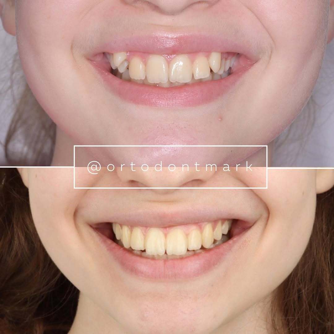
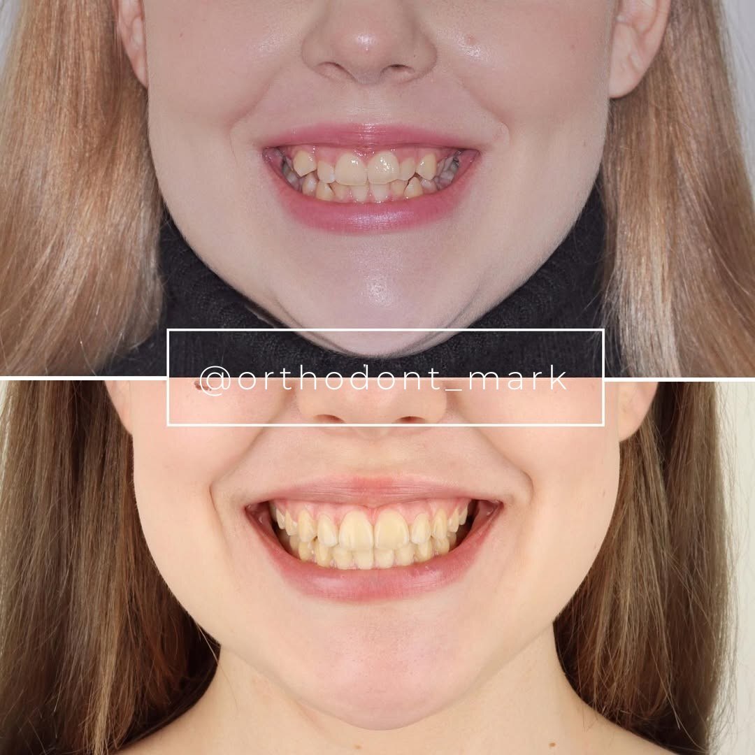
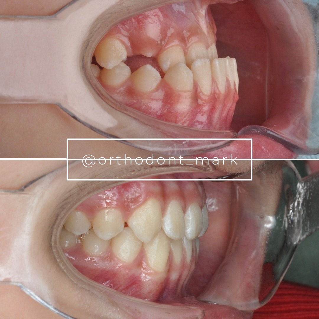
Documented outcomes
Clinical cases from Dr. Mark Radzhabov's practice
Learn the full MARPE protocol from Dr. Mark Radzhabov
Fundamental course covering CBCT patient selection, miniscrew planning, activation protocols, and 60+ clinical cases. Choose the access level that fits your practice.
Essentials of rapid palatal expansion for practicing orthodontists.
- Core RPE concepts and biomechanics
- 6 structured video lessons
- Clinical decision checklists
- Lifetime access to recordings
Deep-dive into MARPE protocol, diagnostics, and clinical execution.
- 6 modules covering MARPE from A to Z
- CBCT case selection walkthroughs
- Miniscrew placement and activation
- 60+ annotated clinical cases
- Direct Q&A with Dr. Mark Radzhabov
5-element medical consultation framework for dentists and orthodontists.
- Trust-building consultation protocol
- 5 lesson modules
- Templates for treatment plan delivery
- Works with any clinical specialty
Clinical FAQ
What are the two primary mechanisms behind expansion relapse after rapid palatal expansion?
Dental rebound — elastic recoil of periodontal ligament, gingival fibers, and buccal alveolar bone — and sutural reclosure driven by intramembranous ossification filling the midpalatal gap. Both operate simultaneously but on different timescales and require distinct retention strategies.
Does MARPE produce less skeletal relapse during consolidation than conventional RPE?
Yes. Chun et al. (2022) found that MARPE demonstrates less decrease in skeletal, dentoalveolar, and periodontal variables during consolidation compared to RPE, attributable to the bone-borne force pathway minimizing elastic strain in dental and periodontal structures.
How do suture separation rates compare between RPE and MARPE in a prospective trial?
Chun et al. (2022) reported midpalatal suture separation in 90% of RPE patients and 95% of MARPE patients, confirming that both appliances reliably open the suture, with MARPE showing a marginally higher separation rate.
What alveolar bone changes does MARPE produce that are relevant to dental rebound risk?
Lim et al. (2017) documented that after MARPE, alveolar bone thickness decreases on the buccal aspect but increases on the palatal aspect. This asymmetry creates an elastic gradient that contributes to dental rebound potential when the appliance is removed.
Does midpalatal surgical splitting improve expansion efficacy and potentially alter relapse trajectory?
Sant'Ana et al. (2016) confirmed that SARME with midpalatal split achieved statistically greater efficacy than SARME without it. More complete initial separation widens the ossification zone, potentially shifting the sutural reclosure timeline and extending the critical retention window.
How does alveolar crest position at the first premolar change after MARPE and why does it matter for stability?
Lim et al. (2017) documented apical movement of the alveolar crest level at the first premolar following MARPE. This shift alters the gingival fiber attachment geometry, which has implications for the elastic rebound component of post-expansion dental relapse.
What role does myofunctional imbalance play in post-expansion transverse relapse?
Girard (2015) identifies perioral and masticatory muscle imbalance as an active compressive force opposing maintained arch width. Contracted lateral muscles must be progressively lengthened alongside skeletal retention to prevent muscle-driven relapse independent of suture biology.
Is myofunctional rehabilitation evidence-based for adult palatal expansion cases?
Yes. Girard (2015) explicitly states that myofunctional rehabilitation is essential in dentofacial orthopedic treatment for both children and adults, framing muscle rebalancing as a structural protocol component, not an optional adjunct.
How should CBCT imaging be used to distinguish dental rebound from sutural reclosure during consolidation?
Sequential CBCT measurements of both basal skeletal width and dentoalveolar width allow independent tracking of osseous infilling at the suture versus buccal root position changes. A decrease in skeletal width with stable arch width suggests active sutural reclosure rather than dental rebound.
What clinical endpoint confirms true skeletal separation rather than dental tipping during MARPE activation?
A radiolucent midpalatal sutural gap on periapical or CBCT imaging, combined with a midline diastema, confirms genuine sutural separation. Arch-width gain measured clinically alone cannot distinguish skeletal from dentoalveolar expansion and should not serve as the sole activation endpoint.
Differentiating dental rebound from sutural reclosure is not academic — it determines your retention duration, appliance choice, and the timing of fixed-appliance mechanics after expansion. The evidence reviewed here, drawn from peer-reviewed imaging studies and prospective trials, supports bone-borne approaches as consistently more favorable during consolidation. Dr. Mark Radzhabov recommends integrating myofunctional rehabilitation alongside retention to address the muscular drivers that literature confirms as essential to lasting skeletal outcomes. To apply these principles in your own case selection, explore structured MARPE training at ortodontmark.com. Key sources: Chun et al. (2022), BMC Oral Health, doi:10.1186/s12903–022-02138-w. Lim et al. (2017), Korean J Orthod, doi:10.4041/kjod.2017.47.5.313.