
Panoramic Radiograph Expansion Screening:
What 2D Reveals
Before You Order the CBCT
A clinical framework for reading the panoramic film as a maturity and bone-quality filter before committing to advanced imaging in expansion case planning.
TL;DR Panoramic radiograph expansion screening gives orthodontists a first-look framework for identifying expansion candidates before committing to CBCT. Root staging, alveolar bone contour, and dental maturity markers on the 2D film narrow your differential and direct the imaging sequence. Suture maturation stages established by Angelieri et al. (2013) across 140 subjects remain the definitive classification reference.
A patient presents with posterior crossbite and crowding, and you reach for the panoramic film already on file before ordering advanced imaging. Panoramic radiograph expansion screening is not a replacement for cone-beam CT, but it is the diagnostic step that prevents unnecessary radiation and sharpens the clinical question before you invest in high-resolution data. At ortodontmark.com, Dr. Mark Radzhabov has structured his expansion planning workflow so that the 2D panoramic assessment dictates what the CBCT must answer — not the other way around. This article walks through the specific radiographic landmarks, maturity indicators, and decision thresholds that make the panoramic film a genuinely productive first-pass tool for miniscrew-assisted and conventional palatal expansion planning.
Which Anatomical Markers on the Panoramic Film Actually Predict
Expansion Readiness
Versus Suture Resistance?
Panoramic radiograph expansion screening is the systematic evaluation of the 2D orthopantomogram for root staging, alveolar morphology, and dental maturity signs that collectively inform whether a patient is a viable candidate for skeletal or dentoalveolar palatal expansion prior to CBCT acquisition. The panoramic film does not resolve the midpalatal suture directly, but it communicates indirectly through proxies — dental root completion stages, alveolar bone height, posterior root angulation, and overall arch width discrepancy — that together form a coherent screening picture. Root apex closure of the maxillary second premolars and first molars provides the most accessible maturity signal. When those roots read as fully formed with closed apices, the practitioner can infer that skeletal development is advanced enough to warrant suture-specific CBCT classification. Conversely, open apices at the first molar suggest a younger skeletal age regardless of chronological age, shifting the protocol toward conventional expansion without the urgency of suture staging. Alveolar bone contour along the buccal shelves is a second panoramic indicator. Thin or tapered alveolar support around the posterior dentition signals limited bone volume for miniscrew anchorage and warrants careful CBCT measurement before finalising the insertion site. The panoramic film cannot quantify bone density in Hounsfield units, but it can flag cases where the buccal plate appears radiographically thin enough to change the miniscrew-assisted expansion protocol entirely.
How Suture Maturation Stages Should Shape
Your Imaging Sequence
After the Panoramic Review
The most widely adopted classification of midpalatal suture maturation divides the developmental arc into five CBCT-defined stages, from a straight high-density line at stage A through to complete osseous fusion at stage E, as Angelieri et al. (2013) established in a sample of 140 subjects ranging from 5.6 to 58.4 years. That span underscores the point: the suture does not age on a fixed calendar, and the panoramic film's maturity proxies must be used to estimate which portion of that lifespan a given patient occupies. Angelieri et al. (2013) found that early stages A and B appear characteristically up to age 13, while the intermediate stage C spans primarily ages 11 to 17. That overlap is clinically significant: a patient who presents at age 15 may carry a stage B suture that responds straightforwardly to conventional expansion, or a stage D suture demanding a bone-borne protocol. The panoramic film cannot distinguish those two scenarios, but it can identify the patients — dense root structure, mature alveolar architecture, late-forming third molars — whose suture is most likely to be advanced, thereby directing the CBCT request efficiently. Sex-based asymmetry in suture fusion compounds the challenge. Angelieri et al. (2013) documented that complete fusion at stages D and E occurred after age 11 only in female patients, whereas 23% of boys aged 14 to 17 showed only partial fusion in the palatine bone. On the panoramic film, this means a 16-year-old male with radiographically immature dentition still has a meaningful probability of an open or partially fused suture — a nuance the 2D screen can flag qualitatively before CBCT confirms it.
When Does the Panoramic Screen Justify
Bone-Borne
Over Tooth-Borne Appliance Selection?
The decision between miniscrew-assisted expansion and conventional RPE is partly a suture question and partly a dental support question — and the panoramic film speaks directly to the latter. Root angulation of the maxillary posterior teeth, the quality of the alveolar bone housing those roots, and the crown-to-root ratio visible on the 2D film collectively determine how much of the expansion force the dentition can safely transmit before buccal tipping overwhelms skeletal response. Chun et al. (2022) reported that midpalatal suture separation was achieved in 90% of RPE patients and 95% of MARPE patients in a prospective randomised trial with identical expansion amounts, with mean patient ages of 14.0 ± 4.5 and 14.1 ± 4.2 years respectively. That differential — five percentage points in favour of bone-borne mechanics — reflects the reduced dental tipping load when miniscrews bypass the posterior dentition entirely. On the panoramic screen, the indicator that predicts whether that five-point gain matters is the angulation of the maxillary first molar roots: excessive mesial root tip already present before expansion indicates the tooth-borne path will amplify the deformation rather than correct it. For miniscrew-assisted expansion treatment planning, the panoramic film also screens for root proximity in the anterior palate region where parasagittal miniscrews are sited. Divergent central incisor roots, visible clearly on the 2D film, provide adequate inter-radicular clearance. Convergent or unusually long lateral incisor roots narrow the safe insertion corridor before a CBCT slice confirms exact dimensions.
What Changing Suture Opening Geometry Means
for Screening
in Older Adolescents
As patients age, the pattern of midpalatal suture separation shifts from a parallel opening to a triangular one in both the frontal and horizontal planes, with the cranial and dorsal dimensions opening progressively less, as Kinzinger et al. (2022) demonstrated through CBCT and dental cast analysis stratifying 60 children and adolescents into three equal groups: under 10 years (n=20), 10–12 years (n=20), and 12 years and older (n=20). The panoramic film cannot resolve that three-dimensional geometry, but it can identify patients old enough and anatomically configured enough that this change in opening pattern becomes clinically relevant. For rapid palatal expander protocol decisions, this geometric shift matters because the posterior suture responds less to mechanical load as skeletal maturity advances. A clinician reading the panoramic film in a 13-year-old patient with fully closed molar apices and dense interproximal bone should already be hypothesising that the cranial and dorsal suture zones will resist separation — a hypothesis the CBCT then quantifies rather than generates from scratch. Interpreting the panoramic film as a directional screen, rather than a definitive answer, protects against two failure modes: ordering CBCT on patients whose open apices and immature alveolar architecture already indicate simple conventional expansion, and under-imaging patients whose mature 2D markers predict a complex bone-borne protocol. The two-stage sequence — panoramic screen followed by targeted CBCT — keeps radiation proportional to clinical complexity.
Which Indications Should Not Rest on the
2D Film
Alone for Expansion Decisions?
Palatal expansion is pursued for a range of indications — transverse skeletal deficiency, posterior crossbite, arch length discrepancy, and airway-related concerns among them. However, Kazmierski (2024) has noted in a narrative review that obstructive sleep apnea alone is not sufficient clinical justification for recommending palatal expansion treatment. This is directly relevant to panoramic radiograph expansion screening because airway-driven referrals often arrive with a strong patient or parent expectation of expansion, yet the 2D film cannot visualise airway dimensions and should not be used to anchor a treatment recommendation built on that indication alone. In those cases, the panoramic screen performs its most useful function negatively: it confirms whether the dentoskeletal anatomy supports expansion on its own merit — adequate bone, appropriate root staging, acceptable angulation — independent of the airway argument. If the 2D markers do not support expansion on transverse grounds, neither does a sleep study. The panoramic film thus acts as an anatomical gatekeeper that separates indication-driven cases from anatomy-supported ones before any further imaging is ordered. The panoramic film is similarly limited in quantifying interradicular bone density, measuring palatal vault depth, or resolving the precise suture stage — all variables that determine whether a miniscrew-assisted expansion protocol is biomechanically sound. These are CBCT tasks. The 2D screen's contribution ends where the three-dimensional anatomy begins, and recognising that boundary is what makes the panoramic-first workflow clinically defensible rather than diagnostically complacent.
Free Clinical Guide
Which Expansion Appliance at Which Age?
Get Dr. Mark Radzhabov's guide to skeletal-expansion appliances — indications, pros & cons, and how to choose by patient age. Free to your inbox.
Prefer video? Watch the free demo lesson on MARPE appliance types, features and trade-offs.
Documented outcomes
Clinical cases from Dr. Mark Radzhabov's practice


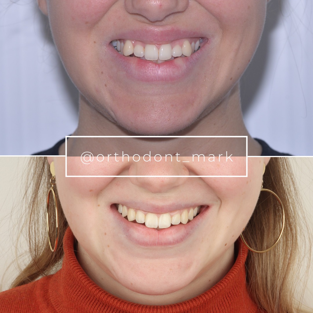
Learn the full MARPE protocol from Dr. Mark Radzhabov
Fundamental course covering CBCT patient selection, miniscrew planning, activation protocols, and 60+ clinical cases. Choose the access level that fits your practice.
Essentials of rapid palatal expansion for practicing orthodontists.
- Core RPE concepts and biomechanics
- 6 structured video lessons
- Clinical decision checklists
- Lifetime access to recordings
Deep-dive into MARPE protocol, diagnostics, and clinical execution.
- 6 modules covering MARPE from A to Z
- CBCT case selection walkthroughs
- Miniscrew placement and activation
- 60+ annotated clinical cases
- Direct Q&A with Dr. Mark Radzhabov
5-element medical consultation framework for dentists and orthodontists.
- Trust-building consultation protocol
- 5 lesson modules
- Templates for treatment plan delivery
- Works with any clinical specialty
Clinical FAQ
What specific panoramic radiograph findings indicate a patient is a strong expansion candidate before CBCT?
Fully closed maxillary molar and premolar apices, adequate alveolar bone height along buccal shelves, and divergent anterior root angulation are the primary 2D signals. Together they suggest skeletal maturity and miniscrew anchorage volume sufficient to warrant detailed CBCT suture staging.
How do dental maturity radiographic signs on the panoramic film correlate with midpalatal suture stage?
They correlate indirectly. Angelieri et al. (2013) established that early suture stages A and B appear characteristically up to age 13, so patients with radiographically immature dentition are more likely to carry an open suture — but CBCT confirmation is required before finalising the expansion protocol.
Can the panoramic radiograph assessment replace CBCT for skeletal expansion planning in adolescents?
No. The panoramic film cannot resolve suture stage, palatal vault depth, or interradicular bone density. Its role is to screen candidates and sharpen the CBCT question, not to substitute for three-dimensional imaging in miniscrew-assisted or surgically assisted expansion planning.
How does biological sex affect panoramic expansion readiness screening in patients aged 14 to 17?
Angelieri et al. (2013) found complete suture fusion occurred after age 11 only in girls, while 23% of boys aged 14 to 17 showed only partial palatine fusion. A male patient with radiographically mature dentition may still carry a partially open suture, making CBCT staging essential before ruling out non-surgical expansion.
What does posterior root angulation on the panoramic film reveal about bone-borne versus tooth-borne appliance selection?
Pre-existing mesial root tip at the maxillary first molar predicts amplified buccal tipping under tooth-borne force. This 2D finding elevates the case toward bone-borne mechanics before CBCT quantifies precise angulation, reducing the risk of dentoalveolar side effects during expansion.
Is obstructive sleep apnea alone a valid indication for ordering palatal expansion based on panoramic screening?
No. Kazmierski (2024) noted that obstructive sleep apnea alone is insufficient clinical justification for palatal expansion. The panoramic film must confirm independent transverse skeletal indication — posterior crossbite, arch length deficit, or buccal bone deficiency — before any expansion recommendation is made.
How does the suture opening pattern change in older adolescents and how does that affect pre-CBCT screening?
Kinzinger et al. (2022) showed the suture opening shifts from parallel to triangular in older patients, with cranial and dorsal zones resisting progressively more. On the panoramic film, patients with mature alveolar architecture should be presumed to have this geometric shift, directing CBCT toward measuring posterior suture resistance specifically.
Which root staging landmarks on the panoramic film are most useful for pre-CBCT expansion screening?
Second premolar and first molar apex closure are the most accessible maturity markers. Open apices indicate younger skeletal age and favour conventional expansion. Closed apices in the posterior quadrant prompt suture-specific CBCT staging to determine whether bone-borne mechanics are necessary.
How does the panoramic radiograph inform miniscrew insertion site planning before CBCT is performed?
Divergent central incisor roots on the panoramic film indicate adequate inter-radicular clearance for parasagittal miniscrew siting. Convergent or unusually long lateral incisor roots visible on the 2D film raise a caution that CBCT must resolve with precise axial slice measurement before finalising the insertion corridor.
What is the clinical rationale for a two-stage radiographic sequence — panoramic first, CBCT second — in expansion case selection?
The panoramic screen filters patients by dental maturity, alveolar morphology, and root angulation, ensuring CBCT is reserved for cases with genuine diagnostic ambiguity. This keeps radiation proportional to complexity and ensures the CBCT answers a pre-specified question rather than serving as a routine first step.
The panoramic film is not a shortcut — it is the disciplined first step that converts a clinical suspicion into a well-formed imaging question for CBCT. Suture maturation staging, root angulation assessment, and alveolar bone contour each narrow the differential before a single miniscrew is planned. Clinicians who build this two-stage radiographic sequence into their workflow consistently ask better CBCT questions and select expansion candidates with greater confidence. Dr. Mark Radzhabov invites orthodontists to submit a case for structured review or explore the miniscrew-assisted expansion training curriculum at ortodontmark.com to refine their radiographic decision protocol. Key sources: Angelieri et al. (2013), Am J Orthod Dentofacial Orthop, doi:10.1016/j.ajodo.2013.04.022. Chun et al. (2022), BMC Oral Health, doi:10.1186/s12903–022-02138-w. Kinzinger et al. (2022), J Orofac Orthop, doi:10.1007/s00056–022-00429-z.

