
RPE Training for Orthodontists
Before
the First Expander
Master screw mechanics, force thresholds, and suture biology — the pre-clinical skills that define safe, predictable palatal expansion.
TL;DR RPE training for orthodontists must begin with screw mechanics, force calibration, and suture histology before the first appliance is placed. Expansion screws generate forces that vary considerably by device, and suture biology dictates timing. Understanding these fundamentals separates predictable outcomes from avoidable complications in daily palatal expansion practice.
A clinician placing a palatal expander without a firm grasp of screw mechanics, force generation, and suture biology is working without a diagnostic map. RPE training for orthodontists and dentists at ortodontmark.com, as structured by Dr. Mark Radzhabov, begins with exactly those fundamentals — not appliance selection, not activation schedules, but the tissue and mechanical principles that determine whether expansion proceeds safely. This article distills the pre-clinical knowledge every practitioner needs before treating the first expansion case, drawing on peer-reviewed imaging studies and in vitro mechanical testing.
Why Suture Histology Comes First in
RPE Training
for Orthodontists
Rapid palatal expansion (RPE) is a fixed orthodontic procedure that applies calibrated transverse force to the midpalatal suture via a jack-screw appliance, separating the two palatal halves to gain arch width. Every decision downstream — activation frequency, retention duration, case selection — is grounded in understanding what is happening inside that suture at the cellular level. Histological evidence provides the most direct window into that process. Caprioglio et al. (2017) obtained histological samples of the midpalatal suture at 7 days and 30 days after RME alongside a control sample. The 7-day specimen reflects the acute tensile response: cellular proliferation, vascular ingrowth, and the earliest matrix deposition along the sutural faces. The 30-day specimen, by contrast, showed newly-formed bone trabeculae oriented perpendicular to the suture axis — a structural signal that early woven bone is being organised along the lines of mechanical stress. For the clinician entering structured RPE training for clinicians, this timeline is not abstract. It directly informs the retention protocol: the suture at 30 days still contains immature, perpendicular trabeculae rather than well-bridged lamellar bone. Removing the appliance or transitioning to a passive retainer before adequate mineralisation creates the conditions for relapse. Suture histology, therefore, is the first chapter of any rigorous RPE curriculum — and understanding it allows practitioners to justify retention durations to patients with mechanistic precision, not habit.
What Does One Turn of an RME Screw
Actually Deliver
to the Suture?
Rapid maxillary expansion training that skips screw mechanics leaves the clinician guessing at cumulative force and displacement. Every activation is a mechanical dose, and the numbers are well-characterised in the literature. Camporesi et al. (2013) established in vitro that one full turn of an RME screw expands 0.8 mm, composed of 4 quarter-turns at 0.2 mm each. This means that a twice-daily quarter-turn protocol delivers 0.4 mm per day — a figure the clinician must track cumulatively against the patient's sutural capacity. Force generation varies substantially by appliance. Camporesi et al. (2013) measured RME appliances producing forces between 16.6 and 34.8 pounds, equivalent to 7.54 to 15.8 kg, depending on device design. More specifically, the Hyrax and A2620 exceeded 20 kg, while the Palatal Split Screw generated approximately 16 kg. These are not interchangeable force profiles. Selecting a Hyrax when a lower-force device is clinically indicated — for example, in a younger patient with an open, compliant suture — is a mechanical error, not a brand preference. For rapid expansion case support, the practical implication is that appliance selection must precede activation planning, and both must be anchored in the patient's sutural resistance as estimated from imaging. Clinicians who treat all RME screws as equivalent will encounter inconsistent outcomes that appear biological but are actually mechanical in origin.
How Does Patient Age Shape the
Diagnostic Protocol
for Palatal Expansion?
Understanding how skeletal age distributes across a clinical population is foundational to RPE patient assessment. Stefan et al. (2022) conducted a dental cast analysis of 60 children examining three age groups — those younger than 10 years, those aged 10 to 12 years, and those 12 years and older — providing a structured lens for understanding how transverse arch dimensions and expansion response differ across developmental windows. The CBCT component of Stefan et al. (2022) included 9 patients aged between 7.3 and 13.8 years, offering three-dimensional imaging correlates of the cast measurements. This dual-modality approach — dental casts combined with cone-beam CT — reflects the diagnostic standard that orthodontic expansion training should build toward: not a single landmark, but convergent evidence from multiple data sources before treatment planning. For clinicians building a palatal expansion diagnostic protocol, the takeaway is methodological. Age brackets are useful for population-level thinking, but the individual patient's sutural maturity — best assessed with CBCT — is what governs appliance choice and expected force response. Orthodontic expansion mentoring at the advanced level therefore focuses on teaching practitioners to read sutural morphology on axial CBCT slices rather than defaulting to age as a proxy. Internal resources on rapid palatal expander case selection at ortodontmark.com extend this framework into a step-by-step clinical workflow.
Free Clinical Guide
Which Expansion Appliance at Which Age?
Get Dr. Mark Radzhabov's guide to skeletal-expansion appliances — indications, pros & cons, and how to choose by patient age. Free to your inbox.
Prefer video? Watch the free demo lesson on MARPE appliance types, features and trade-offs.
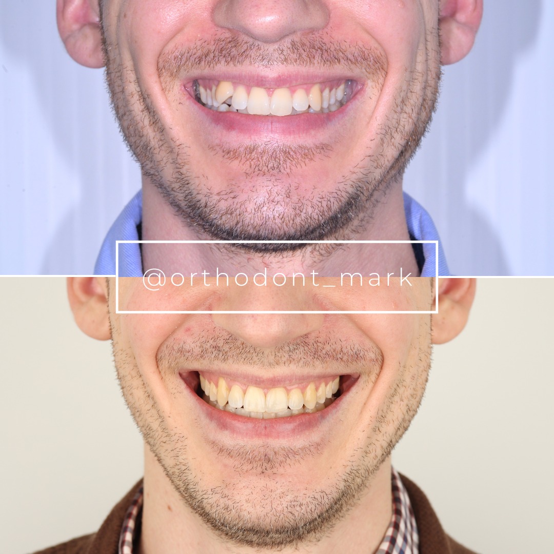
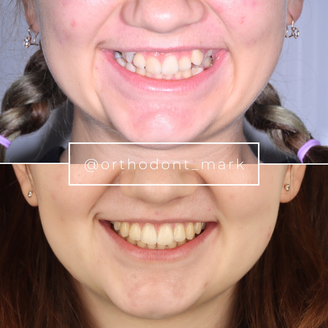
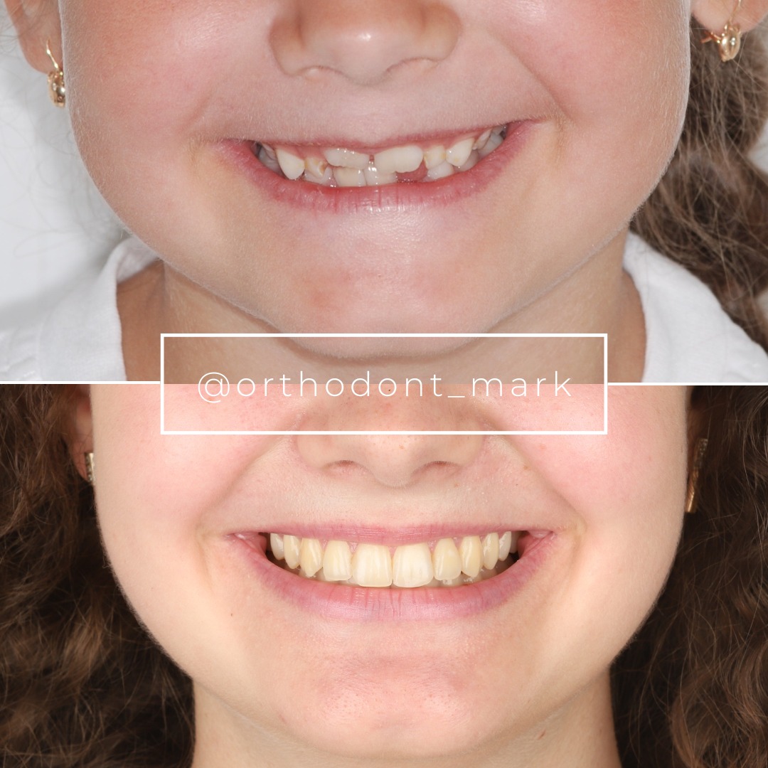
Documented outcomes
Clinical cases from Dr. Mark Radzhabov's practice
Learn the full MARPE protocol from Dr. Mark Radzhabov
Fundamental course covering CBCT patient selection, miniscrew planning, activation protocols, and 60+ clinical cases. Choose the access level that fits your practice.
Essentials of rapid palatal expansion for practicing orthodontists.
- Core RPE concepts and biomechanics
- 6 structured video lessons
- Clinical decision checklists
- Lifetime access to recordings
Deep-dive into MARPE protocol, diagnostics, and clinical execution.
- 6 modules covering MARPE from A to Z
- CBCT case selection walkthroughs
- Miniscrew placement and activation
- 60+ annotated clinical cases
- Direct Q&A with Dr. Mark Radzhabov
5-element medical consultation framework for dentists and orthodontists.
- Trust-building consultation protocol
- 5 lesson modules
- Templates for treatment plan delivery
- Works with any clinical specialty
Clinical FAQ
What mechanical skills should orthodontists master before placing their first RPE appliance?
Before placing an expander, a clinician should be able to calculate cumulative activation in millimetres, select an appliance based on its force output, and interpret midpalatal suture morphology on CBCT. Screw mechanics and suture biology are the two non-negotiable pre-clinical competencies.
How much force does a Hyrax expander generate compared to a Palatal Split Screw?
Camporesi et al. (2013) found the Hyrax and A2620 both exceed 20 kg, while the Palatal Split Screw generates approximately 16 kg. Selecting the wrong device for a given sutural resistance profile creates unpredictable force delivery and inconsistent expansion outcomes.
What does one quarter-turn activation of an RME screw actually deliver to the suture?
According to Camporesi et al. (2013), one quarter-turn delivers 0.2 mm of linear expansion, and a full turn delivers 0.8 mm across 4 activations. Twice-daily quarter-turn protocols therefore deliver 0.4 mm per day cumulatively.
Why is midpalatal suture histology relevant to RPE training for orthodontists?
Caprioglio et al. (2017) showed that at 30 days post-RME, the suture contains newly-formed trabeculae oriented perpendicular to the suture axis — immature bone, not bridged lamellar structure. This directly determines minimum retention duration before appliance removal is safe.
At what post-expansion timepoint does organised bone formation begin in the midpalatal suture?
Caprioglio et al. (2017) obtained histological samples at 7 and 30 days after RME. At 30 days, perpendicular bone trabeculae were already present, indicating that active mineralisation along the expansion axis begins within the first month of retention.
How should CBCT be integrated into a rapid maxillary expansion diagnostic protocol?
CBCT allows direct visualisation of sutural morphology across anterior, middle, and posterior thirds — information a dental cast cannot provide. Stefan et al. (2022) used both modalities together, and that dual-record approach should anchor every pre-expansion workup.
What age groups are clinically relevant when planning RPE case management?
Stefan et al. (2022) structured their cast analysis around three groups: under 10 years, 10 to 12 years, and 12 years and older. These brackets inform population-level expectations, but individual CBCT-based sutural assessment overrides chronological age in clinical decision-making.
How does appliance force range affect RPE protocol and procedures in clinical practice?
Camporesi et al. (2013) measured a force range of 16.6 to 34.8 pounds (7.54 to 15.8 kg) across RME devices. The upper end of that range, generated by the Hyrax, may be excessive for a patient with a compliant, open suture — making appliance selection a force-calibration decision.
What is the learning progression in structured RPE training programs for general dentists and orthodontists?
Effective RPE training moves from suture biology and screw mechanics through imaging interpretation to live case application. Practitioners who skip the mechanical and histological foundations tend to manage complications reactively rather than preventing them through protocol design.
How do expansion therapy outcomes differ across the three developmental age windows studied in CBCT research?
Stefan et al. (2022) examined 60 children across three age groups in dental cast analysis and 9 patients aged 7.3 to 13.8 years with CBCT. Younger patients with open sutures respond with greater skeletal contribution. Older patients show increasing resistance and higher dental tipping risk.
A rigorous RPE training foundation — screw mechanics, suture histology, force calibration, and imaging-guided case selection — is what separates clinicians who expand predictably from those who encounter avoidable setbacks. Before placing a first expander, every practitioner should be able to interpret midpalatal suture histology, match the appliance force output to the patient's skeletal stage, and calculate cumulative activation in millimetres. Dr. Mark Radzhabov's structured RPE training at ortodontmark.com is built around exactly these pre-clinical competencies. Key sources: Stefan et al. (2022), doi:10.1007/s00056–022-00429-z. Caprioglio et al. (2017), doi:10.3390/ijms18030615. Camporesi et al. (2013), BioMedical Engineering OnLine.